Preprint
Article

This version is not peer-reviewed.

Analysis of the Bacterial Microbiota in Wild Populations of Prickly Pear Cochineal, Dactylopius opuntiae in Morocco

A peer-reviewed article of this preprint also exists.

Submitted:

10 October 2025

Posted:

11 October 2025

You are already at the latest version

Abstract

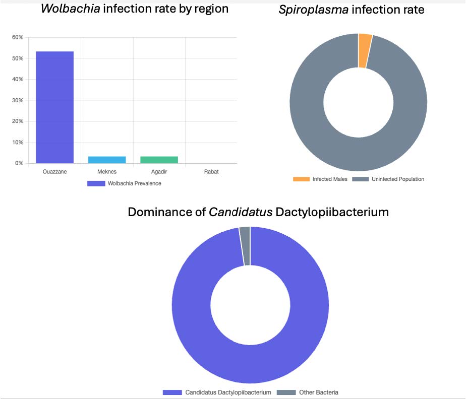
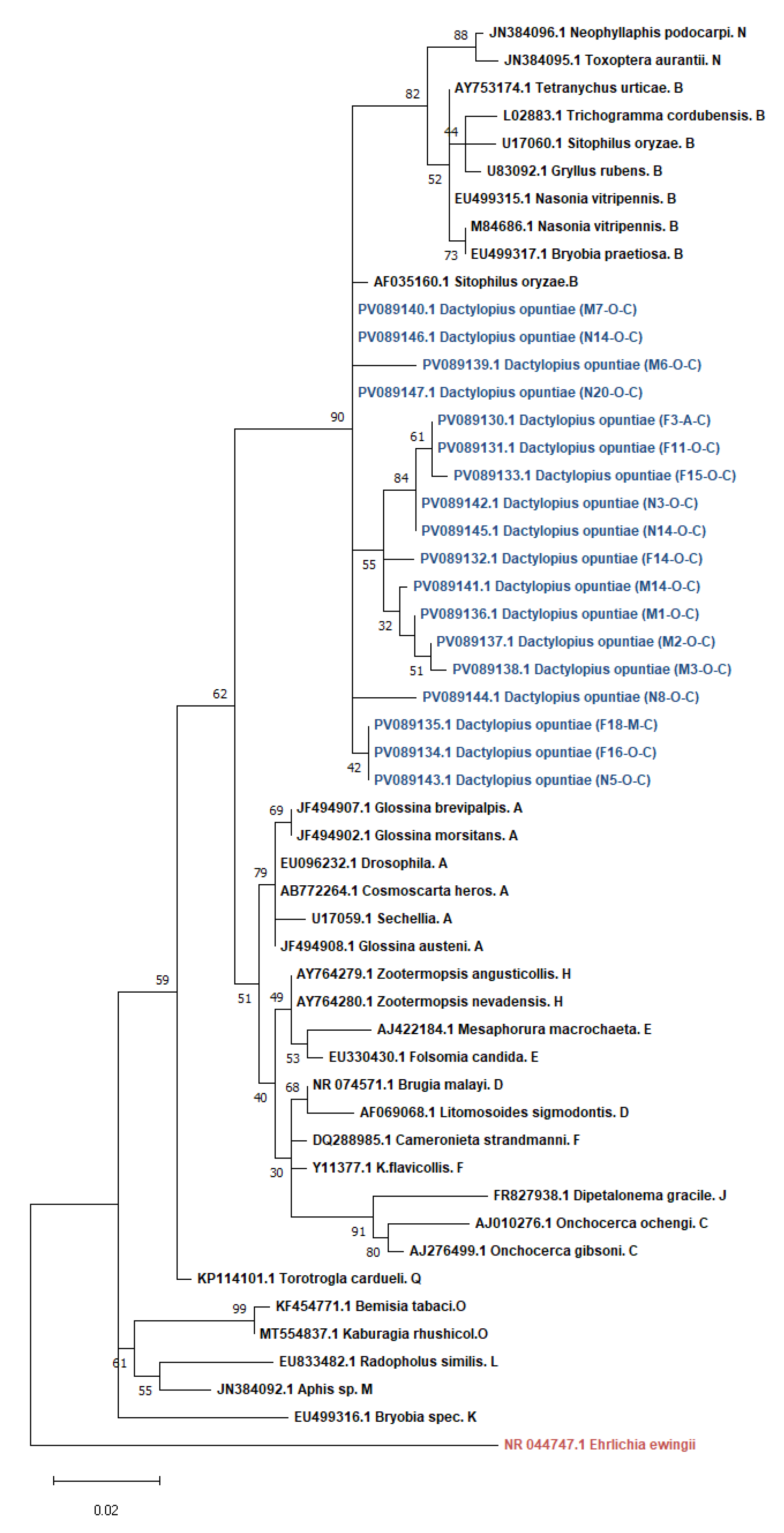
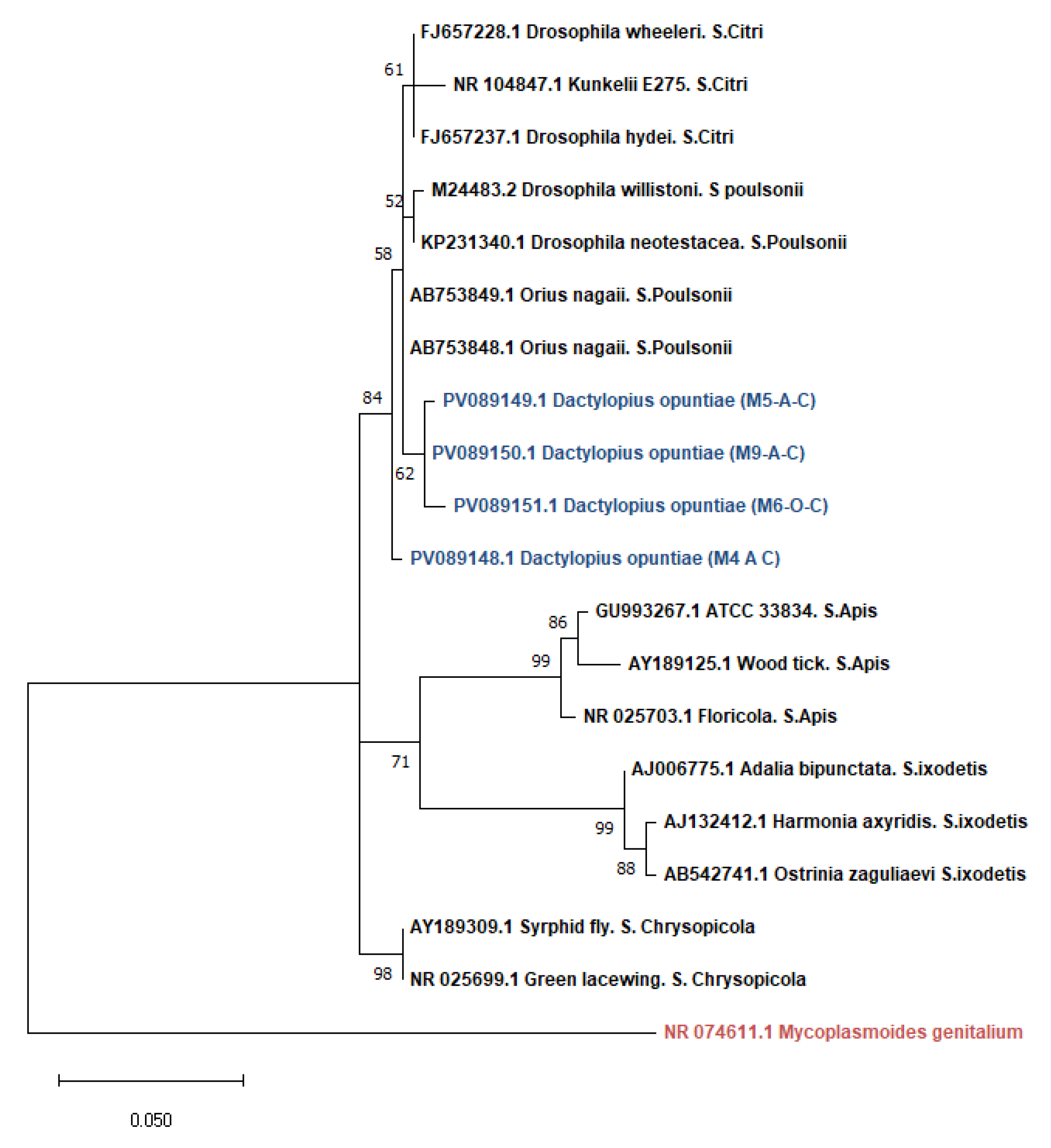
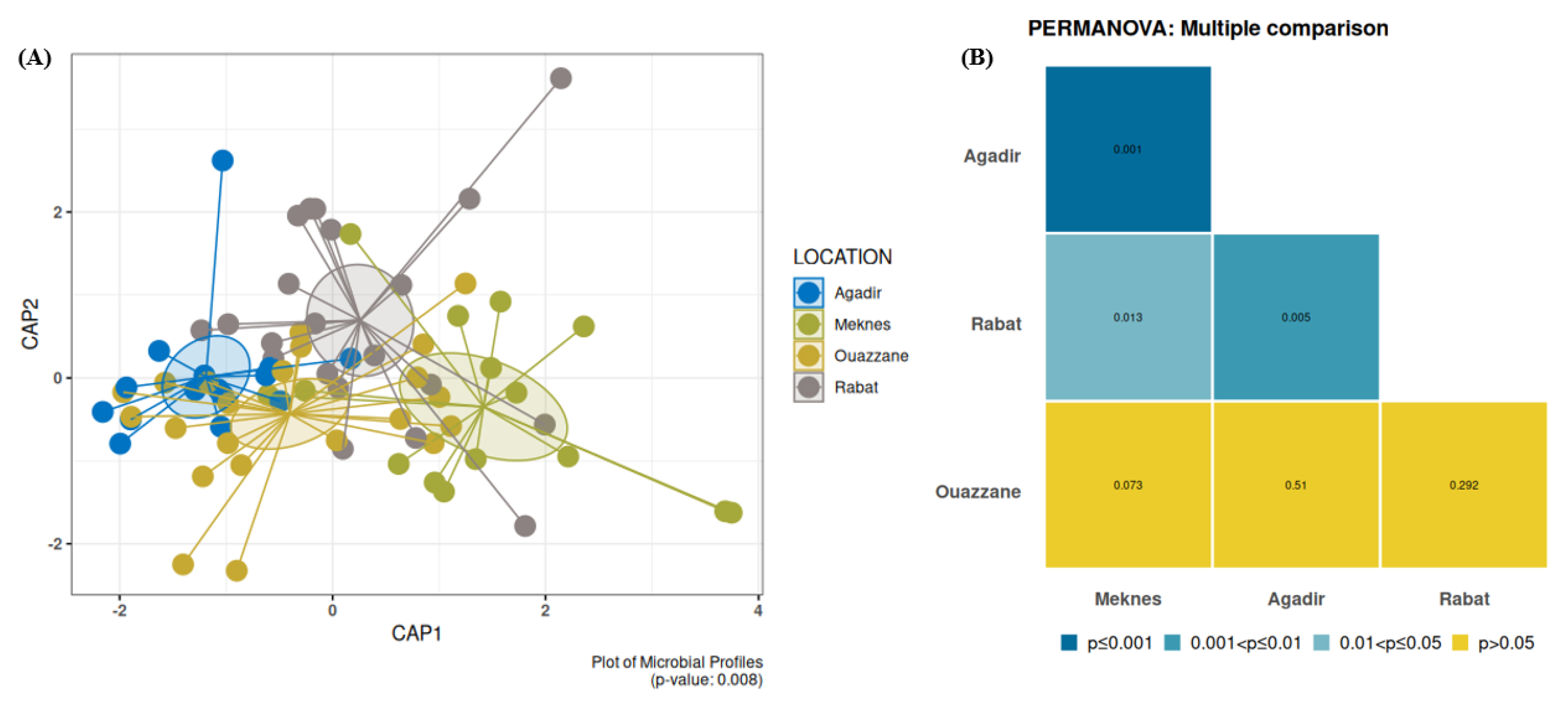
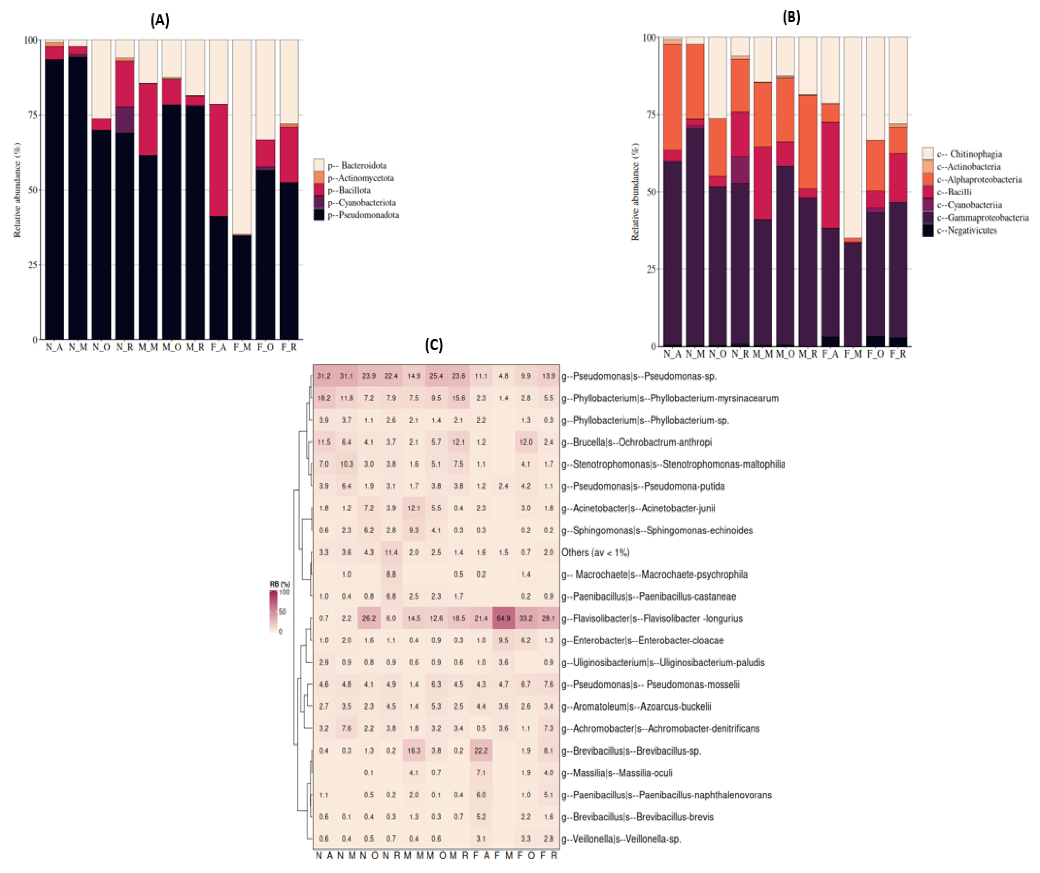
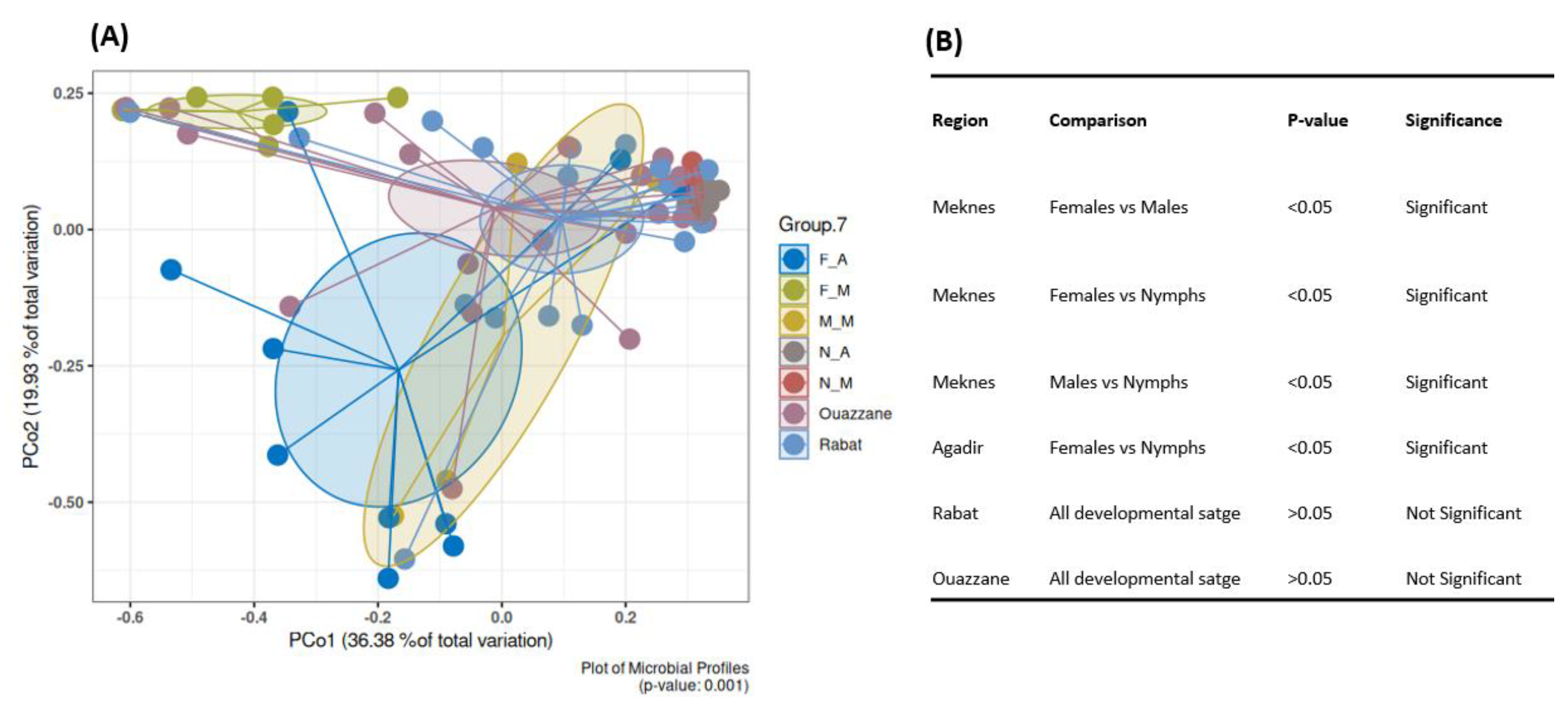
Dactylopius opuntiae (Cockerell) (Hemiptera: Dactylopiidae), the wild cochineal scale, is a major pest of prickly pear crops worldwide. This study characterized the bacterial community structure of D. opuntiae from four Moroccan regions using targeted PCR and full-length 16S rRNA MinION sequencing. We report the first detection of Wolbachia (16.6% prevalence) in D. opuntiae, with infection rates varying geographically from 0% (Rabat) to 53.3% (Ouazzane). Spiroplasma was detected at a lower prevalence (3.3%) and exclusively in males. Phylogenetic analysis placed the Wolbachia strains in supergroup B and Spiroplasma in the poulsonii-citri complex. MinION sequencing revealed Candidatus Dactylopiibacterium as the dominant taxon (97.7%), consistent with its role as an obligate symbiont. After removing this dominant species, we uncovered a diverse bacterial community, including Flavisolibacter, Pseudomonas, Phyllobacterium, Acinetobacter, and Brevibacillus. Beta diversity analysis showed significant geographic variation (PERMANOVA p<0.008), with distinct communities across regions. Females harbored a more specialized microbiome dominated by Flavisolibacter (except in Agadir), whereas males and nymphs showed Pseudomonas dominance. Core microbiome analysis revealed no universal genera across all groups, with females displaying a more restricted core than males and nymphs did. The detection of reproductive symbionts, combined with geographic and sex-specific microbiome patterns, provides insights into the development of microbiome-based pest management strategies. The complementary use of targeted and untargeted sequencing methods is essential for comprehensive microbiome characterization in this economically important pest.

Keywords: 
;  ;  ;  ;  ;  ;  ;  

1. Introduction

Dactylopius opuntiae (Cockerell) (Hemiptera: Dactylopiidae), also known as the wild cochineal scale, is a soft-bodied, flat, and oval-shaped parasitic insect that has gained significant attention in the scientific community. It is recognized by immobile females, is apterous, and can reach a length of up to 5 mm [1,2]. Dactylopius opuntiae is among the most destructive species in the genus Dactylopius [3,4], especially in recently cultivated areas in the Mediterranean region, including France, Spain, Morocco, and Lebanon [5,6,7,8]. Identical attacks by D. opuntiae have been documented in Brazil [9] on the forage cactus species Opuntiae ficus indica, resulting in damage to 100,000 hectares, which was estimated to be worth $25 million [9]. This cochineal infestation reduces productivity and renders fruits and cladodes unsellable [10,11]. Scale insects cause a decline in vigor, yellowing of cladodes, fruit drop, and cactus mortality when they become well-established and cover more than 75% of the cladode surface [12,13,14]. With an average of 150–160 eggs laid by females, which rapidly develop into nymphs, infestations are rapid and erratic [10]. In the field, female life cycles range from 40 to 180 days, depending on the season and weather, whereas males typically complete their life cycles in 35 to 52 days [12,15]. During its life cycle, cochineal releases a white waxy covering that envelops its body, reducing the efficiency of phytosanitary treatments [10]. The proliferation of D. opuntiae in Mediterranean countries has sparked discussions about the best ways to manage this pest. Currently, chemical and biological approaches are the mainstays of D. opuntiae control. Mechanical methods can be used when only a few plants are infested [16,17]. Since its discovery at the end of 2014, D. opuntiae has expanded rapidly and caused significant damage in Morocco, prompting local authorities to launch an emergency intervention by removing and burning approximately 400 ha of crops in the Doukkala region [18]. In Morocco, various integrated pest management approaches for cochineal control have been investigated, including host plant resistance, biological control, and biopesticides [19,20,21,22,23,24].
Dactylopius opuntiae causes significant agricultural damage worldwide, with infestations in Morocco resulting in the destruction of 400 hectares of crops in the Doukkala region alone [18]. The pest reduces productivity through vigor decline, cladode yellowing, fruit drop, and plant mortality when its coverage exceeds 75% of the cladode surface [12,13,14]. Current control methods include mechanical removal, chemical treatments, and emerging biological control approaches [19,20,21,22,23,24].
Symbiotic microorganisms offer promising avenues for pest management through the disruption of required symbionts or manipulation of pest-relevant traits [25]. Insects harbor diverse microbial communities that influence their nutrition, reproduction, fitness, immunity, and pest status [26,27,28]. Among reproductive symbionts, Wolbachia infects 40-75% of arthropod species [29,30] and can manipulate host reproduction through cytoplasmic incompatibility, making it valuable for pest control strategies [31,32]. Spiroplasma, found in 5-10% of insect species [30,33,34,35], can cause male-killing or provide protection against stressors [36,37,38,39,40,41,42,43]. Certain Spiroplasma species, such as Spiroplasma poulsonii, cause male killing in flies, such as Drosophila [40,41], Spiroplasma ixodetis in butterflies [42], and Spiroplasma sp. in ladybird beetles, which alters the sex ratio [43]. Other Spiroplasma species, such as Spiroplasma citri [42] and Spiroplasma apis [65], are known to infect plants and arthropods such as bees [45,46,47]. However, some flies infected with Spiroplasma may become resistant to other infections [43,48,49,50]. Nonetheless, further research revealed that Spiroplasma has various symbiotic relationships [41,48,51,52,53,54].
To the best of our knowledge, this is the first study to examine the bacterial microbiome profile of the full 16S rRNA gene of Dactylopius opuntiae located in Morocco using Oxford Nanopore Technology, which is one of the most cutting-edge and rapidly developing sequencing technologies [55]. Moreover, this study is the first to report Wolbachia infection in D. opuntiae, whereas previous findings have only been reported for Dactylopius coccus. Recently, several studies have been published on the bacterial symbionts associated with the Mexican carmine cochineal Dactylopius coccus (Hemiptera: Coccoidea: Dactylopiidae). This species belongs to the same family (Dactilopiidae) and exhibits morphological characteristics similar to those of Dactylopius opuntiae [56]. According to studies conducted by Ramírez-Puebla et al. (2016) and de León et al. (2017), the metagenomic approaches employed in their research identified two distinct strains of Wolbachia (wDacA and wDacB), a Spiroplasma, and a betaproteobacterium that has been designated as Candidatus Dactylopiibacterium carminicum [57,58]. However, Spiroplasma has been previously reported in this species by Vera-Ponce León et al. (2021), who sequenced and analyzed the genome of a Spiroplasma symbiont associated with both D. opuntiae and D. coccus [59]. Furthermore, researchers have reported the detection of other bacterial species, such as Massilia, Herbaspirillum, Acinetobacter, Mesorhizobium, and Sphingomonas, which may represent transient gut microbiota acquired from the host plant [60].
Despite the growing interest in the microbiomes of Dactylopius species, comprehensive assessments of bacterial diversity across geographical regions remain limited. This study aimed to elucidate the relationship between the bacterial community profiles of D. opuntiae across four distinct regions in Morocco through high-throughput sequencing of the entire 16S rRNA gene, using nanopore technology. Additionally, the presence of reproductive endosymbionts, specifically Spiroplasma and Wolbachia, was examined, as they may hold potential for microbiome-based biocontrol strategies.

2. Materials and Methods

2.1. Dactylopius Opuntiae Collection and DNA Isolation

Dactylopius opuntiae-infected prickly pear cacti were collected from Agadir, Rabat, Meknes, and Ouazzane, four of Morocco's principal-producing regions (Figure S1). During the autumn-winter of 2022, Dactylopius opuntiae nymphs and adults (males and females) were collected, stored in absolute ethanol, and maintained at -20°C until use. At some sites, farmers burned Opuntia spp. to control obvious infestations, as in the Rabat and Meknes regions. During the fieldwork, not every infected plant was destroyed. In other cases, gardeners left "healthy" cactus pads because of insufficient funds, poor visibility of the white wax, or incomplete treatment. These surviving plants were sampled after thorough scrutiny and showed early stage infection. Thus, after burning was used as pest control, samples from "burned areas" were gathered from plants that had living D. opuntiae and accompanying insect fauna (including flies). Additionally, samples were obtained from treated regions, such as Agadir, to assess infestation persistence and related microbial communities, even in areas where chemical control was implemented. Samples were also collected from Ouazzane, a farm that had not been treated (Table 1). Samples were obtained from both insecticide-treated and untreated areas of the farm. This methodology sought to incorporate heterogeneity in pest management approaches to evaluate whether the presence or absence of chemical treatments may affect the bacterial composition of each sample.
Prior to DNA extraction, each sample was surface-sterilized using a 70% v/v ethanol solution, rinsed with sterile deionized water to eliminate any remaining ethanol, and allowed to dry on a sterile surface. The whole individual fly’s DNA was isolated using a modified CTAB (cetyl trimethyl ammonium bromide) protocol [61]. A Q5000 micro-volume UV-Vis spectrophotometer (Quawell Technology, San Jose, CA, USA) was used to measure the amount and quality of the DNA preparations as well as the concentration of double-stranded DNA. The DNA samples were stored in Eppendorf tubes at -20◦C until further examination.
Table 1. Number of Dactylopius opuntiae adults and nymphs used for bacterial community analysis per location.

Region
Coordinates Collection
date

Treatment
No. of insects
Latitude Longitude Temperature N F M
Agadir
30.206132 -9.534147 20◦C Oct. 2022 Insecticide 5 10 -
Rabat 34.002736 -6.748109 22◦C Nov. 2022 Burned 10 6 5
Meknes
33.963659 -5.576012 24◦C Sept. 2022 Burned 4 7 4
Ouazzane 34.807449 -5.658892 18◦C Dec. 2022 No 6 9 7

2.2. Screening and Identification of Bacterial Symbionts

To screen for bacterial symbionts, 120 samples were selected (10 males, 10 females, and 10 nymphs from each region). Wolbachia and Spiroplasma were detected by PCR using primers specific to the 16S rDNA gene (Table S1). During DNA extractions, blank and negative controls were included, and the PCRs were performed under the same conditions. However, these samples did not yield any amplicons. Nested PCR amplification was performed in two stages for screening. Using the bacterial universal primers 27F-1492R, the initial amplification was carried out in a 25 µL reaction that included 2 µL of the template DNA solution, 2.5 µL of KAPA Taq buffer 10X, 0.2 µL of dNTPs (25 mM), 0.2 µL of KAPA Taq DNA polymerase (Roche, Basel, Switzerland), 0.6 µL of forward primer (25 M), 0.6 µL of reverse primer (25 M), and 18.9 µL of water. DNA was first denatured for 3 minutes at 95°C, followed by twenty cycles of 95°C for 30 seconds, 53°C for 30 seconds, and 72°C for 2 minutes, and a final 5 minutes extension at 72°C. The second round of PCR amplification was performed using Wolbachia-specific primers (WspecF-WspecR) and Spiroplasma-specific primers (Spou R1-Spou F1) in a 25 µL reaction mixture containing 2.5 µL of KAPA Taq buffer 10X, 0.2 µL of dNTPs (25 mM), 0.1 µL of KAPA Taq DNA polymerase, 0.4 µL of the forward primer (25 M), 0.4 µL of the reverse primer (25 M), 1 µL of the first-step reaction as template, and 20.4 µL of sterile deionized water. The PCR amplification was conducted with incubation at 95°C for 3 minutes, followed by 20 cycles of 95°C for 30 seconds, primer-specific temperature for 30 seconds, and 72°C for 1 minute, with a final 5 minutes extension at 72°C. The sizes of the amplified fragments were assessed by electrophoresis of the PCR products on a 1.5% agarose gel. Table S1 summarizes the product sizes, annealing temperatures, and primer sequences used in this study. Following purification with polyethylene glycol (20% PEG, 2.5 M NaCl) [62], PCR-positive products were resuspended in 15 µL water. Sanger sequencing was performed on the purified products using the BigDye Terminator v3.1 Cycle Sequencing Kit according to the manufacturer's instructions (Applied Biosystems, Waltham, MA, USA). Reaction products were purified using an ethanol/EDTA process following the manufacturer's recommendations (Applied Biosystems, Waltham, MA, USA) and sequenced on an ABI PRISM 3500 Genetic Analyzer.

2.3. Amplification of the 16S rRNA Gene, Library Preparation, and MinION Sequencing

For MinION amplicon sequencing, 73 samples were selected (Table 1). Using 27F-1429R primers, the full 16S rRNA gene was amplified (Table S1). PCR amplification was conducted in a 25 µL reaction comprising 2.5 µL of KAPA Taq buffer 10X, 0.2 µL of dNTPs (25 mM), 0.2 µL of KAPA Taq DNA polymerase (Roche, Basel, Switzerland), 0.6 µL of forward primer (25 M), 0.6 µL of reverse primer (25 M), 1 µL of the template DNA solution, and 19.9 µL of water. A 5 minutes incubation time at 95 ◦C was used for DNA denaturation, followed by 35 cycles of 95 ◦C for 30 seconds, 54 ◦C for 30 seconds, and 72 ◦C for 2 minutes, with a final 5 minutes extension at 72 ◦C. For the MinION sequencing, the library was prepared using the SQK-NBD114.96 kit from Oxford Nanopore Technologies [63]. Following the manufacturer’s instructions, PCR-purified products were initially diluted to a concentration of 250 fmol in a final volume of 11.5 µL and sequenced. End-preparation was completed by adding a reaction buffer-enzyme mix and 1 µL of a diluted DNA Control Sample (DCS). A thermal cycler was used to incubate the reaction mixture at 20°C for 5 minutes and 65°C for 5 minutes. Next, 0.75 µL of End-prep-DNA was mixed with 3 µL of sterile distilled water (SDW), 1.25 µL of Native Barcode (NB01-96), and 5 µL of Blunt/TA Ligase Master Mix. Regarding the barcoding, a purification process was carried out to eliminate any unincorporated chemicals. The DNA library (5 µL) was combined with 1.5 µL of sequencing buffer and 10 µL of loading beads before loading onto a FLO-MIN114 flow cell. Sequencing was performed using a MinION MK1B device, and data acquisition was controlled using MINKNOW software version 23.11.5 (Oxford Nanopore Technologies).

2.4. Bioinformatics Analysis

The basecalling for the DNA sequence analysis was carried out with Dorado (version 0.8.2) [64], a highly effective tool created by Oxford Nanopore data. The samples were then demultiplexed using Poreshop to identify and remove the barcode sequences from the reads [65]. Raw reads were filtered using NanoFilt according to their length (from 1200 to 1600 bp) and quality (Qscore > 9) [66]. De novo clustering, consensus building, and polishing were performed using the NanoClust pipeline [67]. Taxonomy was performed with Qiime2 with the BLAST + algorithm against the SILVA 138.2 release database [68,69]. Diversity within samples was determined using alpha diversity measures: richness, evenness, Shannon, and Simpson indices. Nonparametric Kruskal-Wallis and Wilcoxon rank-sum tests were used to assess statistical differences in bacterial abundance between populations [70]. To display the similarities between bacterial communities in different areas, beta diversity analysis was conducted based on the Bray-Curtis distance. This was visualized using Canonical Analysis of Principal Coordinates (CAP) [71].To test for significant differences between the studied categories, a permutational multivariate analysis of variance (PERMANOVA) was performed. Statistical significance was considered as a p-value of < 0.05. Additionally, core microbiome analyses were performed to identify the major bacterial taxa in 75% of the samples, using a relative abundance threshold value greater than 0.01%. MetaXplore [72] was used for visualization and downstream analysis, including alpha diversity, beta diversity, relative abundance, and core microbiome. All data supporting the findings of this study are accessible in the NCBI under BioProject PRJNA1293817.

2.5. Phylogenetic Analysis

Phylogenetic analysis was performed using partial 16S rRNA gene sequences derived from specimens infected with Wolbachia and Spiroplasma. Multiple alignments were performed using MUSCLE, as implemented in MEGA 11 software, using standard parameters [73,74]. The sequence length was adjusted by manual editing and trimming of the alignment. The maximum likelihood statistical approach was used to reconstruct the phylogenetic tree using MEGA 11 software. Nucleotide evolution was estimated using the (GTR + G + I) substitution model [75,76]. All 16S rRNA gene sequences generated in this study were uploaded to the NCBI GenBank database with accession numbers for Wolbachia sequences from PV089130-PV089147 and Spiroplasma sequences from PV089148-PV089151.

3. Results

3.1. Infection Status of Reproductive Symbionts in Natural Populations of D. opuntiae

3.1.1. Infection Prevalence

PCR screening was performed to investigate the presence of two reproductive symbionts (Wolbachia and Spiroplasma) in four natural D. opuntiae populations. A total of 120 samples were screened for the presence of reproductive symbionts (Table S2). The screening results indicated that D. opuntiae flies were infected with Wolbachia, with a prevalence of 16,6%. Interestingly, the percentage of Wolbachia infection in natural populations was not evenly distributed across different locations. Only D. opuntiae samples from Ouazzane, Meknes, and Agadir were infected with Wolbachia. In total, 16 flies were infected, six nymphs, four females, and six males out of 30 samples examined from Ouazzane (53,5%), one female out of 30 from Meknes (3,33%), and one female out of 30 samples examined from Agadir (3,33%), while no infection was found in the Rabat region (Table S2). In contrast, the D. opuntiae populations examined were also infected with Spiroplasma, with a prevalence of 3,33% across all regions, equivalent to four out of 120 flies. Three out of 30 males from Agadir (10%) and one male out of 30 samples from Ouazzane (3,33%). In contrast, flies from the Meknes and Rabat populations were not infected with Spiroplasma (Table S2).

3.1.2. Phylogenetic Analysis of Wolbachia and Spiroplasma sequences in D. opuntiae populations

Wolbachia phylogenetic analysis was performed on 18 Wolbachia-infected samples based on partial 16S rRNA gene sequences, using a total of 311 bp of high-quality sequences retained after manual trimming of low-quality ends. The results showed that the Wolbachia sequences detected in D. opuntiae populations belonged to supergroup B, exhibiting a high sequence similarity of pairwise distances (98%) with Wolbachia sequences isolated from Sitophilus oryzae species (Figure 1).
Phylogenetic analysis of Spiroplasma was performed on the four Spiroplasma-infected samples based on partial 16S rRNA gene sequences, using a total of 349 bp of high-quality sequence retained after manual trimming of low-quality ends. According to the results, the Spiroplasma sequences detected in D. opuntiae populations belonged to the Spiroplasma poulsonii–citri complex, exhibiting a high pairwise distance (between 98 and 99%) (Figure 2).

3.4. 16 S rRNA Amplicon Sequencing

The bacterial community composition and diversity of 73 wild D. opuntiae samples from the Agadir, Rabat, Meknes, and Ouazzane regions were investigated using full-length full-16S rRNA gene. After sequencing and quality filtering, 1,103,422 qualified reads were generated, with an average of 13,456 reads/sample. Sixty-two clusters were classified into five phyla, with Pseudomonadota being the most dominant (98,5%), followed by Bacillota, Bacteroidota, Cyanobacteriota, and Actinomycetota. Seven classes were identified: Gammaproteobacteria was the dominant class, comprising 98,2% of the bacterial community, followed by Alphaproteobacteria, Bacilli, Chitinophagia, Actinobacteria, Negativicutes, and Cyanobacteriia. At the genus level, seven genera were identified across all samples, with Uliginosibacterium represented by Candidatus Dactylopiibacterium species as the most abundant genus, representing 97,7% of the bacterial community, whereas the rest of the genera represented less than 1% across all regions (Table S3).

3.4.1. Bacterial Diversity and Composition among D. opuntiae Natural Populations

The bacterial community of D. opuntiae varied across different regions, highlighting the influence of geography on microbial structure. Alpha diversity analysis revealed significant regional variations. Meknes showed the highest Shannon and Simpson diversity indices, which were significantly higher than those of Agadir (Tukey HSD p<0.05). Agadir exhibited slightly higher richness than the other regions, although the differences were not significant (Figure S2). Beta diversity analysis based on Bray-Curtis distances and PERMANOVA revealed significant compositional differences among regions (global p<0.008) (Figure 3A). PERMANOVA analyses revealed significant differences among regions: Agadir and Meknes (p = 0.001), Meknes and Rabat (p = 0.013), and Agadir and Rabat (p = 0.005), indicating spatial variation in microbial communities (Figure 3B). However, Ouazzane showed no significant difference from the other regions (Figure 3B). After excluding the dominant Candidatus Dactylopiibacterium, no significant difference was observed between the regions (Figure S3).
Figure 1. Maximum Likelihood phylogenetic tree of Wolbachia 16S rRNA sequences (353 bp) amplified from Dactylopius opuntiae samples. The sequences obtained in this study (GenBank accession numbers PV089130–PV089147) are highlighted in blue. Samples were coded by gender and developmental stage (M: male, F: female, N: nymph) and collection site (A: Agadir, O: Ouazzane, M: Meknes) (C: Cochineal). Wolbachia reference sequences from GenBank represent supergroup and outgroup sequences in red and all other supergroups (B–H, K–O, Q) in black. The tree was constructed using the Maximum Likelihood method with bootstrap values based on 1,000 replicates (only values above 30% are shown).
Preprints 180363 g001
Figure 2. Maximum Likelihood phylogenetic tree of the four Spiroplasma 16S rRNA gene sequences (349 bp) amplified from D. opuntiae. The sequences obtained in this study (GenBank accession numbers PV089148–PV089151) are highlighted in blue. Sample codes: M (male), A (Agadir), O (Ouazzane), and C (Cochineal). Reference sequences representing the major Spiroplasma groups were included: Poulsonii, Citri, Chrysopicola, Ixodetis, and Apis groups (black). The outgroup sequences are shown in red. GenBank accession numbers and host species are indicated for each sequence in the table. Bootstrap support values (>30%) were based on 1,000 replicates.
Preprints 180363 g002
Figure 3. Diversity of D. opuntiae-associated bacterial communities based on the sites of the Constrained Analysis of Principal Coordinates (CAP) plot using the Bray-Curtis metric (p < 0.008) (A) and pairwise comparison between locations (B).
Preprints 180363 g003
Pseudomonadota was the most prevalent phylum found in the Agadir, Rabat, Meknes and Ouazzane regions (from 99.642 ± 0.11% to 96.172 ± 3.64%, respectively), followed by Bacillota as a second phylum that was found in low abundance in Meknes, Rabat, and Agadir (from 0.543 ± 0.32% to 0.238 ± 0.08%, respectively), while in Ouazzane the second phylum was Bacteroidota (3.77 ± 3.64%), followed by Bacteroidota which was found in lower abundance in Meknes, Agadir, and Rabat (from 0.425 ± 0.12% to 0.085 ± 0.04%, respectively). Other phyla, such as Cyanobacteriota and Actinomycetota, were only found in flies from Rabat, although their abundance was still relatively low (0.464 ± 0.45% and 0.024 ± 0.02%, respectively) (Figure S4A). At the class level (Figure S4B), Gammaproteobacteria was the most dominant class in Agadir, Meknes, Rabat, and Ouazzane (from 99.37 ± 0.11% to 95.986 ± 3.63%). While the remaining classes were distributed with varying prevalences across all regions, the case of Meknes, the second class was Bacilli, followed by Chitinophagia, Alphaproteobacteria, and Negativicutes (from 0.526 ± 0.31% to 0.017 ± 0.01%, respectively). In contrast, the second class in Ouazzane was Chitinophagia, followed by Alphaproteobacteria and Bacilli (from 3.77 ± 3.64% to 0.058 ± 0.04%, respectively). In Agadir, the second class was Alphaproteobacteria, followed by Bacilli and Chitinophagia (from 0.272 ± 0.1% to 0.12 ± 0.07%, respectively). Conversely, in Rabat, the second most abundant class was Cyanobacteria, followed by Bacilli, Alphaproteobacteria, Chitinophagia, Actinobacteria, and Negativicutes (from 0.464 ± 0.45% to 0.012 ± 0.01%, respectively) (Figure 5B). At the genus and species levels, Uliginosibacterium dominated across all locations, represented by Candidatus Dactylopiibacterium (from 98.961 ± 0.21% to 95.544 ± 3.61%), followed by a low abundance of the genus Flavisolibacter, represented by Flavisolibacter longurius in the Ouazzane and Meknes regions (3.77 ± 3.64% and 0.425 ± 0.12%, respectively). In Rabat, Macrochaete, represented by Macrochaete psychrophila, was the second genus, whereas in Agadir, the second genus was Pseudomonas, represented by Pseudomonas sp. (0.464 ± 0.45% and 0.238 ± 0.08%, respectively), while the rest of the genera were found at low prevalence rates across all regions (< 0.1%) (Figure S4C).

3.4.2. Bacterial Diversity and Composition among D. opuntiae Gender and Developmental Stage, Excluding Candidatus Dactylopiibacterium

The dominance of Candidatus Dactylopiibacterium can obscure the presence of other microbial taxa and potentially confound assessments of overall diversity. To analyze the heterogeneity in bacterial structure between D. opuntiae gender and developmental stage, clusters corresponding to the dominant Candidatus Dactylopiibacterium species were removed. A total of 24 clusters were excluded from the investigation, reducing the number of clusters from 62 to 38.

Bacterial Diversity

Alpha diversity indicators such as ACE richness, Shannon diversity, Simpson's index, and Pielou's evenness differed between the seven groups. Interestingly, females from Agadir (F_M) had the lowest richness and diversity across all indices, indicating a limited number of bacterial species. In contrast, males from Meknes (M_M) and nymphs from Rabat (N_R) were more diverse. The data showed that developmental stage and geographic origin shaped the microbial community structure and diversity (Figure S5).
Beta diversity analysis based on Bray-Curtis dissimilarity demonstrated variations in microbial communities across different developmental stages (Figure 4 and Figure S6A). PERMANOVA indicated notable variation in Meknes between females, males, and nymphs (p < 0.05), as well as between females and nymphs in Agadir (p < 0.05). In contrast, Rabat and Ouazzane exhibited no significant differences across the developmental stages and genders (p > 0.05) and were thus combined (Figure S6B). In light of these findings, the samples could be grouped into seven distinct categories: Meknes: F_M, M_M, N_M; Agadir: F_A, N_A; Rabat and Ouazzane (Figure 4).

Bacterial Composition

While the beta diversity analyses supported grouping samples into seven categories, the relative abundance analysis was conducted according to the developmental stage across each region individually. This approach was adopted to uncover stage-specific differences in bacterial composition that might be obscured by grouped data.
At the phylum level, Pseudomonadota emerged as the predominant bacterial phylum, showing significant dominance in the nymph and male samples. The community comprised more than 90% of nymphs from Agadir (N_A: 93.41 ± 0.7%) and Meknes (N_M: 94.24 ± 0.32%), and was notably prevalent in male samples, attaining 78.40 ± 6.68% in M_O and 77.78 ± 6.17% in M_R. In contrast, the samples from females exhibited greater diversity at the phylum level. The second most prevalent phylum was Bacteroidota, which was particularly abundant in female samples, where it constituted 64.88 ± 9.48% in F_M, 33.23 ± 12.96% in F_O, and 28.08 ± 13.11% in F_R. Bacteroidota was detected at significantly reduced levels in male and nymph samples, especially in N_M at 2.15 ± 0.14% and N_A at 0.71 ± 0.37%. Other phyla, such as Bacillota, were significantly present in F_A at 37.41 ± 9.05% and M_M at 23.94 ± 8.06%, whereas Cyanobacteriota was infrequently observed, reaching a peak in N_R at 8.80 ± 8.77%. The abundance of Actinomycetota was generally low, peaking at 1.52 ± 0.59% in N_A (Figure 5A).
At the class level, Gammaproteobacteria was the predominant bacterial class at various developmental stages, especially in nymphs and males. The highest levels were recorded in N_M (69.98 ± 2.09%), M_O (57.77 ± 4.78%), N_R (51.9 ± 7.11%), and N_O (51.249 ± 8.08%), with dominance observed in females from Rabat (F_R: 43.85 ± 11.26%) and Ouazzane (F_O: 40.01 ± 10.68%). In contrast, females from Meknes (F_M) presented Chitinophagia as the predominant class, comprising 64.88 ± 9.48% of the microbial community. This class was the second most abundant in F_O (33.23 ± 12.96%), in F_R (28.08 ± 13.11%), and in F_A (21.38 ± 8.35%), whereas it had a low prevalence in nymphs and males. In the majority of nymphs and males, Alphaproteobacteria was the second most prevalent group, exhibiting significant values of N_A (34.21 ± 3.12%), M_R (30.11 ± 4.3%), and N_M (24.27 ± 2.38%). In females, Bacilli made a significant contribution, following Chitinophagia and Gammaproteobacteria, particularly in F_A at 34.29 ± 8.33%. Classes such as Negativicutes, Actinobacteria, and Cyanobacteria exhibited low or sporadic abundance, with a significant peak of Cyanobacteria observed in N_R at 8.80 ± 8.77% (Figure 5B).
At the genus level, Flavisolibacter (Flavisolibacter longurius) was the most abundant genus in all female groups, except for F_A, reaching 64.88 ± 9.48% in F_M, 33.23 ± 12.96% in F_O, and 28.08 ± 13.11% in F_R. In contrast, Brevibacillus was the most dominant genus in F_A (27.41 ± 6.85%), making it the top genus in this group. The second most abundant genus across female samples varied: Pseudomonas presented by three species, the case for Pseudomonas sp., Pseudomonas mosselii, and Pseudomonas putida was prominent in F_R (23.44 ± 7.06%), F_O (21.13 ± 5.14%), and F_M (13.092 ± 5.24%), respectively. While Massilia (7.13 ± 2.39%) and Paenibacillus (Paenibacillus castaneae) (6.02 ± 1.88%) were notable in F_A. In male and nymph samples, Pseudomonas consistently dominated, especially in N_M (43.46 ± 1.81%), N_A (40.72 ± 3.13%), and M_O (35.95 ± 5.14%), followed by Phyllobacterium represented by two species, the case for Phyllobacterium sp. and Phyllobacterium myrsinacearum, Stenotrophomonas (tenotrophomonas maltophilia), or Brucella (Ochrobactrum anthropi). Phyllobacterium reached 17.73 ± 3.7% in M_R and 22.09 ± 2.99% in N_A, whereas Stenotrophomonas peaked at 10.26 ± 1.45% in N_M. Other genera, such as Acinetobacter (Acinetobacter junii), Achromobacter (Achromobacter denitrificans), and Enterobacter (Enterobacter cloacae), were present in lower abundance but contributed to group-specific differences (Figure 5C).

Core Microbiome

Regarding the results based on developmental stage and region parameters, the core bacterial community was composed of 24 clusters distributed across the regions/developmental stages (Table S4). However, except for Candidatus Dactylopiibacterium, no specific taxon was shared among all the groups. The most significant intersection occurred between M_M and N_A, sharing 8 genera: Acinetobacter [C0], Aromatoleum [C28], Uliginosibacterium [C37], Phyllobacterium [C39], Brucella [C4], Stenotrophomonas [C48], Phyllobacterium (variant) [C54], and Paenibacillus [C63]. M_M alone harbored five unique genera: Sphingomonas [C11], Massilia [C42], Brevibacillus [C67], and two distinct clusters of Flavisolibacter [C79, C83]. F_A contributed to one unique genus, Brevibacillus [C40], whereas F_M had a single core genus, Flavisolibacter [C91]. Three genera of Pseudomonas were shared by F_A, M_M, and N_A in the case of [C22, C75, C31]. One genus, Flavisolibacter [C91], was shared among F_A, F_M, and M_M (Table S4).
Figure 5. Relative abundance of natural Dactylopius opuntiae population microbiota at the phylum (A), class level (B), and heat map (C) of bacterial genera and species identified in D. opuntiae populations after excluding Candidatus Dactylopiibacterium based on their developmental stages and locations.
Preprints 180363 g005

4. Discussion

This study investigated the bacterial diversity in wild populations of D. opuntiae, emphasizing the presence of reproductive symbionts and the influence of geographic origin and developmental stage on bacterial community composition. Samples were collected from four different locations of the prickly pear cactus in Morocco. The bacterial microbiome was analyzed using MinION amplicon sequencing, targeting the full-length 16S rRNA gene. A genus-specific 16S rRNA PCR assay was conducted to identify the presence of the reproductive symbionts Wolbachia and Spiroplasma.

4.1. Infection Prevalence

While Sanger sequencing identified Wolbachia in several individuals of both sexes, Spiroplasma was found solely in a specific group of males. However, neither of these endosymbionts was identified using MinION amplicon sequencing. This disparity likely arises from their irregular distribution and low abundance, which may render species undetectable using amplicon-based community profiling. Recent studies by Marshall et al. (2024) [77] and Nolan et al. (2025) [78] demonstrated that nanopore sequencing surpasses Sanger sequencing in elucidating community diversity, especially in identifying co-occurring taxa within mixed samples. This suggests that dominant and prevalent taxa establish a stable microbial community, whereas rare endosymbionts contribute to individual variation. The data indicate that high-throughput sequencing can identify dominant species; however, without adequate sequencing depth or targeted enrichment, it may fail to detect rare or unevenly distributed symbionts [77,78]. In this study, Sanger sequencing was conducted using genus-specific primers for Wolbachia and Spiroplasma through nested PCR amplification, facilitating the identification of these symbionts, even at low abundance. In contrast, MinION-based 16S rRNA community profiling, which is untargeted and more affected by detection thresholds, failed to identify these endosymbionts. This highlights how a targeted versus untargeted methodological design can strongly influence the sensitivity of detection, especially for rare or inconsistently distributed taxa.
To the best of our knowledge, this is the first report of Wolbachia in D. opuntiae. This finding expands the known host range of symbionts within this genus. Wolbachia infection in scale insects of the genus Dactylopius was first documented in 2007 [79]. Later, Ramírez-Puebla et al. (2016) provided a detailed characterization and identified two different strains, wDacA (Candidatus Wolbachia bourtzisii) and wDacB (Candidatus Wolbachia pipientis), in D. coccus populations using PCR and metagenomic sequencing [57]. The detection of Wolbachia in D. opuntiae samples is therefore aligned with earlier work on D. coccus, yet it represents a novel association for this pest species. Nonetheless, a variety of agricultural pests are recognized as carrying one or more strains of Wolbachia, and the estimated frequency of infection in arthropod species varies between 40 and 75% [29,30], including aphids [61,80,81], multiple species of the Drosophilidae family [82,83,84,85], and fruit flies from the Tephritidae family [86,87,88,89,90,91].
Phylogenetic analyses of Wolbachia sequences identified in this study demonstrated the highest homology to strains from supergroup B. These findings are in contrast to those of Ramírez-Puebla et al. (2016), who reported that metagenome analysis recovered the genome sequences of Candidatus Wolbachia bourtzisii wDacA, identified in Dactylopius coccus, which belongs to supergroup A [57]. This divergence likely reflects host and geographical variation, as the distribution of Wolbachia supergroups differs across regions [92]. Wolbachia density is also known to influence cytoplasmic incompatibility (CI), with low prevalence reducing CI expression and higher densities enhancing it [93,94,95]. Environmental factors such as temperature can further modulate this effect [93]. Given the low infection rate observed, it is unlikely that Wolbachia induces CI or alters the sex ratios of D. opuntiae. The presence of the symbiont in both sexes suggests stable maintenance, potentially supported by horizontal transmission, a phenomenon frequently observed in insects and possibly mediated by parasitoids or host plants [96,97,98,99]. Nonetheless, the ability of D. opuntiae to host Wolbachia provides a basis for potential transinfection strategies, which have already been demonstrated in Drosophila [100,101], Ceratitis capitata [90,102,103,104], Aedes aegypti [105,106,107], Bactrocera oleae [108], and Culex quinquefasciatus [109].
The identification of Spiroplasma in Moroccan D. opuntiae samples aligns with previous research documenting the presence of this symbiont in D. coccus. Ramírez-Puebla et al. (2016) and de León et al. (2017) based on metagenomic techniques [57,58], and recently in both D. opuntiae and D. coccus through genomic analyses that provided high-quality Spiroplasma genomes (Vera Ponce León et al., 2021) [59]. In our study, Spiroplasma was found exclusively in males, and phylogenetic analysis placed the sequences within the poulsonii–citri clade. This contrasts with Vera Ponce León et al. (2021), who identified S. ixodetis in D. opuntiae and D. coccus [59]. The identification of the poulsonii clade is notable, as members are known for reproductive manipulation, including male-killing in Drosophila [110,111,112], while the identification of citri-clade strain is not related to the gender, but represented as a vector insects that activate hexamerin-mediated immunity [113], and in insects like Drosophila, S. citri proliferates could causes death, overriding immune defenses [114]. The absence of documented phenotypes in Dactylopius, coupled with the unique occurrence of Spiroplasma in males, prompts an inquiry into the potential host-symbiont dynamics and ecological significance of this symbiont.

4.2. Dynamics of the Bacterial Communities Associated with D. opuntiae across Geographical Locations

To examine the influence of geography on bacterial populations, 16S rRNA amplicon sequencing was performed on natural D. opuntiae samples. Distinct community clusters were identified across the four populations, marking the initial thorough evaluation of bacterial diversity in D. opuntiae across several areas and at different developmental stages. Analyses both including and excluding Candidatus Dactylopiibacterium further elucidated its impact on diversity patterns. Bacterial assemblages exhibited variability in richness and evenness, with certain locations displaying balanced communities, whereas others were dominated by a limited number of taxa. The disparities were evident in both alpha and beta diversity indices, highlighting the significant influence of geography on the microbial composition. Comparable patterns have been observed in other insects; in the case of aphids belonging to the subfamily Hormaphidinae, the dominance of Buchnera led to reduced alpha diversity [115]. In parasitoids such as D. daci, Wolbachia dominance reduced evenness, yet beta diversity indicated population-level variation [116].
Regarding the bacterial composition of D. opuntiae, our results indicated that Candidatus Dactylopiibacterium (Betaproteobacterium, Rhodocyclaceae) was predominantly present in D. opuntiae, exhibiting an average relative abundance of approximately 97% across all regions, whereas other genera accounted for less than 3%. This consistent prevalence suggests that it is a significant symbiont of D. opuntiae. Prior research has indicated its occurrence in various Dactylopius species, such as D. coccus and D. opuntiae, where it is linked to nitrogen recycling and amino acid biosynthesis [58,60]. Detection has occurred in the ovaries of both species [60], and genomes have been sequenced from both sexes of D. coccus and wild D. opuntiae [58]. It is phylogenetically classified within Betaproteobacteria, order Rhodocyclales, and is closely related to Azoarcus, a nitrogen-fixing plant endophyte [117]. Metatranscriptomic analyses have indicated significant transcriptional activity in the hemolymph, along with additional activity in the gut and ovaries, implying a role in nutrient supply and digestion of cactus polysaccharides [118]. These functional capabilities highlight Candidatus Dactylopiibacterium as a crucial vertically transmitted symbiont essential for host nutrition, development, and ecological adaptation.

4.3. Gender-Based Differences in Bacterial Composition of D. opuntiae

The microbial community of D. opuntiae varied based on developmental stage and geographic location. Rabat and Ouazzane displayed stable profiles across all stages, whereas Meknes and Agadir showed notable differences among females, males, and nymphs. These results highlight the interaction between ontogeny and geography in shaping the microbiota structure. Similar developmental stage-dependent changes have been observed in other insects, such as Bactrocera dorsalis, where pupae and adults harbored distinct communities compared to larvae [119]. These results further support this observation by showing that in D. opuntiae, adult females exhibit significantly different microbial communities compared to males and nymphs, suggesting a sex-specific pattern that may be shaped by host biology.
At the genus level. Females from Meknes, Ouazzane, and Rabat exhibited a predominance of Flavisolibacter, whereas those from Agadir were characterized by Brevibacillus as the most prevalent genus, indicating significant geographical differences. Males and nymphs exhibited a consistent dominance of Pseudomonas, indicating a potential host stage effect. The ecological roles of these taxa likely contribute to their prevalence. For example, Flavisolibacter, a genus associated with soil and plants, exhibits chitin-degrading capabilities and plays a role in organic matter turnover [120,121,122,123,124]. Brevibacillus serves both entomopathogenic and symbiotic functions in insects [125,126,127], and Pseudomonas is recognized for its wide range of habitats and hosts, as well as its notable metabolic diversity [128,129]. This genus has evolved to engage in both beneficial and detrimental interactions, primarily with plants [130,131], insects [132,133], and humans [134]. Less abundant genera, such as Phyllobacterium and Stenotrophomonas, likely indicate associations with plants or the environment. The observed patterns indicate that the D. opuntiae microbiome is influenced by intrinsic factors such as sex and developmental stage, as well as extrinsic factors including geographic region and host plants, demonstrating a flexible and adaptive microbial community.
Our findings also identified Massilia (Oxalobacteraceae), represented by the Massilia oculi species, a genus noted for its extensive ecological distribution. It has been isolated from various environments, including air, aerosols, dust, water, soil, the phyllosphere, the rhizosphere, and the roots of multiple plant species [135,136,137], as well as from insects [60]. Massilia can be selectively enriched through root exudates, which affect plant metabolism by improving nitrogen uptake and auxin-related pathways, consequently promoting plant growth and enhancing seed oil content [138]. Acinetobacter, represented by Acinetobacter junii, is typically recognized as a common free-living saprophyte that exhibits significant metabolic versatility and the ability to adapt to various human-associated and natural environments [168–170]. Reports indicate its presence in agricultural soils, seawater, plants, and insects [142,143,144], where it is functionally associated with the digestion of plant polymers [145], detoxification of plant compounds, and immune defense mechanisms [146]. Sphingomonas, represented by Sphingomonas echinoides species, has been reported to contribute to plant protection and the enhancement of plant growth [147,148], as well as its involvement in the bioremediation of environmental contaminants [149,150] and stress tolerance [151], isolated from a range of sources, including marine water [152], soil [153], and insects [154,155]. Interestingly, these three genera (Massilia, Acinetobacter, and Sphingomonas) were also identified by Ramírez-Puebla et al. (2010) in various Dactylopius species, including D. opuntiae. The observed overlap indicates a possible horizontal transmission of these bacteria from Cactaceae plant sap to Dactylopius spp. during feeding [60].
Core microbiome analysis did not reveal any common genera across all developmental stages and location groups, indicating the lack of a universal core in D. opuntiae. Overlaps were specific to groups, with males from Meknes (M_M) and nymphs from Agadir (N_A) exhibiting the greatest number of shared genera. In contrast, genera such as Flavisolibacter, Brevibacillus, and Sphingomonas were associated with specific stages or regions. The consistent occurrence of Pseudomonas and Flavisolibacter across various groups indicates that these taxa are likely to fulfill important functional roles within the host’s microbiome. The integration of taxonomic and diversity analyses indicated a sex-specific organization of the microbiome, implying that females possess a more specialized and potentially functionally distinct bacterial community. Gao & Wang (2022) observed a comparable sex-specific core microbiome pattern in the wolf spider Pardosa astrigera, with females containing only three unique OTUs, whereas males exhibited over 110 male-specific OTUs, along with 155 shared between the sexes [156]. Similarly, in the invasive mealybug Phenacoccus solenopsis, Wang et al. (2023) observed notably different core microbiomes between females and males [157].
Overall, our results demonstrate the intricate bacterial community structure of D. opuntiae, which is influenced by host sex and geographic origin. Targeted Sanger sequencing uncovered low-abundance symbionts (Wolbachia and Spiroplasma) that remained undetected by MinION amplicon sequencing, highlighting the significance of employing complementary methods. The elimination of the predominant Candidatus Dactylopiibacterium revealed a more diverse microbiota, with Flavisolibacter identified as a significant taxon that varied by region and consistently dominated the bacterial community of females, suggesting pronounced sex- and geography-related structuring. Females demonstrated a more limited core microbiome, indicating a unique microbial pattern that was linked to sex. The microbial variation in D. opuntiae is influenced by developmental stage, sex, geographic origin, environmental exposure, host plant interactions, and the presence of reproductive symbionts, including Wolbachia and Spiroplasma. The considerable influence of D. opuntiae on agriculture in Morocco, coupled with the continuous pursuit of sustainable pest control methods, highlights the ecological importance of concealed microbial diversity. These findings serve as a crucial basis for the advancement of microbiome-oriented integrated pest management strategies [158,159].

5. Conclusions

This study revealed the complex bacterial community structure of D. opuntiae, which is shaped by geography, sex, and developmental stage. Key findings include: (1) first detection of Wolbachia in D. opuntiae with geographic variation in prevalence; (2) male-specific Spiroplasma infection; (3) dominance of Candidatus Dactylopiibacterium across all populations; (4) hidden diversity revealed after removing the dominant symbiont; and (5) sex-specific microbiome specialization, with females harboring more restricted communities. The complementary use of targeted PCR and MinION sequencing proved to be essential for comprehensive characterization. These findings provide a foundation for developing microbiome-based pest management strategies, particularly through potential Wolbachia transinfection approaches or the disruption of obligate symbionts.

Supplementary Materials

The following supporting information can be downloaded at the website of this paper posted on Preprints.org. Figure S1. Graphical map of the regions studied. Figure S2. Species richness and diversity indices of D. opuntiae samples based on geographical location. Figure S3. PCoA of D. opuntiae-associated bacterial communities, excluding Candidatus Dactylopiibacterium, according to the Bray-Curtis metric. Figure S4. Relative abundance of natural D. opuntiae population microbiota at the phylum (A), class (B), and genus/species (C) levels. Figure S5. Species richness and diversity indices for D. opuntiae samples showed significant variations after excluding Candidatus Dactylopiibacterium based on their developmental stages and locations. Figure S6. Diversity of D. opuntiae-associated bacterial communities based on all developmental stages and locations. Table S1. List of bacterial primers and annealing temperatures. Table S2. Prevalence of bacterial endosymbionts screened in populations of D. opuntiae. Table S3: Representation and classification of the identified clusters of D. opuntiae. Table S4: Overall core prevalence based on developmental stages and locations.

Author Contributions

Conceptualization, A.M. and G.T.; methodology, G.T.; formal analysis, I.R. and G.T.; investigation, I.R., N.B., I.G., P.S.; resources, G.T.; data curation, I.R., N.B., G.T.; writing—original draft preparation, I.R.; writing—review and editing, Y.E., A.M., M.B., I.G., N.B., P.S., G.T.

Funding

We would like to express our gratitude to the Food and Agriculture Organization of the United Nations (FAO) and the International Atomic Energy Agency (IAEA), through their Joint FAO/IAEA Centre of Nuclear Techniques in Food and Agriculture. Particularly, the FAO/IAEA Coordinated Research Project “Colony management of insects for sterile insect technique application” allowed to exchange valuable information related to the study of key symbiont species associated to mass-produced insects. Funding was provided by the FAO/IAEA Contract 22662. This research was partially funded by the ERASMUS+ International Mobility Program, KA107. No conflicts of interest have been declared for any of the co-authors.

Data Availability Statement

Data to support this study are available from the National Center for Biotechnology Information (https://www.ncbi.nlm.nih.gov). The GenBank numbers are PV089130-PV089147 and PV089148-PV089151. The BioProject number is PRJNA1293817.

Conflicts of Interest

The authors declare no conflicts of interest

References

  1. Rodríguez, L.; Niemeyer, H.M. Cochineal Production: A Reviving Precolumbian industry. Athena Review 2001, 2(4), 76–78. [Google Scholar]
  2. Pérez Guerra, G. Biosystematics of the Family Dactylopiidae (Homoptera: Coccinea) with Emphasis on the Life Cycle of Dactylopius Coccus Costa. 1991.
  3. Klein, H. Biological Control of Invasive Cactus Species (Family Cactaceae). 2.2. Cochineal Insects (Dactylopius Spp.). PPRI Leaflet Series; Weeds Biocontrol, 2002; Vol. 2.2;
  4. Paterson, I.D.; Hoffmann, J.H.; Klein, H.; Mathenge, C.W.; Neser, S.; Zimmermann, H.G. Biological Control of Cactaceae in South Africa. afen 2011, 19, 230–246. [Google Scholar] [CrossRef]
  5. Foldi, I. Liste des Cochenilles de France (Hemiptera, Coccoidea). 2001. [Google Scholar] [CrossRef]
  6. Ben-Dov, Y.; Sanchez-Garcia, I. New data on several species of scale insect (hemiptera: coccoidea) from southern spain 2015, 313–317.
  7. Bouharroud, R.; Amarraque, A.; Qessaoui, R. First Report of the Opuntia Cochineal Scale Dactylopius Opuntiae (Hemiptera: Dactylopiidae) in Morocco. EPPO Bulletin 2016, 46, 308–310. [Google Scholar] [CrossRef]
  8. Moussa, Z.; Yammouni, D.; Azar, D. Dactylopius Opuntiae (Cockerell, 1896), a New Invasive Pest of the Cactus Plants Opuntia Ficus-Indica in the South of Lebanon (Hemiptera, Coccoidea, Dactylopiidae). 2017. [Google Scholar] [CrossRef]
  9. Lopes, E.; Brito, C.; Albuquerque, I.; Batista, J. Desempenho Do O´leo de Laranja No Controle Da Cochonilhado-Carmim Em Palma Gigante. Engenharia Ambiental 2009, 6: 252-258.
  10. Badii, M.H.; Flores, A.E. Prickly Pear Cacti Pests And Their Control In Mexico. Florida Entomologist 2001, 503–503. [Google Scholar] [CrossRef]
  11. Portillo, M.I.; Vigueras, A.L. A Review On The Cochineal Species In Mexico, Hosts And Natural Enemies. Acta Hortic. 2006, 249–256. [Google Scholar] [CrossRef]
  12. Mann, J. Cactus-Feeding Insects and Mites. 1969.
  13. Vanegas-Rico, J.; Lomeli-Flores, R.; Rodríguez-Leyva, E.; Mora-Aguilera, G.; Valdez, J. Natural Enemies of Dactylopius Opuntiae (Cockerell) on Opuntia Ficus-Indica (L.) Miller in Central Mexico. 2010, 26, 415–433. [Google Scholar]
  14. Vanegas-Rico, J.; Lomeli-Flores, R.; Rodríguez-Leyva, E.; Pérez, A.; González-Hernández, H.; Marín-Jarillo, A. Hyperaspis Trifurcata (Coleoptera: Coccinellidae) y Sus Parasitoides En El Centro de México. Revista colombiana de entomología 2015, 41, 194–199. [Google Scholar]
  15. Ochoa, M.J.; Lobos, E.; Portillo, L.; Vigueras, A.L. Importance Of Biotic Factors And Impact On Cactus Pear Production Systems. Acta Hortic. 2015, 327–333. [Google Scholar] [CrossRef]
  16. Santos, D.; Farias, I.; Lira, M.; Santos, M.; Arruda, G. Manejo e Utilizaçao Da Palma Forrageira (Opuntiae Nopalea) Em Pernambuco. IPA, Recife, 2006.
  17. Torres, J.B.; Giorgi, J.A. Management of the False Carmine Cochineal Dactylopius Opuntiae (Cockerell): Perspective from Pernambuco State, Brazil. Phytoparasitica 2018, 46, 331–340. [Google Scholar] [CrossRef]
  18. Ochoa; Barbera History and Economic and Agroecological Importance. Crop Ecology, Cultivation and Uses of Cactus Pear (Ed. by P Inglese, C Mondragon, A Nefzaoui & C Saenz)Pp. 1–11. FAO,; Rome, Italy, 2017. ISBN 978-92-5-109860-8.
  19. Bouharroud, R.; Sbaghi, M.; Boujghagh, M.; El Bouhssini, M. Biological Control of the Prickly Pear Cochineal Dactylopius Opuntiae Cockerell (Hemiptera: Dactylopiidae). EPPO Bulletin 2018, 48, 300–306. [Google Scholar] [CrossRef]
  20. El Aalaoui, M.; Bouharroud, R.; Sbaghi, M.; El Bouhssini, M.; Hilali, L.; Dari, K. Comparative Toxicity of Different Chemical and Biological Insecticides against the Scale Insect Dactylopius Opuntiae and Their Side Effects on the Predator Cryptolaemus Montrouzieri. Archives of Phytopathology and Plant Protection 2019, 52, 155–169. [Google Scholar] [CrossRef]
  21. El Aalaoui, M.; Bouharroud, R.; Sbaghi, M.; El Bouhssini, M.; Hilali, L. Predatory Potential of Eleven Native Moroccan Adult Ladybird Species on Different Stages of Dactylopius Opuntiae (Cockerell) (Hemiptera: Dactylopiidae). EPPO Bulletin 2019, 49, 374–379. [Google Scholar] [CrossRef]
  22. Sbaghi, M.; Bouharroud, R.; Boujghagh, M.; Bouhssini, M.E. Sources de Résistance d’Opuntia Spp. Contre La Cochenille à Carmin, Dactylopius Opuntiae, Au Maroc. EPPO Bulletin 2019, 49, 585–592. [Google Scholar] [CrossRef]
  23. Ramdani, C.; Bouharroud, R.; Sbaghi, M.; Mesfioui, A.; El Bouhssini, M. Field and Laboratory Evaluations of Different Botanical Insecticides for the Control of Dactylopius Opuntiae (Cockerell) on Cactus Pear in Morocco. Int J Trop Insect Sci 2021, 41, 1623–1632. [Google Scholar] [CrossRef]
  24. Ramdani, C.; El Fakhouri, K.; Sbaghi, M.; Bouharroud, R.; Boulamtat, R.; Aasfar, A.; Mesfioui, A.; El Bouhssini, M. Chemical Composition and Insecticidal Potential of Six Essential Oils from Morocco against Dactylopius Opuntiae (Cockerell) under Field and Laboratory Conditions. Insects 2021, 12, 1007. [Google Scholar] [CrossRef]
  25. Douglas, A.E. Symbiotic Microorganisms: Untapped Resources for Insect Pest Control. Trends in Biotechnology 2007, 25, 338–342. [Google Scholar] [CrossRef]
  26. Baumann, P. Biology Of Bacteriocyte-Associated Endosymbionts Of Plant Sap-Sucking Insects. Annual Review of Microbiology 2005, 59, 155–189. [Google Scholar] [CrossRef]
  27. Zchori-Fein, E.; Bourtzis, K. Manipulative Tenants: Bacteria Associated with Arthropods; CRC Press, 2011. ISBN 978-1-4398-2749-9.
  28. Buchner, P. Endosymbiosis of Animals with Plant Microorganisms; Wiley, 1965. ISBN 978-0-470-11517-6.
  29. Zug, R.; Hammerstein, P. Still a Host of Hosts for Wolbachia: Analysis of Recent Data Suggests That 40% of Terrestrial Arthropod Species Are Infected. PLoS ONE 2012, 7, e38544. [Google Scholar] [CrossRef]
  30. Hilgenboecker, K.; Hammerstein, P.; Schlattmann, P.; Telschow, A.; Werren, J.H. How Many Species Are Infected with Wolbachia? – A Statistical Analysis of Current Data: Wolbachia Infection Rates. FEMS Microbiology Letters 2008, 281, 215–220. [Google Scholar] [CrossRef]
  31. Gong, J.-T.; Li, T.-P.; Wang, M.-K.; Hong, X.-Y. Wolbachia-Based Strategies for Control of Agricultural Pests. Current Opinion in Insect Science 2023, 57, 101039. [Google Scholar] [CrossRef] [PubMed]
  32. Nikolouli, K.; Colinet, H.; Renault, D.; Enriquez, T.; Mouton, L.; Gibert, P.; Sassu, F.; Cáceres, C.; Stauffer, C.; Pereira, R.; et al. Sterile Insect Technique and Wolbachia Symbiosis as Potential Tools for the Control of the Invasive Species Drosophila Suzukii. J Pest Sci 2018, 91, 489–503. [Google Scholar] [CrossRef]
  33. Goryacheva, I.; Blekhman, A.; Andrianov, B.; Romanov, D.; Zakharov, I. Spiroplasma Infection in Harmonia Axyridis - Diversity and Multiple Infection. PLOS ONE 2018, 13, e0198190. [Google Scholar] [CrossRef]
  34. Duron, O.; Bouchon, D.; Boutin, S.; Bellamy, L.; Zhou, L.; Engelstädter, J.; Hurst, G.D. The Diversity of Reproductive Parasites among Arthropods: Wolbachia Do Not Walk Alone. BMC Biol 2008, 6, 27. [Google Scholar] [CrossRef] [PubMed]
  35. Hackett, K.; Clark, T. Ecology of Spiroplasmas.In: Whitcomb RF, Tully JG:Editors. The Mycoplasmas. New York: Academic Press 1979, 113–200.
  36. Bolaños, L.; Servín-Garcidueñas, L.; Martínez-Romero, E. Arthropod-Spiroplasma Relationship in the Genomic Era. FEMS microbiology ecology 2015, 91. [Google Scholar] [CrossRef] [PubMed]
  37. Heyworth, E.R.; Ferrari, J. A Facultative Endosymbiont in Aphids Can Provide Diverse Ecological Benefits. J Evol Biol 2015, 28, 1753–1760. [Google Scholar] [CrossRef] [PubMed]
  38. Frago, E.; Mala, M.; Weldegergis, B.T.; Yang, C.; McLean, A.; Godfray, H.C.J.; Gols, R.; Dicke, M. Symbionts Protect Aphids from Parasitic Wasps by Attenuating Herbivore-Induced Plant Volatiles. Nat Commun 2017, 8, 1860. [Google Scholar] [CrossRef]
  39. Guidolin, A.; Cataldi, T.; Labate, C.; Francis, F.; Cônsoli, F. Spiroplasma Affects Host Aphid Proteomics Feeding on Two Nutritional Resources. Scientific reports 2018, 8. [Google Scholar] [CrossRef]
  40. Williamson, D.L.; Sakaguchi, B.; Hackett, K.J.; Whitcomb, R.F.; Tully, J.G.; Carle, P.; Bové, J.M.; Adams, J.R.; Konai, M.; Henegar, R.B. Spiroplasma Poulsonii Sp. Nov., a New Species Associated with Male-Lethality in Drosophila Willistoni, a Neotropical Species of Fruit Fly. International Journal of Systematic and Evolutionary Microbiology 1999, 49, 611–618. [Google Scholar] [CrossRef]
  41. Anbutsu, H.; Fukatsu, T. Tissue-Specific Infection Dynamics of Male-Killing and Nonmale-Killing Spiroplasmas in Drosophila Melanogaster. FEMS Microbiol Ecol 2006, 57, 40–46. [Google Scholar] [CrossRef]
  42. Jiggins, F.M.; Hurst, G.D.; Jiggins, C.D.; v d Schulenburg, J.H.; Majerus, M.E. The Butterfly Danaus Chrysippus Is Infected by a Male-Killing Spiroplasma Bacterium. Parasitology 2000, 120 (Pt 5), 439–446. [Google Scholar] [CrossRef]
  43. Majerus, T.; Graf von der Schulenburg, J.; Majerus, M.; Hurst, G. Molecular Identification of a Male-Killing Agent in the Ladybird Harmonia Axyridis (Pallas) (Coleoptera: Coccinellidae). Insect molecular biology 1999, 8. [Google Scholar] [CrossRef]
  44. Piché-Mongeon, V.; Guzman-Novoa, E. Pathogen Spillover from Honey Bees (Apis Mellifera L.) to Wild Bees in North America. Discov Anim 2024, 1, 33. [Google Scholar] [CrossRef]
  45. Whitcomb, R.F.; Chen, T.A.; Williamson, D.L.; Liao, C.; Tully, J.G.; Bové, J.M.; Mouches, C.; Rose, D.L.; Coan, M.E.; Clark, T.B. Spiroplasma Kunkelii Sp. Nov.: Characterization of the Etiological Agent of Corn Stunt Disease. International Journal of Systematic and Evolutionary Microbiology 1986, 36, 170–178. [Google Scholar] [CrossRef]
  46. Bastian, F.O.; Elzer, P.H.; Wu, X. Spiroplasma Spp. Biofilm Formation Is Instrumental for Their Role in the Pathogenesis of Plant, Insect and Animal Diseases. Exp Mol Pathol 2012, 93, 116–128. [Google Scholar] [CrossRef]
  47. C, M.; Jm, B.; J, A.; Tb, C.; Jg, T. A Spiroplasma of Serogroup IV Causes a May-Disease-like Disorder of Honeybees in Southwestern France. Microbial ecology 1982, 8. [Google Scholar] [CrossRef]
  48. Hamilton, P.; Leong, L.; Koop, B.; Perlman, S. Transcriptional Responses in a Drosophila Defensive Symbiosis. Molecular ecology 2014, 23. [Google Scholar] [CrossRef] [PubMed]
  49. Takeo, Y.; Ayumi, T. Illustrations of Common Adult Ticks in the Mainland Japan. Bull. Hoshizaki Green Found 18, 2015, 287–305.
  50. Ml, K. A Checklist of the Ticks (Acari: Argasidae, Ixodidae) of Japan. Experimental & applied acarology 2018, 75. [Google Scholar] [CrossRef]
  51. H, M.; Vn, S.; Lb, K.; Gd, H. Male-Killing Spiroplasma Naturally Infecting Drosophila Melanogaster. Insect molecular biology 2005, 14. [Google Scholar] [CrossRef]
  52. Ballinger, M.J.; Perlman, S.J. Generality of Toxins in Defensive Symbiosis: Ribosome-Inactivating Proteins and Defense against Parasitic Wasps in Drosophila. PLoS Pathogens 2017, 13, e1006431. [Google Scholar] [CrossRef]
  53. Łukasik, P.; Guo, H.; Van Asch, M.; Ferrari, J.; Godfray, H. Protection against a Fungal Pathogen Conferred by the Aphid Facultative Endosymbionts Rickettsia and Spiroplasma Is Expressed in Multiple Host Genotypes and Species and Is Not Influenced by Co-Infection with Another Symbiont. Journal of evolutionary biology 2013, 26. [Google Scholar] [CrossRef]
  54. Xie, J.; Vilchez, I.; Mateos, M. Spiroplasma Bacteria Enhance Survival of Drosophila Hydei Attacked by the Parasitic Wasp Leptopilina Heterotoma. PLoS One 2010, 5, e12149. [Google Scholar] [CrossRef]
  55. Laver, T.; Harrison, J.; O’Neill, P.A.; Moore, K.; Farbos, A.; Paszkiewicz, K.; Studholme, D.J. Assessing the Performance of the Oxford Nanopore Technologies MinION. Biomolecular Detection and Quantification 2015, 3, 1–8. [Google Scholar] [CrossRef]
  56. Chávez-Moreno, C.K.; Tecante, A.; Casas, A. The Opuntia (Cactaceae) and Dactylopius (Hemiptera: Dactylopiidae) in Mexico: A Historical Perspective of Use, Interaction and Distribution. Biodivers Conserv 2009, 18, 3337–3355. [Google Scholar] [CrossRef]
  57. Ramírez-Puebla, S.T.; Ormeño-Orrillo, E.; Vera-Ponce de León, A.; Lozano, L.; Sanchez-Flores, A.; Rosenblueth, M.; Martínez-Romero, E. Genomes of Candidatus Wolbachia Bourtzisii wDacA and Candidatus Wolbachia Pipientis wDacB from the Cochineal Insect Dactylopius Coccus (Hemiptera: Dactylopiidae). G3 Genes|Genomes|Genetics 2016, 6, 3343–3349. [Google Scholar] [CrossRef] [PubMed]
  58. Vera-Ponce De León, A.; Ormeño-Orrillo, E.; Ramírez-Puebla, S.T.; Rosenblueth, M.; Degli Esposti, M.; Martínez-Romero, J.; Martínez-Romero, E. Candidatus Dactylopiibacterium Carminicum, a Nitrogen-Fixing Symbiont of Dactylopius Cochineal Insects (Hemiptera: Coccoidea: Dactylopiidae). Genome Biology and Evolution 2017, 9, 2237–2250. [Google Scholar] [CrossRef]
  59. Vera-Ponce León, A.; Dominguez-Mirazo, M.; Bustamante-Brito, R.; Higareda-Alvear, V.; Rosenblueth, M.; Martínez-Romero, E. Functional Genomics of a Spiroplasma Associated with the Carmine Cochineals Dactylopius Coccus and Dactylopius Opuntiae. BMC Genomics 2021, 22, 240. [Google Scholar] [CrossRef] [PubMed]
  60. Ramírez-Puebla, S.T.; Rosenblueth, M.; Chávez-Moreno, C.K.; Catanho Pereira de Lyra, M.C.; Tecante, A.; Martínez-Romero, E. Molecular Phylogeny of the Genus Dactylopius (Hemiptera: Dactylopiidae) and Identification of the Symbiotic Bacteria. Environ Entomol 2010, 39, 1178–1183. [Google Scholar] [CrossRef]
  61. Augustinos, A.A.; Santos-Garcia, D.; Dionyssopoulou, E.; Moreira, M.; Papapanagiotou, A.; Scarvelakis, M.; Doudoumis, V.; Ramos, S.; Aguiar, A.F.; Borges, P.A.V.; et al. Detection and Characterization of Wolbachia Infections in Natural Populations of Aphids: Is the Hidden Diversity Fully Unraveled? PLoS ONE 2011, 6, e28695. [Google Scholar] [CrossRef] [PubMed]
  62. Hartley, J. PEG Precipitation for Selective Removal of Small DNA Fragments. Focus 1996, 18, 27. [Google Scholar]
  63. Ligation Sequencing Amplicons - Native Barcoding Kit 96 V14 (SQK-NBD114.96) (NBA_9170_v114_revN_15Sep2022). Available online: https://nanoporetech.com/document/ligation-sequencing-amplicons-native-barcoding-v14-sqk-nbd114-96 (accessed on 21 October 2024).
  64. Releases · Nanoporetech/Dorado. Available online: https://github.com/nanoporetech/dorado/releases (accessed on 29 October 2024).
  65. Wick, R.R.; Judd, L.M.; Gorrie, C.L.; Holt, K.E. Completing Bacterial Genome Assemblies with Multiplex MinION Sequencing. Microbial Genomics 2017, 3. [Google Scholar] [CrossRef]
  66. De Coster, W.; D’Hert, S.; Schultz, D.T.; Cruts, M.; Van Broeckhoven, C. NanoPack: Visualizing and Processing Long-Read Sequencing Data. Bioinformatics 2018, 34, 2666–2669. [Google Scholar] [CrossRef] [PubMed]
  67. Rodríguez-Pérez, H.; Ciuffreda, L.; Flores, C. NanoCLUST: A Species-Level Analysis of 16S rRNA Nanopore Sequencing Data. Bioinformatics 2021, 37, 1600–1601. [Google Scholar] [CrossRef]
  68. Camacho, C.; Coulouris, G.; Avagyan, V.; Ma, N.; Papadopoulos, J.; Bealer, K.; Madden, T.L. BLAST+: Architecture and Applications. BMC Bioinformatics 2009, 10, 421. [Google Scholar] [CrossRef]
  69. Quast, C.; Pruesse, E.; Yilmaz, P.; Gerken, J.; Schweer, T.; Yarza, P.; Peplies, J.; Glöckner, F.O. The SILVA Ribosomal RNA Gene Database Project: Improved Data Processing and Web-Based Tools. Nucleic Acids Research 2013, 41, D590–D596. [Google Scholar] [CrossRef]
  70. Hollander, M.; Wolfe, D.A.; Chicken, E. Nonparametric Statistical Methods; Third edition.; John Wiley & Sons, Inc: Hoboken, New Jersey, 2014; ISBN 978-0-470-38737-5. [Google Scholar]
  71. Anderson, M.J.; Willis, T.J. CANONICAL ANALYSIS OF PRINCIPAL COORDINATES: A USEFUL METHOD OF CONSTRAINED ORDINATION FOR ECOLOGY. Ecology 2003, 84, 511–525. [Google Scholar] [CrossRef]
  72. Bel Mokhtar, N.; Asimakis, E.; Galiatsatos, I.; Maurady, A.; Stathopoulou, P.; Tsiamis, G. Development of MetaXplore: An Interactive Tool for Targeted Metagenomic Analysis. Current Issues in Molecular Biology 2024, 46, 4803–4814. [Google Scholar] [CrossRef]
  73. Edgar, R.C. MUSCLE: Multiple Sequence Alignment with High Accuracy and High Throughput. Nucleic Acids Res 2004, 32, 1792–1797. [Google Scholar] [CrossRef]
  74. Kumar, S.; Stecher, G.; Li, M.; Knyaz, C.; Tamura, K. MEGA X: Molecular Evolutionary Genetics Analysis across Computing Platforms. Molecular biology and evolution 2018, 35. [Google Scholar] [CrossRef]
  75. Lefort, V.; Longueville, J.-E.; Gascuel, O. SMS: Smart Model Selection in PhyML. Mol Biol Evol 2017, 34, 2422–2424. [Google Scholar] [CrossRef]
  76. Akaike, H. Autoregressive Model Fitting for Control. Ann Inst Stat Math 1971, 23, 163–180. [Google Scholar] [CrossRef]
  77. Marshall, A.; T. Fuller, D.; Dougall, P.; Kumaragama, K.; Dhaniyala, S.; Sur, S. Application of Nanopore Sequencing for Accurate Identification of Bioaerosol-Derived Bacterial Colonies. Environmental Science: Atmospheres 2024, 4, 754–766. [CrossRef]
  78. Nolan, J.M.; Skujina, I.; Hurpy, G.; Tighe, A.J.; Whelan, C.; Teeling, E.C. Evaluation of Oxford Nanopore Technologies MinION Sequencer as a Novel Short Amplicon Metabarcoding Tool Using Arthropod Mock Sample and Irish Bat Diet Characterisation. Ecology and Evolution 2025, 15, e71333. [Google Scholar] [CrossRef]
  79. Pankewitz, F.; Zöllmer, A.; Hilker, M.; Gräser, Y. Presence of Wolbachia in Insect Eggs Containing Antimicrobially Active Anthraquinones. Microb Ecol 2007, 54, 713–721. [Google Scholar] [CrossRef]
  80. Jeyaprakash, A.; Hoy, M.A. Long PCR Improves Wolbachia DNA Amplification: Wsp Sequences Found in 76% of Sixty-Three Arthropod Species. Insect Mol Biol 2000, 9, 393–405. [Google Scholar] [CrossRef]
  81. Zytynska, S.E.; Weisser, W.W. The Natural Occurrence of Secondary Bacterial Symbionts in Aphids. Ecological Entomology 2016, 41, 13–26. [Google Scholar] [CrossRef]
  82. Richardson, K.M.; Schiffer, M.; Griffin, P.C.; Lee, S.F.; Hoffmann, A.A. Tropical Drosophila Pandora Carry Wolbachia Infections Causing Cytoplasmic Incompatibility or Male Killing: MULTIPLE Wolbachia IN A TROPICAL Drosophila. Evolution 2016, 70, 1791–1802. [Google Scholar] [CrossRef] [PubMed]
  83. Hamm, C.A.; Begun, D.J.; Vo, A.; Smith, C.C.R.; Saelao, P.; Shaver, A.O.; Jaenike, J.; Turelli, M. Wolbachia Do Not Live by Reproductive Manipulation Alone: Infection Polymorphism in Drosophila Suzukii and D. Subpulchrella. Molecular Ecology 2014, 23, 4871–4885. [Google Scholar] [CrossRef] [PubMed]
  84. Mateos, M.; Castrezana, S.J.; Nankivell, B.J.; Estes, A.M.; Markow, T.A.; Moran, N.A. Heritable Endosymbionts of Drosophila. Genetics 2006, 174, 363–376. [Google Scholar] [CrossRef]
  85. Bourtzis, K.; Nirgianaki, A.; Markakis, G.; Savakis, C. Wolbachia Infection and Cytoplasmic Incompatibility in Drosophila Species. Genetics 1996, 144, 1063–1073. [Google Scholar] [CrossRef] [PubMed]
  86. Asimakis, E.D.; Doudoumis, V.; Hadapad, A.B.; Hire, R.S.; Batargias, C.; Niu, C.; Khan, M.; Bourtzis, K.; Tsiamis, G. Detection and Characterization of Bacterial Endosymbionts inSoutheast Asian Tephritid Fruit Fly Populations. BMC Microbiol 2019, 19, 290. [Google Scholar] [CrossRef]
  87. Mateos, M.; Martinez, H.; Lanzavecchia, S.B.; Conte, C.; Guillén, K.; Morán-Aceves, B.M.; Toledo, J.; Liedo, P.; Asimakis, E.D.; Doudoumis, V.; et al. Wolbachia Pipientis Associated to Tephritid Fruit Fly Pests: From Basic Research to Applications 2018, 358333.
  88. Yong, H.-S.; Song, S.-L.; Chua, K.-O.; Lim, P.-E. Predominance of Wolbachia Endosymbiont in the Microbiota across Life Stages of Bactrocera Latifrons (Insecta: Tephritidae). Meta Gene 2017, 14, 6–11. [Google Scholar] [CrossRef]
  89. Schuler, H.; Arthofer, W.; Riegler, M.; Bertheau, C.; Krumböck, S.; Köppler, K.; Vogt, H.; Teixeira, L.A.F.; Stauffer, C. Multiple Wolbachia Infections in Rhagoletis Pomonella. Entomologia Experimentalis et Applicata 2011, 139, 138–144. [Google Scholar] [CrossRef]
  90. Sarakatsanou, A.; Diamantidis, A.D.; Papanastasiou, S.A.; Bourtzis, K.; Papadopoulos, N.T. Effects of Wolbachia on Fitness of the Mediterranean Fruit Fly (Diptera: Tephritidae). Journal of Applied Entomology 2011, 135, 554–563. [Google Scholar] [CrossRef]
  91. Sun, X.; Cui, L.; Li, Z. Diversity and Phylogeny of Wolbachia Infecting Bactrocera Dorsalis (Diptera: Tephritidae) Populations from China. Environmental Entomology 2007, 36, 1283–1289. [Google Scholar] [CrossRef] [PubMed]
  92. Torres-Llamas, A.; Díaz-Sáez, V.; Morales-Yuste, M.; Ibáñez-De Haro, P.; López-López, A.E.; Corpas-López, V.; Morillas-Márquez, F.; Martín-Sánchez, J. Assessing Wolbachia Circulation in Wild Populations of Phlebotomine Sand Flies from Spain and Morocco: Implications for Control of Leishmaniasis. Parasites Vectors 2025, 18, 155. [Google Scholar] [CrossRef]
  93. Bordenstein, S.R.; Bordenstein, S.R. Temperature Affects the Tripartite Interactions between Bacteriophage WO, Wolbachia, and Cytoplasmic Incompatibility. PLoS ONE 2011, 6, e29106. [Google Scholar] [CrossRef]
  94. Breeuwer, J.A.; Werren, J.H. Cytoplasmic Incompatibility and Bacterial Density in Nasonia Vitripennis. Genetics 1993, 135, 565–574. [Google Scholar] [CrossRef]
  95. Dutton, T.J.; Sinkins, S.P. Strain-specific Quantification of Wolbachia Density in Aedes Albopictus and Effects of Larval Rearing Conditions. Insect Molecular Biology 2004, 13, 317–322. [Google Scholar] [CrossRef]
  96. Ahmed, M.Z.; De Barro, P.J.; Ren, S.-X.; Greeff, J.M.; Qiu, B.-L. Evidence for Horizontal Transmission of Secondary Endosymbionts in the Bemisia Tabaci Cryptic Species Complex. PLoS ONE 2013, 8, e53084. [Google Scholar] [CrossRef]
  97. Huigens, M.E.; de Almeida, R.P.; Boons, P.A.H.; Luck, R.F.; Stouthamer, R. Natural Interspecific and Intraspecific Horizontal Transfer of Parthenogenesis–Inducing Wolbachia in Trichogramma Wasps. Proc. R. Soc. Lond. B 2004, 271, 509–515. [Google Scholar] [CrossRef]
  98. Ahmed, M.Z.; Li, S.-J.; Xue, X.; Yin, X.-J.; Ren, S.-X.; Jiggins, F.M.; Greeff, J.M.; Qiu, B.-L. The Intracellular Bacterium Wolbachia Uses Parasitoid Wasps as Phoretic Vectors for Efficient Horizontal Transmission. PLoS Pathog 2015, 11, e1004672. [Google Scholar] [CrossRef]
  99. Li, S.-J.; Ahmed, M.Z.; Lv, N.; Shi, P.-Q.; Wang, X.-M.; Huang, J.-L.; Qiu, B.-L. Plant–Mediated Horizontal Transmission of Wolbachia between Whiteflies. The ISME Journal 2017, 11, 1019–1028. [Google Scholar] [CrossRef]
  100. Hoffmann, A.A.; Ross, P.A.; Rašić, G. Wolbachia Strains for Disease Control: Ecological and Evolutionary Considerations. Evol Appl 2015, 8, 751–768. [Google Scholar] [CrossRef]
  101. Hughes, G.L.; Rasgon, J.L. Transinfection: A Method to Investigate Wolbachia -Host Interactions and Control Arthropod-Borne Disease: Transinfection of Arthropods. Insect Mol Biol 2014, 23, 141–151. [Google Scholar] [CrossRef]
  102. Kyritsis, G.A.; Koskinioti, P.; Bourtzis, K.; Papadopoulos, N.T. Effect of Wolbachia Infection and Adult Food on the Sexual Signaling of Males of the Mediterranean Fruit Fly Ceratitis Capitata. Insects 2022, 13, 737. [Google Scholar] [CrossRef]
  103. Zabalou, S.; Apostolaki, A.; Livadaras, I.; Franz, G.; Robinson, A.S.; Savakis, C.; Bourtzis, K. Incompatible Insect Technique: Incompatible Males from a Ceratitis Capitata Genetic Sexing Strain. Entomologia Experimentalis et Applicata 2009, 132, 232–240. [Google Scholar] [CrossRef]
  104. Zabalou, S.; Riegler, M.; Theodorakopoulou, M.; Stauffer, C.; Savakis, C.; Bourtzis, K. Wolbachia-Induced Cytoplasmic Incompatibility as a Means for Insect Pest Population Control. Proc. Natl. Acad. Sci. U.S.A. 2004, 101, 15042–15045. [Google Scholar] [CrossRef]
  105. Schmidt, T.L.; Barton, N.H.; Rašić, G.; Turley, A.P.; Montgomery, B.L.; Iturbe-Ormaetxe, I.; Cook, P.E.; Ryan, P.A.; Ritchie, S.A.; Hoffmann, A.A.; et al. Local Introduction and Heterogeneous Spatial Spread of Dengue-Suppressing Wolbachia through an Urban Population of Aedes Aegypti. PLoS Biol 2017, 15, e2001894. [Google Scholar] [CrossRef]
  106. McMeniman, C.J.; Lane, R.V.; Cass, B.N.; Fong, A.W.C.; Sidhu, M.; Wang, Y.-F.; O’Neill, S.L. Stable Introduction of a Life-Shortening Wolbachia Infection into the Mosquito Aedes Aegypti. Science 2009, 323, 141–144. [Google Scholar] [CrossRef] [PubMed]
  107. Hoffmann, A.A.; Montgomery, B.L.; Popovici, J.; Iturbe-Ormaetxe, I.; Johnson, P.H.; Muzzi, F.; Greenfield, M.; Durkan, M.; Leong, Y.S.; Dong, Y.; et al. Successful Establishment of Wolbachia in Aedes Populations to Suppress Dengue Transmission. Nature 2011, 476, 454–457. [Google Scholar] [CrossRef] [PubMed]
  108. Apostolaki, A.; Livadaras, I.; Saridaki, A.; Chrysargyris, A.; Savakis, C.; Bourtzis, K. Transinfection of the Olive Fruit Fly Bactrocera Oleae with Wolbachia: Towards a Symbiont-Based Population Control Strategy: Transinfection of Bactrocera Oleae with Wolbachia. Journal of Applied Entomology 2011, 135, 546–553. [Google Scholar] [CrossRef]
  109. Ant, T.H.; Herd, C.; Louis, F.; Failloux, A.B.; Sinkins, S.P. Wolbachia Transinfections in Culex Quinquefasciatus Generate Cytoplasmic Incompatibility. Insect Mol Biol 2020, 29, 1–8. [Google Scholar] [CrossRef] [PubMed]
  110. Mateos, M.; Winter, L.; Winter, C.; Higareda-Alvear, V.M.; Martinez-Romero, E.; Xie, J. Independent Origins of Resistance or Susceptibility of Parasitic Wasps to a Defensive Symbiont. Ecology and Evolution 2016, 6, 2679–2687. [Google Scholar] [CrossRef] [PubMed]
  111. Jaenike, J.; Unckless, R.; Cockburn, S.N.; Boelio, L.M.; Perlman, S.J. Adaptation via Symbiosis: Recent Spread of a Drosophila Defensive Symbiont. Science 2010, 329, 212–215. [Google Scholar] [CrossRef] [PubMed]
  112. Harumoto, T.; Lemaitre, B. Male-Killing Toxin in a Bacterial Symbiont of Drosophila. Nature 2018, 557, 252–255. [Google Scholar] [CrossRef]
  113. Eliautout, R.; Dubrana, M.-P.; Vincent-Monégat, C.; Vallier, A.; Braquart-Varnier, C.; Poirié, M.; Saillard, C.; Heddi, A.; Arricau-Bouvery, N. Immune Response and Survival of Circulifer Haematoceps to Spiroplasma Citri Infection Requires Expression of the Gene Hexamerin. Developmental & Comparative Immunology 2016, 54, 7–19. [Google Scholar] [CrossRef]
  114. Herren, J.K.; Lemaitre, B. Spiroplasma and Host Immunity: Activation of Humoral Immune Responses Increases Endosymbiont Load and Susceptibility to Certain Gram-Negative Bacterial Pathogens in Drosophila Melanogaster: Spiroplasma and Drosophila Host Immunity. Cellular Microbiology 2011, 13, 1385–1396. [Google Scholar] [CrossRef]
  115. Xu, T.; Chen, J.; Jiang, L.; Qiao, G. Diversity of Bacteria Associated with Hormaphidinae Aphids (Hemiptera: Aphididae). Insect Science 2021, 28, 165–179. [Google Scholar] [CrossRef]
  116. Towett-Kirui, S.; Morrow, J.L.; Close, S.; Royer, J.E.; Riegler, M. Bacterial Communities Are Less Diverse in a Strepsipteran Endoparasitoid than in Its Fruit Fly Hosts and Dominated by Wolbachia. Microb Ecol 2023, 86, 2120–2132. [Google Scholar] [CrossRef]
  117. Hurek, T.; Reinhold-Hurek, B. Azoarcus Sp. Strain BH72 as a Model for Nitrogen-Fixing Grass Endophytes. Journal of Biotechnology 2003, 106, 169–178. [Google Scholar] [CrossRef]
  118. Bustamante-Brito, R.; Vera-Ponce De León, A.; Rosenblueth, M.; Martínez-Romero, J.; Martínez-Romero, E. Metatranscriptomic Analysis of the Bacterial Symbiont Dactylopiibacterium Carminicum from the Carmine Cochineal Dactylopius Coccus (Hemiptera: Coccoidea: Dactylopiidae). Life 2019, 9, 4. [Google Scholar] [CrossRef]
  119. Zhao, X.; Zhang, X.; Chen, Z.; Wang, Z.; Lu, Y.; Cheng, D. The Divergence in Bacterial Components Associated with Bactrocera Dorsalis across Developmental Stages. Front. Microbiol. 2018, 9, 114. [Google Scholar] [CrossRef]
  120. Lee, J.-J.; Kang, M.-S.; Kim, G.S.; Lee, C.S.; Lim, S.; Lee, J.; Roh, S.H.; Kang, H.; Ha, J.M.; Bae, S.; et al. Flavisolibacter Tropicus Sp. Nov., Isolated from Tropical Soil. International Journal of Systematic and Evolutionary Microbiology 2016, 66, 3413–3419. [Google Scholar] [CrossRef]
  121. Li, Y.-D.; Zhou, X.-K.; Mo, M.-H.; Jiao, J.-Y.; Yang, D.-Q.; Li, W.-J.; Zhang, T.-K.; Qin, S.-C.; Duan, Y.-Q. Flavisolibacter nicotiana Nov. , Isolated from Rhizosphere Soil of Nicotiana Tabacum L. International Journal of Systematic and Evolutionary Microbiology 2019, 69, 2082–2088. [Google Scholar] [CrossRef] [PubMed]
  122. Maeng, S.; Park, Y.; Lee, S.E.; Han, J.H.; Bai, J.; Kim, M.K. Flavisolibacter Longurius Sp. Nov., Isolated from Soil. Arch Microbiol 2021, 203, 2825–2830. [Google Scholar] [CrossRef] [PubMed]
  123. Baik, K.S.; Kim, M.S.; Lee, J.H.; Lee, S.S.; Im, W.-T.; Seong, C.N. Flavisolibacter rigui sp. Nov., Isolated from Freshwater of an Artificial Reservoir and Emended Description of the Genus Flavisolibacter. International Journal of Systematic and Evolutionary Microbiology 2014, 64, 4038–4042. [Google Scholar] [CrossRef]
  124. Tran, D.T.; Mitchum, M.G.; Zhang, S.; Wallace, J.G.; Li, Z. Soybean Microbiome Composition and the Impact of Host Plant Resistance. Front Plant Sci 2023, 14, 1326882. [Google Scholar] [CrossRef]
  125. Nicholson, W.L. Roles of Bacillus Endospores in the Environment: CMLS, Cell. Mol. Life Sci. 2002, 59, 410–416. [Google Scholar] [CrossRef]
  126. Shida, O.; Takagi, H.; Kadowaki, K.; Komagata, K. Proposal for Two New Genera, Brevibacillus Gen. Nov. and Aneurinibacillus Gen. Nov. International Journal of Systematic Bacteriology 1996, 46, 939–946. [Google Scholar] [CrossRef] [PubMed]
  127. De Oliveira, E.J.; Rabinovitch, L.; Monnerat, R.G.; Passos, L.K.J.; Zahner, V. Molecular Characterization of Brevibacillus Laterosporus and Its Potential Use in Biological Control. Appl Environ Microbiol 2004, 70, 6657–6664. [Google Scholar] [CrossRef] [PubMed]
  128. Silby, M.W.; Winstanley, C.; Godfrey, S.A.C.; Levy, S.B.; Jackson, R.W. Pseudomonas Genomes: Diverse and Adaptable. FEMS Microbiol Rev 2011, 35, 652–680. [Google Scholar] [CrossRef]
  129. Crone, S.; Vives-Flórez, M.; Kvich, L.; Saunders, A.M.; Malone, M.; Nicolaisen, M.H.; Martínez-García, E.; Rojas-Acosta, C.; Catalina Gomez-Puerto, M.; Calum, H.; et al. The Environmental Occurrence of Pseudomonas Aeruginosa. APMIS 2020, 128, 220–231. [Google Scholar] [CrossRef] [PubMed]
  130. Mercado-Blanco, J.; Bakker, P.A.H.M. Interactions between Plants and Beneficial Pseudomonas Spp.: Exploiting Bacterial Traits for Crop Protection. Antonie van Leeuwenhoek 2007, 92, 367–389. [Google Scholar] [CrossRef] [PubMed]
  131. Xin, X.-F.; Kvitko, B.; He, S.Y. Pseudomonas Syringae: What It Takes to Be a Pathogen. Nat Rev Microbiol 2018, 16, 316–328. [Google Scholar] [CrossRef] [PubMed]
  132. Ramos, J.-L. Pseudomonas: Volume 7: New Aspects of Pseudomonas Biology; Goldberg, J.B., Filloux, A., Eds.; SpringerLink Bücher; Springer Netherlands: Dordrecht s.l, 2015; ISBN 978-94-017-9554-8. [Google Scholar]
  133. Kim, H.R.; Lee, H.M.; Yu, H.C.; Jeon, E.; Lee, S.; Li, J.; Kim, D.-H. Biodegradation of Polystyrene by Pseudomonas Sp. Isolated from the Gut of Superworms (Larvae of Zophobas Atratus ). Environ. Sci. Technol. 2020, 54, 6987–6996. [Google Scholar] [CrossRef]
  134. Huszczynski, S.M.; Lam, J.S.; Khursigara, C.M. The Role of Pseudomonas Aeruginosa Lipopolysaccharide in Bacterial Pathogenesis and Physiology. Pathogens 2019, 9, 6. [Google Scholar] [CrossRef]
  135. Hrynkiewicz, K.; Baum, C.; Leinweber, P. Density, Metabolic Activity, and Identity of Cultivable Rhizosphere Bacteria on Salix Viminalis in Disturbed Arable and Landfill Soils. Z. Pflanzenernähr. Bodenk. 2010, 173, 747–756. [Google Scholar] [CrossRef]
  136. Grönemeyer, J.L.; Burbano, C.S.; Hurek, T.; Reinhold-Hurek, B. Isolation and Characterization of Root-Associated Bacteria from Agricultural Crops in the Kavango Region of Namibia. Plant Soil 2012, 356, 67–82. [Google Scholar] [CrossRef]
  137. Ulrich, K.; Ulrich, A.; Ewald, D. Diversity of Endophytic Bacterial Communities in Poplar Grown under Field Conditions: Endophytic Bacteria in Poplar. FEMS Microbiology Ecology 2008, 63, 169–180. [Google Scholar] [CrossRef]
  138. Han, Q.; Zhu, G.; Qiu, H.; Li, M.; Zhang, J.; Wu, X.; Xiao, R.; Zhang, Y.; Yang, W.; Tian, B.; et al. Quality Traits Drive the Enrichment of Massilia in the Rhizosphere to Improve Soybean Oil Content. Microbiome 2024, 12, 224. [Google Scholar] [CrossRef]
  139. Towner, K. The Genus Acinetobacter. In The Prokaryotes; Dworkin, M., Falkow, S., Rosenberg, E., Schleifer, K.-H., Stackebrandt, E., Eds.; Springer New York: New York, NY, 2006; pp. 746–758. ISBN 978-0-387-25496-8. [Google Scholar]
  140. Doughari, H.J.; Ndakidemi, P.A.; Human, I.S.; Benade, S. The Ecology, Biology and Pathogenesis of Acinetobacter Spp.: An Overview. Microb. Environ. 2011, 26, 101–112. [Google Scholar] [CrossRef]
  141. Sand, M.; De Berardinis, V.; Mingote, A.; Santos, H.; Göttig, S.; Müller, V.; Averhoff, B. Salt Adaptation in Acinetobacter Baylyi: Identification and Characterization of a Secondary Glycine Betaine Transporter. Arch Microbiol 2011, 193, 723–730. [Google Scholar] [CrossRef]
  142. Xie, R.; Dong, C.; Wang, S.; Danso, B.; Dar, M.A.; Pandit, R.S.; Pawar, K.D.; Geng, A.; Zhu, D.; Li, X.; et al. Host-Specific Diversity of Culturable Bacteria in the Gut Systems of Fungus-Growing Termites and Their Potential Functions towards Lignocellulose Bioconversion. Insects 2023, 14, 403. [Google Scholar] [CrossRef]
  143. Pourramezan, Z.; Ghezelbash, G.R.; Romani, B.; Ziaei, S.; Hedayatkhah, A. Screening and Identification of Newly Isolated Cellulose-Degrading Bacteria from the Gut of Xylophagous Termite Microcerotermes Diversus (Silvestri). Mikrobiologiia 2012, 81, 796–802. [Google Scholar] [CrossRef] [PubMed]
  144. Van Dexter, S.; Boopathy, R. Biodegradation of Phenol by Acinetobacter Tandoii Isolated from the Gut of the Termite. Environ Sci Pollut Res 2019, 26, 34067–34072. [Google Scholar] [CrossRef] [PubMed]
  145. Banerjee, S.; Maiti, T.K.; Roy, R.N. Production, Purification, and Characterization of Cellulase from Acinetobacter Junii GAC 16.2, a Novel Cellulolytic Gut Isolate of Gryllotalpa Africana, and Its Effects on Cotton Fiber and Sawdust. Ann Microbiol 2020, 70, 28. [Google Scholar] [CrossRef]
  146. Zhao, M.; Lin, X.; Guo, X. The Role of Insect Symbiotic Bacteria in Metabolizing Phytochemicals and Agrochemicals. Insects 2022, 13, 583. [Google Scholar] [CrossRef]
  147. Andrews, M.; Andrews, M.E. Specificity in Legume-Rhizobia Symbioses. IJMS 2017, 18, 705. [Google Scholar] [CrossRef]
  148. Asaf, S.; Numan, M.; Khan, A.L.; Al-Harrasi, A. Sphingomonas: From Diversity and Genomics to Functional Role in Environmental Remediation and Plant Growth. Critical Reviews in Biotechnology 2020, 40, 138–152. [Google Scholar] [CrossRef] [PubMed]
  149. Yu, F.B.; Shan, S.D.; Luo, L.P.; Guan, L.B.; Qin, H. Isolation and Characterization of a Sphingomonas Sp. Strain F-7 Degrading Fenvalerate and Its Use in Bioremediation of Contaminated Soil. Journal of Environmental Science and Health, Part B 2013, 48, 198–207. [Google Scholar] [CrossRef]
  150. Chen, J.; Wong, M.H.; Wong, Y.S.; Tam, N.F.Y. Multi-Factors on Biodegradation Kinetics of Polycyclic Aromatic Hydrocarbons (PAHs) by Sphingomonas Sp. a Bacterial Strain Isolated from Mangrove Sediment. Marine Pollution Bulletin 2008, 57, 695–702. [Google Scholar] [CrossRef]
  151. Halo, B.A.; Khan, A.L.; Waqas, M.; Al-Harrasi, A.; Hussain, J.; Ali, L.; Adnan, M.; Lee, I.-J. Endophytic Bacteria ( Sphingomonas Sp. LK11) and Gibberellin Can Improve Solanum Lycopersicum Growth and Oxidative Stress under Salinity. Journal of Plant Interactions 2015, 10, 117–125. [Google Scholar] [CrossRef]
  152. Gilewicz, M.; Not Available, N.A.; Nadalig, T.; Budzinski, H.; Doumenq, P.; Michotey, V.; Bertrand, J.C. Isolation and Characterization of a Marine Bacterium Capable of Utilizing 2-Methylphenanthrene. Applied Microbiology and Biotechnology 1997, 48, 528–533. [Google Scholar] [CrossRef] [PubMed]
  153. Daane, L.L.; Harjono, I.; Zylstra, G.J.; Häggblom, M.M. Isolation and Characterization of Polycyclic Aromatic Hydrocarbon-Degrading Bacteria Associated with the Rhizosphere of Salt Marsh Plants. Appl Environ Microbiol 2001, 67, 2683–2691. [Google Scholar] [CrossRef] [PubMed]
  154. Lv, N.; Li, R.; Cheng, S.; Zhang, L.; Liang, P.; Gao, X. The Gut Symbiont Sphingomonas Mediates Imidacloprid Resistance in the Important Agricultural Insect Pest Aphis Gossypii Glover. BMC Biol 2023, 21, 86. [Google Scholar] [CrossRef]
  155. Peng, Y.; Zhang, X.; Wang, G.; Li, Z.; Lai, X.; Yang, B.; Chen, B.; Du, G. The Gut Microbial Community Structure of the Oriental Armyworm Mythimna Separata (Walker) (Lepidoptera: Noctuidae) Affects the the Virulence of the Entomopathogenic Fungus Metarhizium Rileyi. BMC Microbiology 2025, 25, 232. [Google Scholar] [CrossRef]
  156. Gao, Y.; Wu, P.; Cui, S.; Ali, A.; Zheng, G. Divergence in Gut Bacterial Community between Females and Males in the Wolf Spider Pardosa Astrigera. Ecology and Evolution 2022, 12, e8823. [Google Scholar] [CrossRef] [PubMed]
  157. Wang, L.; Liu, X.; Ruan, Y. Sex-Specific Differences in Symbiotic Microorganisms Associated with an Invasive Mealybug ( Phenacoccus Solenopsis Tinsley) Based on 16S Ribosomal DNA. PeerJ 2023, 11, e15843. [Google Scholar] [CrossRef]
  158. Toju, H.; Fukatsu, T. Diversity and Infection Prevalence of Endosymbionts in Natural Populations of the Chestnut Weevil: Relevance of Local Climate and Host Plants: Endosymbionts In Weevil Populations. Molecular Ecology 2011, 20, 853–868. [Google Scholar] [CrossRef]
  159. Simhadri, R.K.; Fast, E.M.; Guo, R.; Schultz, M.J.; Vaisman, N.; Ortiz, L.; Bybee, J.; Slatko, B.E.; Frydman, H.M. The Gut Commensal Microbiome of Drosophila Melanogaster Is Modified by the Endosymbiont Wolbachia. mSphere 2017, 2, e00287-17. [Google Scholar] [CrossRef] [PubMed]
Figure 4. Principal Coordinates Analysis (PCoA) plot of bacterial communities linked with D. opuntiae based on their developmental stages and Pairwise PERMANOVA results, excluding Candidatus-Dactylopiibacterium and samples from Agadir region according to the Bray-Curtis metric (p < 0.001).
Preprints 180363 g004
Disclaimer/Publisher’s Note: The statements, opinions and data contained in all publications are solely those of the individual author(s) and contributor(s) and not of MDPI and/or the editor(s). MDPI and/or the editor(s) disclaim responsibility for any injury to people or property resulting from any ideas, methods, instructions or products referred to in the content.
Copyright: This open access article is published under a Creative Commons CC BY 4.0 license, which permit the free download, distribution, and reuse, provided that the author and preprint are cited in any reuse.
Prerpints.org logo

Preprints.org is a free preprint server supported by MDPI in Basel, Switzerland.

Subscribe

Disclaimer

Terms of Use

Privacy Policy

Privacy Settings

© 2026 MDPI (Basel, Switzerland) unless otherwise stated