Preprint
Article

This version is not peer-reviewed.

Oral Administration of Bovine Lactoferrin Modulates the Effects of Chronic Stress on the Lung Immune Response

A peer-reviewed article of this preprint also exists.

Submitted:

29 September 2025

Posted:

30 September 2025

You are already at the latest version

Abstract

Stress is a predisposing factor for pulmonary diseases, but its effects on the lungs of healthy individuals have not been elucidated yet. Since bovine lactoferrin (bLf) is a powerful immunomodulator, in this study, we focused on evaluating whether lactoferrin is able to modulate the effects of chronic stress on humoral and cellular immunity of the lung. We performed a chronic restraint stress (RS) and oral administration of bLf BALB/c model and we evaluated serum corticosterone, body weight and some parameters of lung immunity, such as immunoglobulin concentrations in serum and tracheobronchial lavages (TBL), secretory IgA (S-IgA) levels in TBL, IgA-secreting plasma cells, pIgR relative expression, CD4+ lymphocyte Th1 and Th2 populations and Antigen Presenting Cells (APC) populations on the lung. Our results demonstrate that stress increases corticosterone, production of total IgA and IgG, while decreasing the levels of IgM and S-IgA, promotes a Th1/Th2 profile imbalance, and decreases APC populations. Interestingly, bLf modulates serum corticosterone levels and stress-induced weight loss, and it also modulates humoral and cellular effects produced by chronic stress. These results demonstrated that bLf should be considered as a new therapeutic target for further studies to focus on the prophylactic and co-therapeutic administration to treat and prevent respiratory diseases.

Keywords: 
;  ;  ;  ;  

1. Introduction

In recent decades, stress has been studied and defined by various authors, highlighting the importance of the nature of the stressful stimulus (physical or psychological), its duration (acute or chronic), and the individual's response in an attempt to maintain homeostasis [1]. This response involves various organs and systems, among which the release of catecholamines and hormones through activation of the hypothalamic-pituitary-adrenal axis and the action of mediators on immune cells [2] stand out, which can act systemically or locally, mainly in the mucous membranes that are in constant interaction with the environment [3], one of the most constantly exposed to environmental antigens is the lung. The lower respiratory airways have various immune mechanisms that play an important role in their proper functioning. The lower respiratory tract is considered an immunologically privileged site since only low molecular weight particles have access, but immune cells are also involved in maintaining proper conditions for gas exchange [4,5]. Evidence suggests that stress could be a predisposing factor for respiratory pathologies [6], such as infectious diseases [7], by promoting an increase in the number of inflammatory infiltrate cells and prompting defects in cellular remodeling in models of chronic inflammation similar to bronchitis [8,9]. It has been reported that stress also produces exacerbation of diseases such as asthma [10], Chronic Obstructive Pulmonary Disease (COPD), and allergic rhinitis [11]. Recently, some research has focused on the use of adjuvants in the treatment and prevention of respiratory diseases; thus, the administration of bovine lactoferrin (bLf) has been proposed [12,13]. Lactoferrin is a glycoprotein present in the secretions of all mammals, with homology in its structure between different species and direct recognition by receptors in different cell types, which allows its heterologous administration; therefore, bovine lactoferrin (bLf) is one of the most widely used in experimental and clinical trials [14,15,16]. It has been demonstrated that bLf has antioxidant, antiviral, antifungal, and antimicrobial activity, as well as immunomodulatory effects, including the reduction of the inflammatory infiltrate in infected tissues. Studies performed on different models and anatomical sites have reported that bLf promotes the production of IgM and IgA antibodies, it has anti-inflammatory properties in sepsis processes, and immunomodulatory properties to promote the Th1-Th2 balance [17,18,19,20,21,22]. Despite this, there are no studies focused on elucidating the effect of chronic stress in the respiratory airways of healthy individuals, and little is currently known about the modulation of bLf on the lung response to stress. The present study demonstrated that bLf modulates weight loss-stress-induced, immunoglobulin concentrations in serum and tracheobronchial lavages, S-IgA concentration, and IgA+ plasma cell populations of the lung, Th1 and Th2 CD4+ lymphocyte percentages, and APC populations in a corticosterone-dependent manner. This work provides a first overview to elucidate the immunomodulatory effects of lactoferrin in the respiratory immunological response during chronic stress, thus contributing to developing a novel therapeutic target to treat and prevent infectious and chronic lung diseases.

2. Results

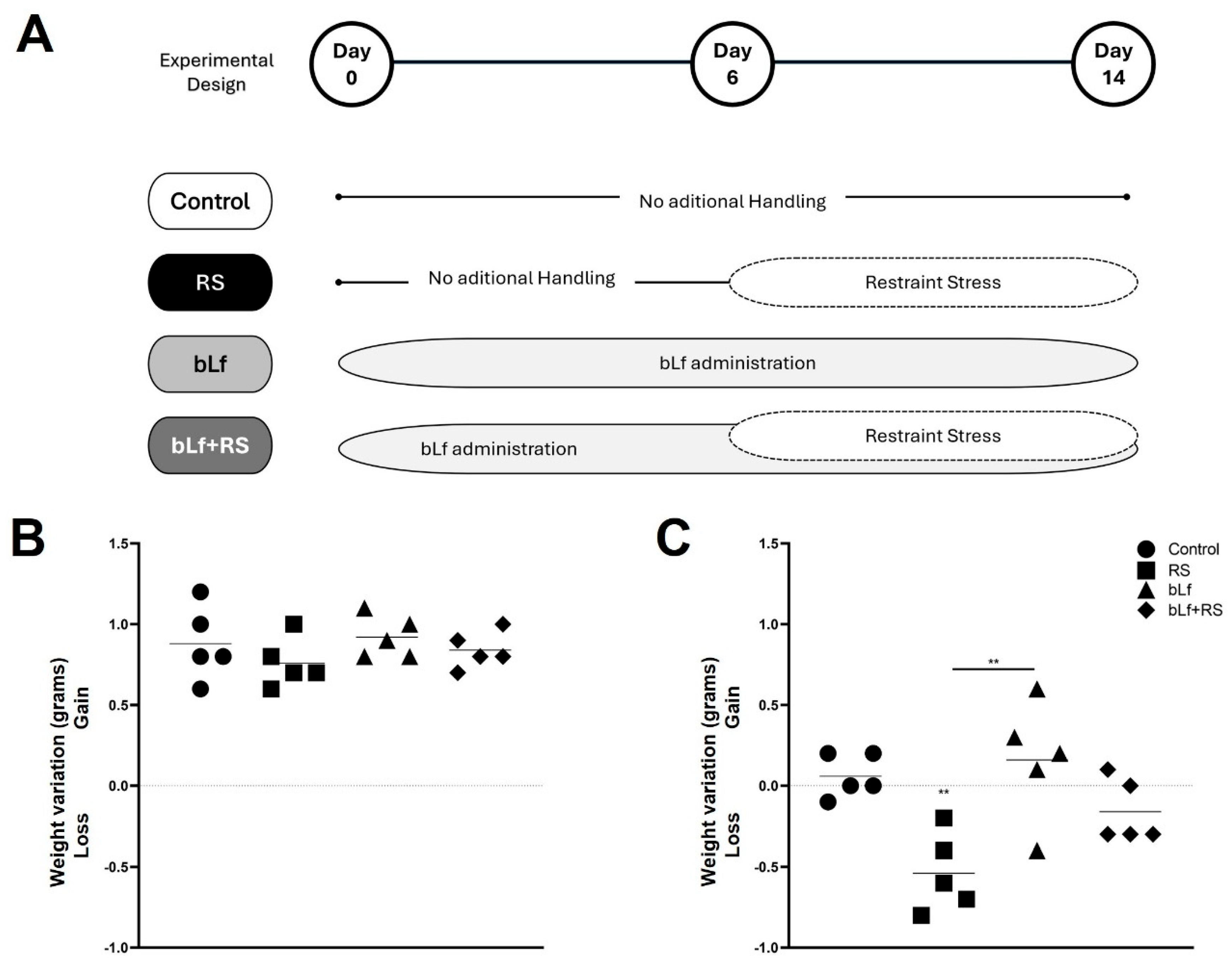
2.1. Bovine Lactoferrin Counteracts Weight Loss Induced by Stress

Mice were aleatory separated into four experimental groups (Figure 1A). They gained weight during the homing week, before the stress protocol (Figure 1B. Once the stress protocol concluded (Figure C), RS lost 0.5±0.24g (p value ≤0.01).

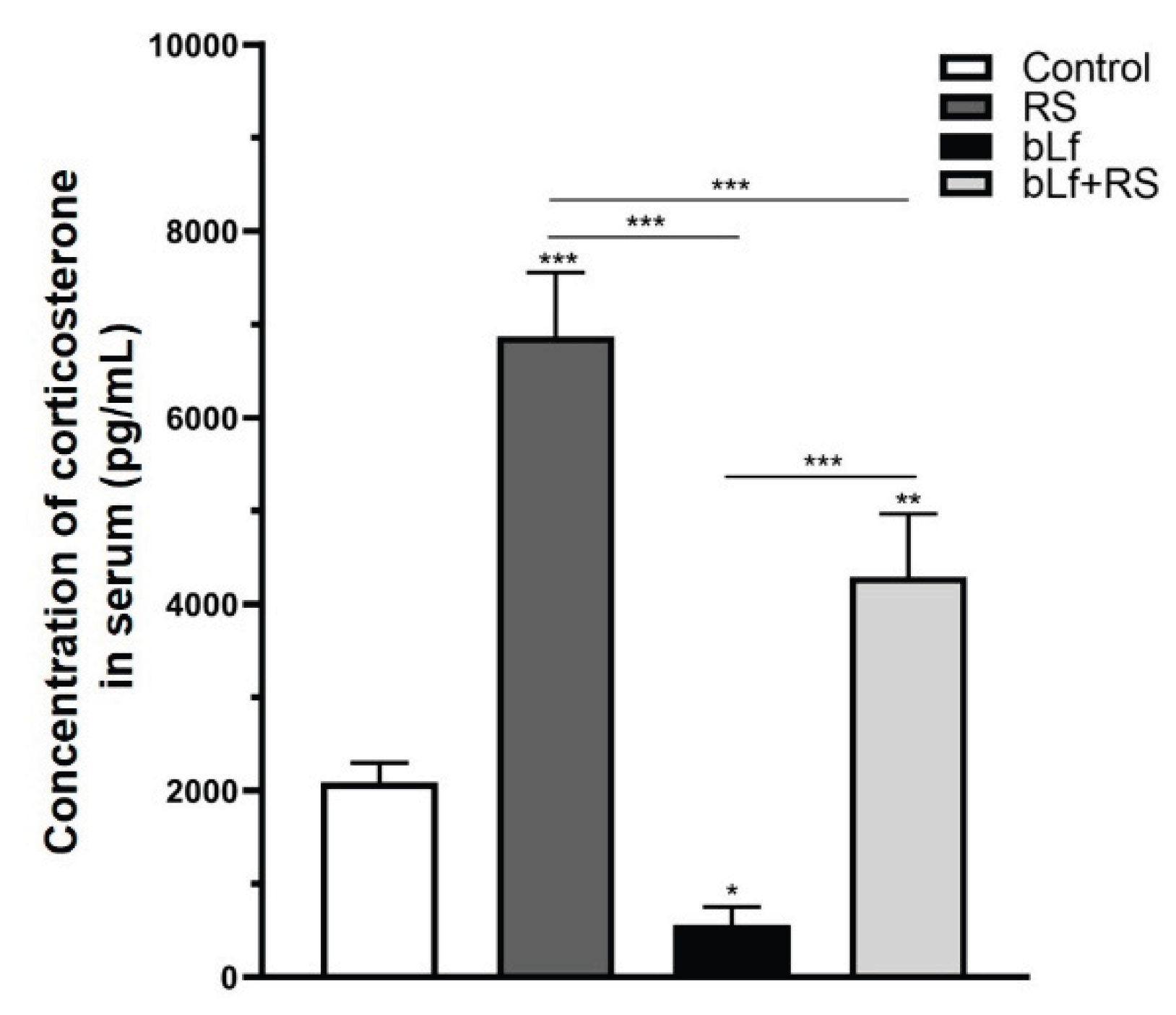
2.2. Bovine Lactoferrin Modulates Corticosterone

Chronic stress increased serum corticosterone (Figure 2) in RS (6869.30±684.94 pg/mL; p≤0.001), while in bLf these levels were decreased (561.12±191.36 pg/mL; p≤0.05) compared to CTL (2086.64±214.68 pg/mL). Interestingly, in bLf+RS mice group corticosterone was increased (4288.41±680.55 pg/mL; p≤0.01) in a lower concentration than RS group. These results suggest that oral bLf administration before and during chronic stress modulates corticosterone release.

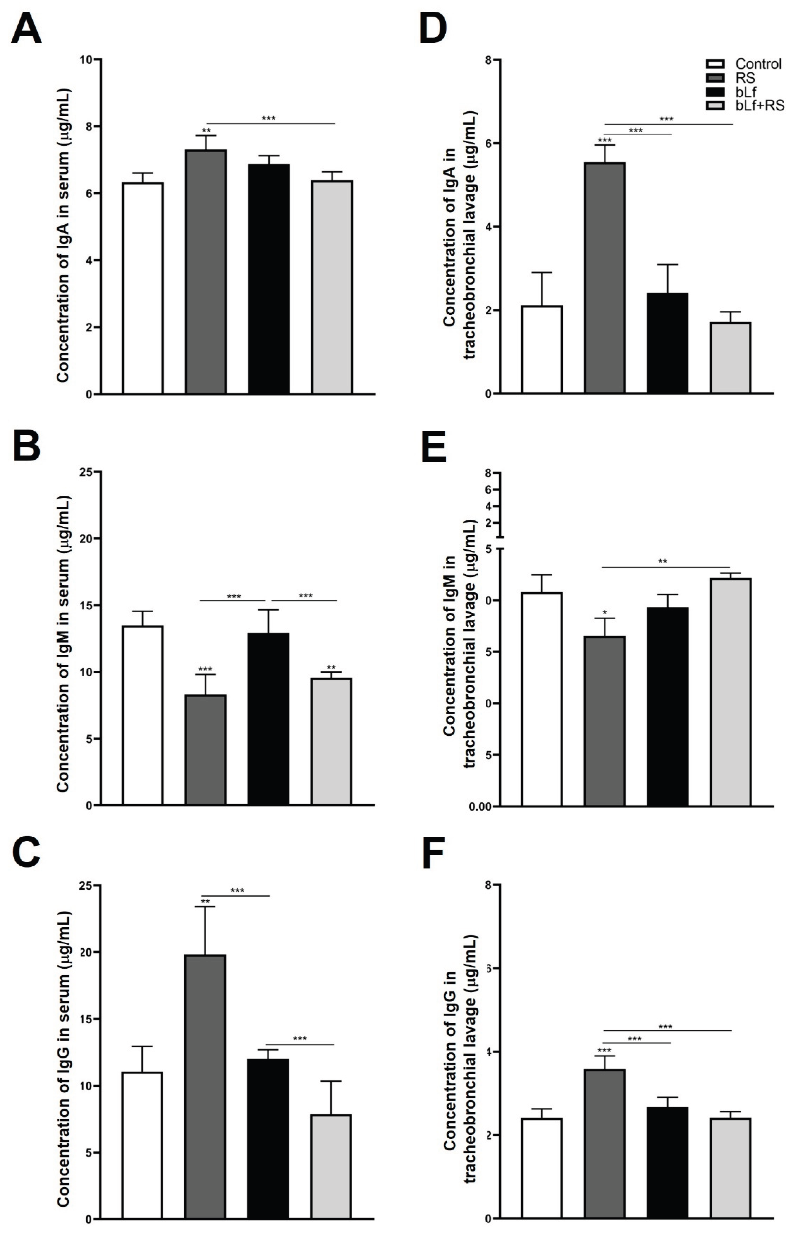
2.3. Bovine Lactoferrin Modulates the Effect of Chronic Stress in Immunoglobulin Concentrations

Immunoglobulin concentrations of serum and TBL were modified by chronic stress (Figure 3). Total IgA was increased in serum (Figure 3A; 5.55±0.41 µg/mL, p ≤0.01) and TBL (Figure 3B; 5.55±0.41 µg/mL, p ≤0.001) versus CTL group. Serum IgM (Figure 3 B) was diminished in RS (8.32±1.49 µg/mL, p ≤0.001) and bLf+RS (9.57±0.41, p ≤0.01), as well as IgM TBL levels of the RS group (Figure 3E; 0.16±0.01 µg/mL, p ≤0.05), in comparison to CTL group. IgG was increased in serum of RS (Figure 3C; 19.83±3.57 µg/mL, p ≤0.01) and TBL (Figure 3F; 3.58±0.31 µg/mL, p ≤0.001) versus CTL group. These results demonstrate bLf immunomodulatory properties by up- or downregulating immunoglobulin concentrations of both serum and TBL during stress.

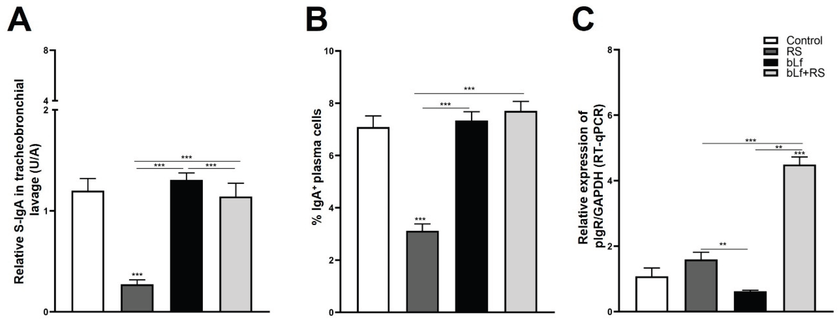
2.4. Bovine Lactoferrin Modulates Stress-Induced Diminished S-IgA Release in Tracheobronchial Lavage and Serum

The analysis of S-IgA TBL concentration (Figure 4A; 0.27±0.04 U/A), and the IgA+ plasma cell population (Figure 4B; 3.12%±0.25%, p≤0.001) was diminished both in the RS group versus CTL. Besides this, relative expression of pIgR is expressed four times more in bLf+RS (Figure 3C; 4.49±0.24, p≤0.001) versus CTL group. These results suggest that chronic stress decreases TBL S-IgA levels, and this might be a consequence of the decrease of IgA+ plasma cell populations in the lung, but it seems not to be related to pIgR relative expression. Meanwhile oral administration of bLf before and during stress modulates S-IgA concentrations and IgA+ plasma cell population similar to CTL group and promotes overexpression of pIgR.

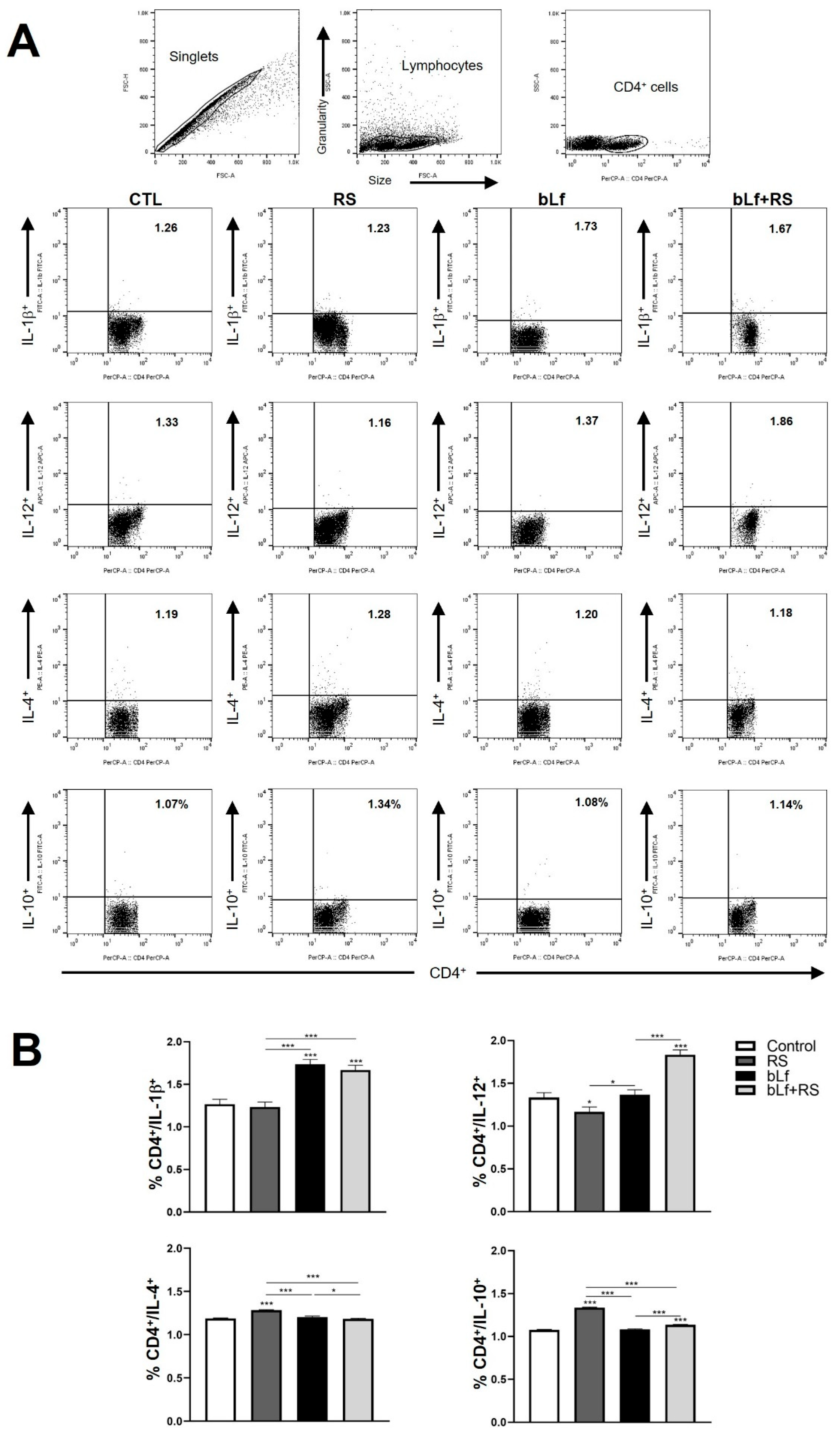
2.5. bLf Promotes Th1 Response and Decreases Th2 Established During Stress

Representative dot plots of CD4+ T cell populations are shown in Figure 5A. By analyzing cytokine profile of the lung lymphocytes (Figure 5B), we found that CD4+/IL-12+ population decreased (1.16%±0.06%), and upregulated CD4+/IL-4+ (1.28%±0.01%) and CD4+/IL-10+ (1.34%±0.01%) in RS group. In mice administrated with bLf CD4+/IL-1β+ population was increased (1.73%±0.05%). In bLf+RS group, CD4+/IL-1β+ (1.67±0.05%) and CD4+/IL-12+ (1.86±0.06%) percentages were increased, remarkably CD4+/IL-4+ (1.18±0.01) and CD4+/IL-10+ (1.14%±0.01%) decreased to levels similar to CTL group. These results demonstrate that chronic stress upregulates Th2 cytokines, while bLf administration modulates by reestablishing Th1/Th2 balance in the cytokine profile.

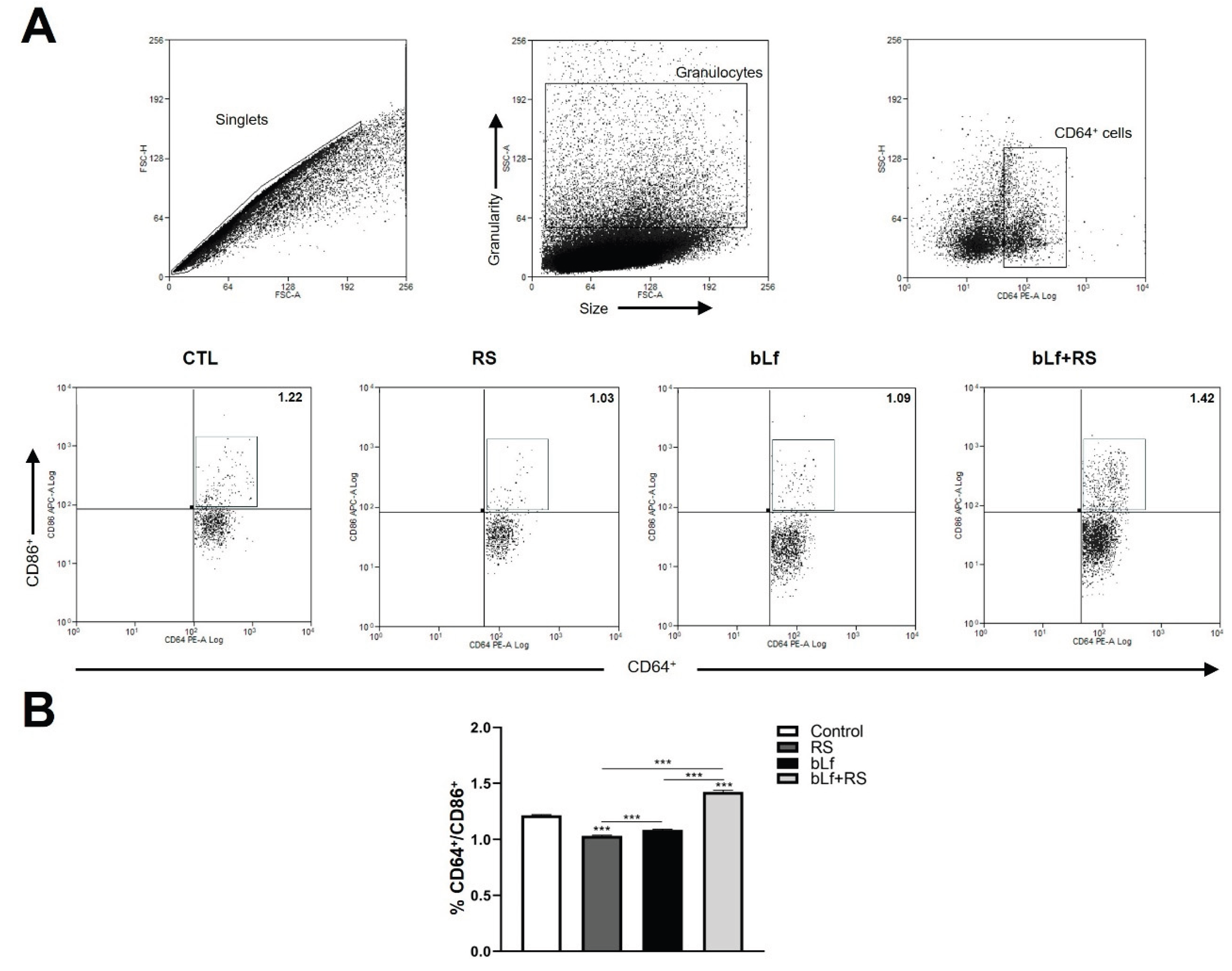
2.6. bLf Modulates CD64+/CD86+ Cell Populations Promoted by Stress

Representative dot plots of APC cell populations are shown in Figure 6A. Analysis of CD64+/CD86+ cell populations (Figure 6B) diminished (1.03%±0.01%, p≤0.001) in RS, while it was increased in bLf+RS (1.42%±0.02%). These results demonstrate bLf capability to modulate APC cell percentages during chronic stress.

3. Discussion

Among many other effects related to exposure to stress, metabolic deregulation is one of the most studied. Despite contradictory effects on weight variation, it seems to be dependent on the duration of exposure to the stimulus. Weight lost in our group subjected to chronic stress is consequently to previous reports [23] and evidence suggests that this might be consequent to Hypothalamic-Pituitary Axis (HPA) activation, which mediators promotes leptin reduction [24], resulting in lower body fat [25] and diminished energy metabolism [26], disturbing metabolism of carbohydrates, lipids and food intake related hormones [27]. In accordance with our results in the bLf+RS group, studies refer that lactoferrin administration ameliorates weight loss related to chronic diseases such as hypertension [28] and influenza [12]. Controversially, it has been reported that lactoferrin administration facilitates weight loss in patients with obesity [29] or predisposed to obesity [30]. However, in our experimental model, we propose that the main mechanism by which bLf prevents body weight loss is related to the modulation of stress-related hormones.
We demonstrated that prophylactic and therapeutic administration of bLf modulates serum corticosterone during chronic restraint stress. As it is known, one of the first events in response to stress stimulus is the activation of the HPA axis, releasing neurotransmitters and hormones such as norepinephrine and corticosterone[2,31], which in turn promote changes in almost every body tissue, including the immune system [3,32] and the lung [7,8,11]. In this regard, bLf is capable of modulating corticosterone in a manner that depends on the stressor, dose, and timing [33,34]. It has been proposed that bLf is endocytosed by enterocytes and transported to plasma, passing through the blood-brain barrier to cerebrospinal fluid of the choroidal plexus [35,36,37]. Afterwards, bLf modulates the increase of nitric oxide production by upregulation of all nitric oxide synthase isoforms [38]. Then, the anti-stress effect of opioid µ receptors decreases HPA activity to Corticoid Release Hormone (CRH) release, but it doesn’t modify other mediators such as Adrenocorticotropic Hormone (ACTH) [39], epinephrine, and glucagon [40,41]. Several models of chronic stress and administration of lactoferrin performed in rodents are similar to the findings of the present study, which provide strong evidence that oral bLf administration modulates serum corticosterone levels in response to stress stimuli [40,42,43].
On the other hand, one of the main determinants of the correct functioning of the lung immunological system is the immunoglobulins present in tracheobronchial secretions. There are many studies of how stress promotes immunoglobulin production via HPA axis activation in healthy individuals, but most of the research is about the intestine [44]. In accordance with this, it has been proposed that stress can have diverse effects in serum immunoglobulins in a duration and intensity of the stimuli dependent manner. The first immunoglobulin secreted by plasma cells is IgM. It has been described that in the respiratory IgM acts mainly due to the deficiency of other immunoglobulins, since it is found in low concentrations because the IgM-producing cells are found less frequently in the bronchial tree, and due to its size, its transudation from the bloodstream to the bronchial lavage is too complicated [45]. Nevertheless, it has been reported that IgM concentrations may be increased or decreased depending on the model disease in which it has been evaluated, some of them being fibrosis (barely detectable), pneumonitis and transplant rejection (increased) [46]. In this study, chronic stress promoted a decrease in IgM concentrations, but this effect was modulated by bLf administration, which could be promoting a proper functioning of the defense against potential pathogens in the lung.
Furthermore, IgA has a determining role in lung pathologies such as asthma and COPD, since it can determine tolerance to certain antigens; besides, it has been reported that in the bronchial secretions of asthmatic patients, the concentration of IgA is increased, and in patients with COPD, it is decreased [47]. Previously an increase in IgA concentrations in intestinal lavages from the duodenum and ileum during chronic stress has been reported [48]. In contrast, Jarillo-Luna [49] found decreased IgA concentrations in small intestine lavages in a mouse model of chronic stress. Otherwise, both authors reported that the intestine is one of the sites whose immune system is most resistant to stress. In our model, an increase in IgA levels in serum and TBL suggests that chronic stress might be modulating the isotype change of IgA-producing cells in the lung, favoring their secretion and transudation from the serum, probably as protection against potential pathogens or particles that could threaten the homeostasis of this mucosa. To clarify the mechanism by which there is such an increase in IgA concentrations, it would be necessary to determine the difference in the concentrations of monomeric IgA (mIgA) and dimeric IgA (dIgA).
Since there are not studies focused on elucidating the role of bLf on immunoglobulin modulation in the respiratory secretions, our results demonstrate that bLf does not modify the total IgA, IgG or IgM in serum nor TBL concentration, in contrast to the increase reported in distal small intestine [50]. This could be explained by the nature of both mucous tissues. Strikingly, the modulation of these immunoglobulins in the stressed groups is remarkable. bLf antibodies modulation during stress found in our results are similar to those found in the intestinal lavage on a chronic immobilization model [43]. In the lower respiratory airways, it has only been reported previously that bLf increased IL-17 producing cells and enhances IFN- mediated responses [51], reduced lung consolidation and infiltration in bronchial lavage during influenza virus infection [12] , and it is also capable of regulating cytokine genes related to IgA production [52]. Even in human trials, bLf has been reported to contribute to protection against viral infections and modulate respiratory immunity [53], but there is no further information about the mechanisms of IgM and IgG secretion. Since IgG levels in serum and TBL showed a similar pattern to IgA concentrations, these results suggest that chronic stress modifies IgG change of isotype and transudation mechanisms, which are also modulated by bLf. Nowadays, we are far from clearly understanding how the allostasis mechanisms are made, and this study provides an initial overview of how chronic stress modifies immunoglobulin levels in a healthy individual, while demonstrating that lactoferrin modulates the effects of stress in the humoral immunity of the lungs.
It has been well established that one of the most important mechanisms to maintain mucosal homeostasis is the S-IgA. The deficiency of this immunoglobulin is related to pathogen colonization, asthma, allergic diseases and chronic obstructive respiratory disease progression [54,55,56]. In concordance with these reports, our results showed decreased S-IgA levels in the TBL and percentages of IgA+ plasma cells in the RS group. Interestingly, these parameters in the bLf+RS group are similar to control, demonstrating the immunomodulatory effect of bLf. Since S-IgA production and secretion depend on the polymeric immunoglobulin receptor expression in the lung epithelial cells, we evaluated the expression of pIgR gene in the lung epithelial cells. pIgR is related to the process of transcytosis and the release of the secretory component (SC) to form S-IgA complex [57]. Several studies have demonstrated that pIgR expression is regulated by some cytokines, such as TNF- , IFN- , TGF- , as well as signal pathways activated by glucocorticoid receptors [58]. Hence, it has been shown that IL-4 is capable of downregulating bronchial epithelial pIgR expression [59]. Therefore, the relative expression of the pIgR gene had no difference in the stress group but was overexpressed in the bLf+RS group. Despite that, studies on cultures of primary human bronchial epithelial cells from smoke patients demonstrated that pIgR protein expression is not necessarily related to its gene expression, and this could be related to post-transcriptional mechanisms [60,61].
In previous studies, the bLf administration modulates IgA+ plasma cell populations of the distal small intestine [50]. This is the first report focused on the effect of stress on the lung immunoglobulins and demonstrated bLf’s capability of modulating the decrease of IgA+ plasma cell population and S-IgA low expression produced by stress. Nevertheless, further studies are required to clarify if it is related to pIgR expression and the mechanisms implicated. These findings seem to be another protective mechanism of lactoferrin as a prophylactic and adjuvant therapeutic agent in lower respiratory tract diseases.
Many studies have focused on the role of stress in pulmonary diseases. It has been widely reported that it induces airway inflammation [9] via HPA axis activation [10]. In consequence, asthma symptoms are exacerbated by promoting an increment of CD4+ Th2 cells [62] and the further secretion of cytokines, such as IL-4, IL-5, and IL-13 [63,64,65,66]. At the same time, Th1 cytokines are diminished [67,68]. Asthma symptoms improve by administering a glucocorticoid receptor antagonist [69]. There are also findings in infectious diseases. In a model of influenza virus, which demonstrates that stress promotes a Th2 profile, decreasing the antiviral defense and worsening the development of the disease [70]. Lafuse and cols. has reported that stress improves IL-10 levels by inducing psychological stress in mice infected with Mycobacterium tuberculosis (MTB), which promotes the pathogenicity [71]. We found that T CD4+/IL-4+ and T CD4+/IL-10+ populations are highly increased in the RS and our results are consistent with these reports, suggesting that chronic stress might be promoting lung susceptibility to infectious and respiratory diseases.
On the other hand, there are many studies that demonstrate the immunomodulatory effect of lactoferrin in multiple respiratory pathologies by promoting or downregulating inflammatory responses [72]. It has been reported that oral administration of bLf diminished viral load in BALB/c mice infected with the influenza virus, and it also promotes tissue repair response by decreasing cell infiltration [12]. Furthermore, some findings suggest bLf is capable of downregulating asthma symptoms and ovalbumin-induced lung inflammation [73]. In an MTB infection, bLf demonstrated its capability of increasing the number of CD4+ Th1 cytokine-producing cells in the lung, thus promoting the amelioration of pathological response and preventing the formation of granuloma [51,74]. Some authors hypothesized that bLf could be a novel prophylactic and therapeutic agent to ameliorate COVID-19 and other infectious lung diseases [75]. Our results demonstrated that bLf is modulating the increase of Th2 lymphocyte populations during chronic stress and promoting a Th1 profile, thus balancing the cytokine profile in the lung. These effects may be able to avoid exacerbation of chronic pulmonary or allergic diseases and prevent infectious diseases of the lower respiratory airways, but more studies are required to clearly understand the mechanisms involved in the signaling of these immunological events.
The role of macrophages and antigen-presenting cells is a remarkable finding in recent studies focused on elucidating the mechanisms of pulmonary homeostasis management. Among other functions, these are the first cells to protect the lower respiratory tract against pathogens and coordinate and regulate respiratory secretions [76], and its presence is related to the prevention of asthma exacerbation [77]. There is evidence that stress diminishes APC population, such as dendritic cells [69], and this might be related to decrease in IL-12 release in response to glucocorticoids [78]. In this work, chronic stress diminished CD64+/CD86+ cell populations, but this effect was avoided by the prophylactic administration of bLf.
The evidence provided in the present study demonstrated that oral administration of bLf modulates the effect of chronic stress in the serum and TBL immunoglobulins, prevents the decrease of S-IgA and IgA+ plasma cell populations promoted by stress, and counterbalances Th1-Th2 lymphocyte profiles. Even when there are more studies to clearly understand these findings, such as protein expression of pIgR and the role of macrophages in lung immunity, we hypothesize that this may be a protective effect of bLf by maintaining the innate immune system fit to mount a response. This assertion is supported by the fact that bLf administration had no changes without exposure to stress stimulus. Thus, the present study provides a first overview of the potential prophylactic and co-therapeutic administration of bLf to prevent low respiratory infectious diseases and amelioration of chronic lung diseases.

4. Materials and Methods

4.1. Animals

BALB/c mice (age 10-12 weeks old and weight 25-30 g) were used. Mice were housed in transparent polycarbonate boxes with sterile shavings bed and kept on a 12-h light/dark cycle (lights on at 6:00 a.m.) at room temperature at 20˚C, with relative humidity of 55% and provided with water and Purina Lab Diet 5001. Animals were handled according to a protocol (ESM-CICUAL-03/06-09-2020) in accordance with the Mexican federal regulations for animal experimentation and care (NOM-062-ZOO-1999, Ministry of Agriculture, SAGARPA, Mexico City, Mexico) and the experiments were approved by the Institutional Animal Care and Use Committee of the Escuela Superior de Medicina, Instituto Politécnico Nacional.

4.2. Stress and bLf Administration Protocol

Mice were randomly divided into four experimental groups with n=10: a) Control (CTL): kept for 14 days in housing with water and food ad libitum; b) Restraint Stress (RS): mice were maintained for six more days with minimal manipulation and then, they were introduced into movement restriction chambers with adequate ventilation for four hours, from 8:00 a.m. to 12:00 p.m., during eight consecutive days [79]; c) bLf: 5 mg of bovine lactoferrin diluted in 100µL of vehicle (sterile water) were administered by oral deposition daily, during 14 consecutive days [50,80]; d) bLf+RS: 5 mg of bovine lactoferrin diluted in 100µL of sterile water were administered daily, for a period of six days before stress induction. Administration of bLf continued for eight more days (for a total of 14 days), wherein the mice survived, simultaneously subjected to movement restriction stress for four hours (Figure 1A).

4.3. Mice Weight

Mice were weighed first upon arrival at the laboratory, then, before the stress protocol, and finally, a third measure was made after the experimental model, right before euthanasia. The weight registered at the beginning of each week was subtracted from the next measure to analyze weight gain or loss (Figure 1B and C).

4.4. Blood Collection

Each mouse was euthanized by an intraperitoneal injection of a lethal dose of 100 mg/kg body weight pentobarbital sodium salt (cat. P3761, Sigma-Aldrich) and exsanguinated by cardiac puncture. Approximately 1 mL of blood was collected and centrifuged at 3000 rpm for 10 min to obtain the serum, and it was stored at -20°C for further use.

4.5. Corticosterone Assay

The corticosterone concentration in the plasma was determined using a commercially available ELISA kit according to the manufacturer's instructions (cat. no. 501320 Cayman Chemical). Plasma samples’ corticosterone concentrations were calculated based on a standard curve and were expressed in pg/mL (Figure 2).

4.6. Tracheobronchial Lavage

Once exsanguination was performed, the mouse was fixed on a dissection table, and the thoracic cavity was exposed. An incision was made in the trachea by inserting a metal cannula, which was fixed to introduce 1 mL of 1X PBS to the lungs, performing a gentle massage and aspiration, recovering the lavage, which was placed in a microtube. The lavage was centrifuged at 1500 rpm at 4ºC for 10min. The supernatant was collected and stored at -20 ºC with briefly modification [81].

4.7. Immunoglobulin Measurement

Concentrations of total IgM, IgG, and IgA, as well as secretory IgA (S-IgA), were determined in the tracheobronchial lavages by performing a sandwich ELISA with briefly modifications [50]. The plate was coated with the capture antibody (anti-IgA, anti-IgM, anti-IgG) and incubated. HRP-coupled antibody was added: anti-IgA (HRP goat anti-mouse cat. 626720 Life technologies); anti-IgM (HRP goat anti-mouse cat. M31507Life technologies), anti-IgG (HRP goat anti-mouse cat. 626520), and anti-Secretory Component (HRP goat anti-mouse cat. sc-374343, Santacruz). Absorbance was measured at 490 nm by using an enzyme immunoassay reader (Sigma).

4.8. Lung Lymphocyte Cells Purification

Lungs were removed and incubated in 15mL of 1X RPMI 1640 (cat. 23400-062, GIBCO) supplemented with 1% fetal bovine serum (FBS), with 100 µM ethylenediaminetetraacetic acid (8993-01 J.T. Baker) and 1mM dithiothreitol (D9779, Sigma Chemical) for 30 min at 37ºC. After incubation, tissue dissociation was made with a plunger and a steel mesh, then filtered and centrifuged. The cell button was taken to 75%/40% Percoll gradients to obtain leukocytes, and 40%/20% to obtain epithelial cells. Interphase cell rings were collected and washed to obtain a pellet [82].

4.9. Flow Cytometry Assay

Suspension of 1 x10⁶ cells was incubated with corresponding extracellular markers for plasma cells (anti-CD19/PE cat. 553786, anti-CD138/APC cat. 558626 BD Biosciences), APC (anti-CD64/PE cat. 558455, BD Biosciences, anti-CD86 cat. 105012, BioLegend) and T CD4+ cells (anti-CD4/PerCP cat. 100538, BioLegend). Subsequently, APC were fixed with 4% paraformaldehyde. T lymphocytes and plasma cells were fixed and permeabilized by incubating for 20 min in the dark with Cytofix/Cytoperm (cat. 554722, BD Biosciences) and centrifuged at 1500 rpm at 4°C for 5 min. Then, cells were washed with Perm Wash (cat. 554723, BD Biosciences) and incubated with antibody cocktails respectively: anti-IgA/FITC (Cat. 559354, BD Biosciences) for plasma cells, and Th1(anti-IL-1β/FITC cat. IC413F, R&D Systems, anti-IL-12/APC cat. 554480, BD Biosciences) and Th2 (anti-IL-4/PE cat. 554435, BD Biosciences, anti-IL-10/FITC cat. 505005, BioLegend) for each analysis of intracellular cytokines. After incubation, the cell pellet was washed with Perm Wash, and then the samples were fixed and filtered [83]. Samples were stored at 4°C in the dark until analysis on the FACS ARIA flow cytometer (Beckton, Dickinson Company) with BD FACSDIVA™️ v6.1 software (BD Biosciences), acquiring 20,000 gated events from each sample. The data were analyzed using FlowJo v10.10.0 (BD Life Sciences). Cell percentages were reported as the mean ± SD.

4.10. Real-Time qPCR of pIgR

4.10.1. RNA Extraction

The extraction of RNA from leukocytes was carried out by a gradient of TRIzol reagent (cat. Invitrogen™, 15596026, Life Technologies, Carlsbad, CA, USA) and the chloroform technique [84].

4.10.2. cDNA Synthesis

RNA was treated with the RQ1 RNase-Free Dnase Kit (cat. M6101, ThermoScientific) following the manufacturer's instructions. The synthesis of the cDNA was made using a commercial kit (RevertAid First Strand cDNA Synthesis kit, cat. K1622, ThermoScientific) by following the manufacturer’s instructions. cDNA samples were stored at −70°C.

4.10.3. Quantitative Real-Time Polymerase Chain Reaction (qRT-PCR)

The primers for pIgR and GAPDH were designed with Primer Express v.3.0.1 (Applied Biosystems) software and synthesized by UNIPARTS S.A. of C.V. (Table 1). For the PCR reactions, each well was filled with: 10µL of SYBR™️ Green PCR Master Mix (cat. 4309155, Applied Biosystems™️, Thermo Fischer Scientific); 0.2 µL Primer F; 0.2 µL Primer R; 7.6 µL of Water; and 2 µL of the sample. The amplification was carried out in the Step One Real-Time PCR System (Applied Biosystems™️) and was analyzed with the help of StepOne™️ v2.3 software. Data normalization was carried out using the constitutive gene GAPDH for determination.

4.11. Statistical Analysis

The results were analyzed by a one-way ANOVA and Turkey’s multiple comparisons test with the software GraphPad Prism v. 8.0.2. Data is presented as the mean ± standard deviation (SD). Differences of P≤0.05 were considered significant.

Author Contributions

Conceptualization I.M.A.-M., A.A.R.-A and M.Y.-O.; methodology M.Y.-O., E.J.Z.-A., C.A.G.-C., B.M.-A., M.V.-T., D.R.-V., M.A.G.-R. and U.A.G.-S; software M.Y.-O., I.M.A.-M., A.A.R.-A and M.V.-T.; validation I.M.A.-M., A.A.R.-A, M.V.-T. and J.P.-Y.; formal analysis I.M.A.-M., A.A.R.-A and M.Y.-O; investigation M.Y.-O., E.J.Z.-A., C.A.G.-C., B.M.-A., M.V.-T., D.R.-V., M.A.G.-R. and U.A.G.S.; resources I.M.A.-M., A.A.R.-A and J.P.-Y; data curation I.M.A.-M., A.A.R.-A and M.Y.-O.; writing—original draft preparation M.Y.-O., I.M.A.-M. and A.A.R.-A; writing—review and editing M.Y.-O., I.M.A.-M., A.A.R.-A., M.V.-T., D.R.-V.; visualization, M.Y.-O., I.M.A.-M., A.A.R.-A. and M.A.G.-R.; supervision I.M.A.-M., A.A.R.-A and J.P.-Y.; project administration I.M.A.-M. and A.A.R.-A.; funding acquisition I.M.A.-M. and A.A.R.-A. All authors have read and agreed to the published version of the manuscript.

Funding

This research was funded by Sistema de Administración de Programa y Proyectos de Investigación (SAPPI-IPN). Multidisciplinary Project, grant number 20230994. Mariazell Yépez-Ortega received financial support through the scholarship 20240519 by BEIFI and the scholarship 993755 granted by the Secretaría de Ciencia, Humanidades, Tecnología e Innovación (SECIHTI).

Institutional Review Board Statement

The animal study protocol was approved by the Ethics in Research Committee of the Escuela Superior de Medicina of the Instituto Politecnico Nacional (ESM-CICUAL-03/06-09-2020) and according to the technical specifications for the production, care, and use of laboratory animals (Regulation-062-ZOO-1999, Ministry of Agriculture, SAGARPA, México City, México.

Conflicts of Interest

None of the authors has any financial or other interest in the products, devices, or methods mentioned in this article.

Abbreviations

The following abbreviations are used in this manuscript:
bLf Bovine lactoferrin
RS Restraint Stress
TBL Tracheobronchial lavages
S-IgA Secretory Immunoglobulin A
APC Antigen Presenting Cells
COPD Chronic Obstructive Pulmonary Disease
pIgR Polymeric Immunoglobulin Receptor
mIgA Monomeric ImmunoglobulinA
dIgA Dimeric ImmunoglobulinA
SC Secretory component
MTB Mycobacterium tuberculosis

References

  1. Dhabhar, F.S. Effects of Stress on Immune Function: The Good, the Bad, and the Beautiful. Immunol Res 2014, 58, 193–210. [CrossRef]
  2. Chrousos, G.P. The Hypothalamic–Pituitary–Adrenal Axis and Immune-Mediated Inflammation. New England Journal of Medicine 1995, 332, 1351–1363. [CrossRef]
  3. Dhabhar, F.S.; Malarkey, W.B.; Neri, E.; McEwen, B.S. Stress-Induced Redistribution of Immune Cells—From Barracks to Boulevards to Battlefields: A Tale of Three Hormones – Curt Richter Award Winner. Psychoneuroendocrinology 2012, 37, 1345–1368. [CrossRef]
  4. Vittwrakis, S.; Gaga, M.; Oikonomidou, E.; Samitas, K.; Xwrianopoulos, D. Immunological Mechanisms in the Lung. Pneumon 2007, 20, 274–278.
  5. Ardain, A.; Marakalala, M.J.; Leslie, A. Tissue-resident Innate Immunity in the Lung. Immunology 2020, 159, 245–256. [CrossRef]
  6. Agorastos, A.; Chrousos, G.P. The Neuroendocrinology of Stress: The Stress-Related Continuum of Chronic Disease Development. Mol Psychiatry 2022, 27, 502–513. [CrossRef]
  7. Cohen, S. Psychological Stress and Susceptibility to Upper Respiratory Infections. Am J Respir Crit Care Med 1995, 152, S53–S58. [CrossRef]
  8. Leick, E.A.; Reis, F.G.; Honorio-Neves, F.A.; Almeida-Reis, R.; Prado, C.M.; Martins, M.A.; Tibério, I.F.L.C. Effects of Repeated Stress on Distal Airway Inflammation, Remodeling and Mechanics in an Animal Model of Chronic Airway Inflammation. Neuroimmunomodulation 2012, 19, 1–9. [CrossRef]
  9. Reis, F.G.; Marques, R.H.; Starling, C.M.; Almeida-Reis, R.; Vieira, R.P.; Cabido, C.T.; Silva, L.F.F.; Lanças, T.; Dolhnikoff, M.; Martins, M.A.; et al. Stress Amplifies Lung Tissue Mechanics, Inflammation and Oxidative Stress Induced by Chronic Inflammation. Exp Lung Res 2012, 38, 344–354. [CrossRef]
  10. Miyasaka, T.; Dobashi-Okuyama, K.; Takahashi, T.; Takayanagi, M.; Ohno, I. The Interplay between Neuroendocrine Activity and Psychological Stress-Induced Exacerbation of Allergic Asthma. Allergology International 2018, 67, 32–42. [CrossRef]
  11. Lee, L.; Yu, J. Sensory Nerves in Lung and Airways. In Comprehensive Physiology; Wiley, 2014; pp. 287–324.
  12. Shin, K.; Wakabayashi, H.; Yamauchi, K.; Teraguchi, S.; Tamura, Y.; Kurokawa, M.; Shiraki, K. Effects of Orally Administered Bovine Lactoferrin and Lactoperoxidase on Influenza Virus Infection in Mice. J Med Microbiol 2005, 54, 717–723. [CrossRef]
  13. Han, N.; Li, H.; Li, G.; Shen, Y.; Fei, M.; Nan, Y. Effect of Bovine Lactoferrin as a Novel Therapeutic Agent in a Rat Model of Sepsis-Induced Acute Lung Injury. AMB Express 2019, 9, 177. [CrossRef]
  14. Kell, D.B.; Heyden, E.L.; Pretorius, E. The Biology of Lactoferrin, an Iron-Binding Protein That Can Help Defend Against Viruses and Bacteria. Front Immunol 2020, 11. [CrossRef]
  15. Legrand, D. Overview of Lactoferrin as a Natural Immune Modulator. J Pediatr 2016, 173, S10–S15. [CrossRef]
  16. Masson, P.L.; Heremans, J.F. Lactoferrin in Milk from Different Species. Comparative Biochemistry and Physiology Part B: Comparative Biochemistry 1971, 39, 119-IN13. [CrossRef]
  17. Kanwar, J.; Roy, K.; Patel, Y.; Zhou, S.-F.; Singh, M.; Singh, D.; Nasir, M.; Sehgal, R.; Sehgal, A.; Singh, R.; et al. Multifunctional Iron Bound Lactoferrin and Nanomedicinal Approaches to Enhance Its Bioactive Functions. Molecules 2015, 20, 9703–9731. [CrossRef]
  18. Drago-Serrano, M.E.; Flores-Romo, L.; Oliver-Aguillón, G.; Jarillo-Luna, R.A.; Reyna-Garfias, H.; Barbosa-Cabrera, E.; Campos-Rodríguez, R. La Lactoferrina Como Modulador de La Respuesta Inmunitaria. Bioquimia 2008, 33, 71–82.
  19. González-Chávez, S.A.; Arévalo-Gallegos, S.; Rascón-Cruz, Q. Lactoferrin: Structure, Function and Applications. Int J Antimicrob Agents 2009, 33, 301.e1-301.e8. [CrossRef]
  20. Rodríguez-Franco D Actividad Antimicrobiana de La Lactoferrina: Mecanismos y Aplicaciones Clínicas Potenciales; Revista Latinoamericana de Microbiología, 2005; Vol. 47.
  21. Baveye, S.; Elass, E.; Mazurier, J.; Spik, G.; Legrand, D. Lactoferrin: A Multifunctional Glycoprotein Involved in the Modulation of the Inflammatory Process. cclm 1999, 37, 281–286. [CrossRef]
  22. Kruzel, M.L.; Zimecki, M.; Actor, J.K. Lactoferrin in a Context of Inflammation-Induced Pathology. Front Immunol 2017, 8. [CrossRef]
  23. Huang, Y.; Wen, J.; Chen, G. Role and Mechanism of Chronic Restraint Stress in Regulating Energy Metabolism and Reproductive Function Through Hypothalamic Kisspeptin Neurons. J Endocr Soc 2021, 5, A551–A552. [CrossRef]
  24. Harris, R.B.S.; Mitchell, T.D.; Simpson, J.; Redmann, S.M.; Youngblood, B.D.; Ryan, D.H. Weight Loss in Rats Exposed to Repeated Acute Restraint Stress Is Independent of Energy or Leptin Status. American Journal of Physiology-Regulatory, Integrative and Comparative Physiology 2002, 282, R77–R88. [CrossRef]
  25. Michel, C.; Duclos, M.; Cabanac, M.; Richard, D. Chronic Stress Reduces Body Fat Content in Both Obesity-Prone and Obesity-Resistant Strains of Mice. Horm Behav 2005, 48, 172–179. [CrossRef]
  26. Huang, Y.; Liu, Q.; Huang, G.; Wen, J.; Chen, G. Hypothalamic Kisspeptin Neurons Regulates Energy Metabolism and Reproduction Under Chronic Stress. Front Endocrinol (Lausanne) 2022, 13. [CrossRef]
  27. López López, A. Chronic Unpredictable Mild Stress Progressively Disturbs Glucose Metabolism and Appetite Hormones In Rats. Acta Endocrinologica (Bucharest) 2018, 14, 16–23. [CrossRef]
  28. Safaeian, L.; Zabolian, H. Antioxidant Effects of Bovine Lactoferrin on Dexamethasone-Induced Hypertension in Rat. ISRN Pharmacol 2014, 2014, 1–6. [CrossRef]
  29. Ono, T.; Murakoshi, M.; Uchiyama, A. Anti-Obesity Effect of Lactoferrin; Subgroup Analysis Excluding Subjects with Obese and/or Hyper-LDL Cholesterolemia. Immunol Endocr Metab Agents Med Chem 2018, 18, 105–109. [CrossRef]
  30. Jańczuk-Grabowska, A.; Czernecki, T.; Brodziak, A. Gene–Diet Interactions: Viability of Lactoferrin-Fortified Yoghurt as an Element of Diet Therapy in Patients Predisposed to Overweight and Obesity. Foods 2023, 12, 2929. [CrossRef]
  31. Spencer, R.L.; Deak, T. A Users Guide to HPA Axis Research. Physiol Behav 2017, 178, 43–65. [CrossRef]
  32. Zefferino, R.; Di Gioia, S.; Conese, M. Molecular Links between Endocrine, Nervous and Immune System during Chronic Stress. Brain Behav 2021, 11. [CrossRef]
  33. Godínez-Victoria, M.; Cruz-Hernández, T.R.; Reyna-Garfias, H.; Barbosa-Cabrera, R.E.; Drago-Serrano, M.E.; Sánchez-Gómez, M.C.; Campos-Rodríguez, R. Modulation by Bovine Lactoferrin of Parameters Associated with the IgA Response in the Proximal and Distal Small Intestine of BALB/c Mice. Immunopharmacol Immunotoxicol 2017, 39, 66–73. [CrossRef]
  34. Guzmán-Mejía, F.; Vega-Bautista, A.; Molotla-Torres, D.E.; Aguirre-Garrido, J.F.; Drago-Serrano, M.E. Bovine Lactoferrin as a Modulator of Neuroendocrine Components of Stress. Curr Mol Pharmacol 2021, 14, 1037–1045. [CrossRef]
  35. Harada, E.; Itoh, Y.; Sitizyo, K.; Takeuchi, T.; Araki, Y.; Kitagawa, H. Characteristic Transport of Lactoferrin from the Intestinal Lumen into the Bile via the Blood in Piglets. Comp Biochem Physiol A Mol Integr Physiol 1999, 124, 321–327. [CrossRef]
  36. Talukder, Md.J.R.; Takeuchi, T.; Harada, E. Receptor-Mediated Transport of Lactoferrin into the Cerebrospinal Fluid via Plasma in Young Calves. Journal of Veterinary Medical Science 2003, 65, 957–964. [CrossRef]
  37. Kamemori, N.; Takeuchi, T.; Sugiyama, A.; Miyabayashi, M.; Kitagawa, H.; Shimizu, H.; Ando, K.; Harada, E. Trans-Endothelial and Trans-Epithelial Transfer of Lactoferrin into the Brain through BBB and BCSFB in Adult Rats. Journal of Veterinary Medical Science 2008, 70, 313–315. [CrossRef]
  38. Kamemori, N.; Takeuchi, T.; Hayashida, K.; Harada, E. Suppressive Effects of Milk-Derived Lactoferrin on Psychological Stress in Adult Rats. Brain Res 2004, 1029, 34–40. [CrossRef]
  39. Aleshina, G.M.; Yankelevich, I.A.; Zhakharova, E.T.; Kokryakov, V.N. Rossiiskii Fiziologicheskii Zhurnal Imeni I.M. Sechenova. 2016, 102, 846–851.
  40. Takeuchi, T.; Hayashida, K.; Inagaki, H.; Kuwahara, M.; Tsubone, H.; Harada, E. Opioid Mediated Suppressive Effect of Milk-Derived Lactoferrin on Distress Induced by Maternal Separation in Rat Pups. Brain Res 2003, 979, 216–224. [CrossRef]
  41. Maekawa, Y.; Sugiyama, A.; Takeuchi, T. Lactoferrin Ameliorates Corticosterone-Related Acute Stress and Hyperglycemia in Rats. Journal of Veterinary Medical Science 2017, 79, 412–417. [CrossRef]
  42. Peña-Juárez, Ma.C.; Campos-Rodríguez, R.; Godínez-Victoria, M.; Cruz-Hernández, T.R.; Reyna-Garfias, H.; Barbosa-Cabrera, R.E.; Drago-Serrano, M.E. Effect of Bovine Lactoferrin Treatment Followed by Acute Stress on the IgA-Response in Small Intestine of BALB/c Mice. Immunol Invest 2016, 45, 652–667. [CrossRef]
  43. Cruz-Hernández, T.; Gómez-Jiménez, D.; Campos-Rodríguez, R.; Godínez-Victoria, M.; Drago-Serrano, M. Analysis of the Intestinal IgA Response in Mice Exposed to Chronic Stress and Treated with Bovine Lactoferrin. Mol Med Rep 2020, 23, 126. [CrossRef]
  44. Campos-Rodríguez, R.; Godínez-Victoria, M.; Abarca-Rojano, E.; Pacheco-Yépez, J.; Reyna-Garfias, H.; Barbosa-Cabrera, R.E.; Drago-Serrano, M.E. Stress Modulates Intestinal Secretory Immunoglobulin A. Front Integr Neurosci 2013, 7. [CrossRef]
  45. Silberman, D. Acute and Chronic Stress Exert Opposing Effects on Antibody Responses Associated with Changes in Stress Hormone Regulation of T-Lymphocyte Reactivity. J Neuroimmunol 2003, 144, 53–60. [CrossRef]
  46. Reynolds, H.Y. Immunoglobulin G and Its Function in the Human Respiratory Tract. Mayo Clin Proc 1988, 63, 161–174. [CrossRef]
  47. Pilette, C. Mucosal Immunity in Asthma and Chronic Obstructive Pulmonary Disease: A Role for Immunoglobulin A? Proc Am Thorac Soc 2004, 1, 125–135. [CrossRef]
  48. Reyna-Garfias, H.; Miliar, A.; Jarillo-Luna, A.; Rivera-Aguilar, V.; Pacheco-Yepez, J.; Baeza, I.; Campos-Rodríguez, R. Repeated Restraint Stress Increases IgA Concentration in Rat Small Intestine. Brain Behav Immun 2010, 24, 110–118. [CrossRef]
  49. Jarillo-Luna, A.; Rivera-Aguilar, V.; Garfias, H.R.; Lara-Padilla, E.; Kormanovsky, A.; Campos-Rodríguez, R. Effect of Repeated Restraint Stress on the Levels of Intestinal IgA in Mice. Psychoneuroendocrinology 2007, 32, 681–692. [CrossRef]
  50. Arciniega-Martínez, I.M.; Campos-Rodríguez, R.; Drago-Serrano, M.E.; Sánchez-Torres, L.E.; Cruz-Hernández, T.R.; Reséndiz-Albor, A.A. Modulatory Effects of Oral Bovine Lactoferrin on the IgA Response at Inductor and Effector Sites of Distal Small Intestine from BALB/c Mice. Arch Immunol Ther Exp (Warsz) 2016, 64, 57–63. [CrossRef]
  51. Welsh, K.J.; Hwang, S.-A.; Boyd, S.; Kruzel, M.L.; Hunter, R.L.; Actor, J.K. Influence of Oral Lactoferrin on Mycobacterium Tuberculosis Induced Immunopathology. Tuberculosis 2011, 91, S105–S113. [CrossRef]
  52. Yamauchi, K.; Wakabayashi, H.; Shin, K.; Takase, M. Bovine Lactoferrin: Benefits and Mechanism of Action against Infections. Biochemistry and Cell Biology 2006, 84, 291–296. [CrossRef]
  53. Van Splunter, M.E. Immunomodulation by Raw Bovine Milk and Its Ingredients: Effects in Nutritional Intervention, Oral Vaccination and Trained Immunity, Wageningen University, 2018.
  54. Corthésy, B. Multi-Faceted Functions of Secretory IgA at Mucosal Surfaces. Front Immunol 2013, 4. [CrossRef]
  55. de Fays, C.; Carlier, F.M.; Gohy, S.; Pilette, C. Secretory Immunoglobulin A Immunity in Chronic Obstructive Respiratory Diseases. Cells 2022, 11, 1324. [CrossRef]
  56. Wang, X.; Zhang, J.; Wu, Y.; Xu, Y.; Zheng, J. SIgA in Various Pulmonary Diseases. Eur J Med Res 2023, 28, 299. [CrossRef]
  57. Johansen, F.E.; Kaetzel, C.S. Regulation of the Polymeric Immunoglobulin Receptor and IgA Transport: New Advances in Environmental Factors That Stimulate PIgR Expression and Its Role in Mucosal Immunity. Mucosal Immunol 2011, 4, 598–602. [CrossRef]
  58. Jang, Y.S.; Seo, G.-Y.; Lee, J.-M.; Seo, H.-Y.; Han, H.-J.; Kim, S.-J.; Jin, B.-R.; Kim, H.-J.; Park, S.-R.; Rhee, K.-J.; et al. Lactoferrin Causes IgA and IgG2b Isotype Switching through Betaglycan Binding and Activation of Canonical TGF-β Signaling. Mucosal Immunol 2015, 8, 906–917. [CrossRef]
  59. Ladjemi, M.Z.; Gras, D.; Dupasquier, S.; Detry, B.; Lecocq, M.; Garulli, C.; Fregimilicka, C.; Bouzin, C.; Gohy, S.; Chanez, P.; et al. Bronchial Epithelial IgA Secretion Is Impaired in Asthma. Role of IL-4/IL-13. Am J Respir Crit Care Med 2018, 197, 1396–1409. [CrossRef]
  60. Gohy, S.T.; Detry, B.R.; Lecocq, M.; Bouzin, C.; Weynand, B.A.; Amatngalim, G.D.; Sibille, Y.M.; Pilette, C. Polymeric Immunoglobulin Receptor Down-Regulation in Chronic Obstructive Pulmonary Disease. Persistence in the Cultured Epithelium and Role of Transforming Growth Factor-β. Am J Respir Crit Care Med 2014, 190, 509–521. [CrossRef]
  61. Carlier, F.M.; Detry, B.; Lecocq, M.; Collin, A.M.; Planté-Bordeneuve, T.; Gérard, L.; Verleden, S.E.; Delos, M.; Rondelet, B.; Janssens, W.; et al. The Memory of Airway Epithelium Damage in Smokers and COPD Patients. Life Sci Alliance 2024, 7, e202302341. [CrossRef]
  62. Cameron, L.; Palikhe, N.S.; Laratta, C.; Vliagoftis, H. Elevated Circulating Th2 Cells in Women With Asthma and Psychological Morbidity: A New Asthma Endotype? Clin Ther 2020, 42, 1015–1031. [CrossRef]
  63. Ouchi, R.; Kawano, T.; Yoshida, H.; Ishii, M.; Miyasaka, T.; Ohkawara, Y.; Takayanagi, M.; Takahashi, T.; Ohno, I. Maternal Separation as Early-Life Stress Causes Enhanced Allergic Airway Responses by Inhibiting Respiratory Tolerance in Mice. Tohoku J Exp Med 2018, 246, 155–165. [CrossRef]
  64. Rinshō Byōri Abstracts of the 57th Meeting of the Japanese Society of Clinical Pathology 1953, 58.
  65. Dragunas, G.; De Oliveira, M.A.; Tavares-De-Lima, W.; Gosens, R.; Munhoz, C.D. Chronic Unpredictable Stress Exacerbates Allergic Airway Inflammation in Mice. In Proceedings of the Airway pharmacology and treatment; European Respiratory Society, March 10 2022; p. 28.
  66. Okuyama, K.; Ohwada, K.; Sakurada, S.; Sato, N.; Sora, I.; Tamura, G.; Takayanagi, M.; Ohno, I. The Distinctive Effects of Acute and Chronic Psychological Stress on Airway Inflammation in a Murine Model of Allergic Asthma. Allergology International 2007, 56, 29–35. [CrossRef]
  67. Okuyama, K.; Dobashi, K.; Miyasaka, T.; Yamazaki, N.; Kikuchi, T.; Sora, I.; Takayanagi, M.; Kita, H.; Ohno, I. The Involvement of Glucocorticoids in Psychological Stress-Induced Exacerbations of Experimental Allergic Asthma. Int Arch Allergy Immunol 2014, 163, 297–306. [CrossRef]
  68. Sato, S.; Kawano, T.; Ike, E.; Takahashi, K.; Sakurai, J.; Miyasaka, T.; Miyauchi, Y.; Ishizawa, F.; Takayanagi, M.; Takahashi, T. IL-1β Derived Th17 Immune Responses Are a Critical Factor for Neutrophilic-Eosinophilic Airway Inflammation on Psychological Stress-Induced Immune Tolerance Breakdown in Mice. Int Arch Allergy Immunol 2023, 184, 797–807. [CrossRef]
  69. Kawano, T.; Ouchi, R.; Ishigaki, T.; Masuda, C.; Miyasaka, T.; Ohkawara, Y.; Ohta, N.; Takayanagi, M.; Takahashi, T.; Ohno, I. Increased Susceptibility to Allergic Asthma with the Impairment of Respiratory Tolerance Caused by Psychological Stress. Int Arch Allergy Immunol 2018, 177, 1–15. [CrossRef]
  70. Dobbs, C.M.; Feng, N.; Beck, F.M.; Sheridan, J.F. Neuroendocrine Regulation of Cytokine Production during Experimental Influenza Viral Infection: Effects of Restraint Stress-Induced Elevation in Endogenous Corticosterone. The Journal of Immunology 1996, 157.
  71. Lafuse, W.P.; Wu, Q.; Kumar, N.; Saljoughian, N.; Sunkum, S.; Ahumada, O.S.; Turner, J.; Rajaram, M.V.S. Psychological Stress Creates an Immune Suppressive Environment in the Lung That Increases Susceptibility of Aged Mice to Mycobacterium Tuberculosis Infection. Front Cell Infect Microbiol 2022, 12. [CrossRef]
  72. Drago-Serrano, M.E.; Campos-Rodríguez, R.; Carrero, J.C.; Delagarza, M. Lactoferrin: Balancing Ups and Downs of Inflammation Due to Microbial Infections. Int J Mol Sci 2017, 18.
  73. Artym, J.; Kocięba, M.; Zaczyńska, E.; Adamik, B.; Kübler, A.; Zimecki, M.; Kruzel, M. Immunomodulatory Properties of Human Recombinant Lactoferrin in Mice: Implications for Therapeutic Use in Humans. Advances in Clinical and Experimental Medicine 2018, 27, 391–399. [CrossRef]
  74. Actor, J.K. Lactoferrin: A Modulator for Immunity against Tuberculosis Related Granulomatous Pathology. Mediators Inflamm 2015, 2015. [CrossRef]
  75. Zimecki, M.; Actor, J.K.; Kruzel, M.L. The Potential for Lactoferrin to Reduce SARS-CoV-2 Induced Cytokine Storm. Int Immunopharmacol 2021, 95, 107571. [CrossRef]
  76. Bain, C.C.; MacDonald, A.S. The Impact of the Lung Environment on Macrophage Development, Activation and Function: Diversity in the Face of Adversity. Mucosal Immunol 2022, 15, 223–234. [CrossRef]
  77. de Heer, H.J.; Hammad, H.; Soullié, T.; Hijdra, D.; Vos, N.; Willart, M.A.M.; Hoogsteden, H.C.; Lambrecht, B.N. Essential Role of Lung Plasmacytoid Dendritic Cells in Preventing Asthmatic Reactions to Harmless Inhaled Antigen. J Exp Med 2004, 200, 89–98. [CrossRef]
  78. DeKruyff, R.H.; Fang, Y.; Umetsu, D.T. Corticosteroids Enhance the Capacity of Macrophages to Induce Th2 Cytokine Synthesis in CD4+ Lymphocytes by Inhibiting IL-12 Production. J Immunol 1998, 160, 2231–2237.
  79. Oros-Pantoja, R.; Jarillo-Luna, A.; Rivera-Aguilar, V.; Sánchez-Torres, L.E.; Godinez-Victoria, M.; Campos-Rodríguez, R. Effects of Restraint Stress on NALT Structure and Nasal IgA Levels. Immunol Lett 2011, 135, 78–87. [CrossRef]
  80. Drago-Serrano, M.E.; Rivera-Aguilar, V.; Reséndiz-Albor, A.A.; Campos-Rodríguez, R. Lactoferrin Increases Both Resistance to Salmonella Typhimurium Infection and the Production of Antibodies in Mice. Immunol Lett 2010, 134, 35–46. [CrossRef]
  81. Van Hoecke, L.; Job, E.R.; Saelens, X.; Roose, K. Bronchoalveolar Lavage of Murine Lungs to Analyze Inflammatory Cell Infiltration. Journal of Visualized Experiments 2017. [CrossRef]
  82. Reséndiz-Albor, A.A.; Reina-Garfias, H.; Rojas-Hernández, S.; Jarillo-Luna, A.; Rivera-Aguilar, V.; Miliar-García, A.; Campos-Rodríguez, R. Regionalization of PIgR Expression in the Mucosa of Mouse Small Intestine. Immunol Lett 2010, 128, 59–67. [CrossRef]
  83. Arciniega-Martínez, I.; Reséndiz Albor, A.; Cárdenas Jaramillo, L.; Gutiérrez-Meza, J.; Falfán-Valencia, R.; Arroyo, B.; Yépez-Ortega, M.; Pacheco-yépez, J.; Abarca-rojano, E. CD4+/IL-4+ Lymphocytes of the Lamina Propria and Substance P Promote Colonic Protection during Acute Stress. Mol Med Rep 2021, 25, 63. [CrossRef]
  84. Velásquez-Torres, M.; Trujillo-Ferrara, J.G.; Godínez-Victoria, M.; Jarillo-Luna, R.A.; Tsutsumi, V.; Sánchez-Monroy, V.; Posadas-Mondragón, A.; Cuevas-Hernández, R.I.; Santiago-Cruz, J.A.; Pacheco-Yépez, J. Riluzole, a Derivative of Benzothiazole as a Potential Anti-Amoebic Agent against Entamoeba Histolytica. Pharmaceuticals 2023, 16, 896. [CrossRef]
Figure 1. Experimental design and weight monitoring. Mice were divided into four experimental groups: Control (CTL), Restraint Stress (RS), bLf (bLf administered orally), and (bLf+RS). (A) Graphics represent mean and ±SD of weight variation since arrival to before experimental model (B) and during experimental model performance (C). Mice groups had no difference in weight gain or loss during the first week, but after stress protocol, the RS group lost around 0.5 grams (**p≤0.01).
Preprints 178831 g001
Figure 2. Effect of stress and bLf administration on serum corticosterone. Corticosterone was increased almost four times basal in the RS group (***p≤0.001), but it was increased only two times in the bLf+RS group (**p≤0.01). bLf diminished corticosterone by half of the control group (*p ≤0.05).
Preprints 178831 g002
Figure 3. Levels of immunoglobulins in serum and TBL. Total IgA was increased in serum (A) and TBL (D) of the RS groups; total IgM diminished in the RS groups of both serum (B) and TBL (E); and IgG concentrations were increased in serum (C) and TBL (F) of the RS group. These modifications in immunoglobulin levels of stressed mice were modulated by the administration of bLf (bLf+RS groups). (*p≤0.05; ** p≤0.01; ***p≤0.001).
Preprints 178831 g003
Figure 4. Analysis of S-IgA levels in TBL, secretion, and transport-related mechanisms. RS decreased S-IgA in TBL (A) and the percentage of IgA+ plasma cell populations (***p≤0.001) (B), but these levels are not modified in bLf nor bLf+RS groups. Relative expression of pIgR was increased in bLf+RS (***p≤0.001) (C).
Preprints 178831 g004
Figure 5. Effect of stress and bLf administration on Th1 and Th2 populations in lung. Representative dot plots of CD4+ cell populations are shown in Figure 5A. In RS group, CD4+/IL-12+ population decreased, while CD4+/IL-4+ and CD4+/IL-10+ cell percentages increased. bLf administration upregulated CD4+/IL-1β+ populations. In bLf+RS group, CD4+/IL-1β+ and CD4+/IL-12+ percentages were increased, but CD4+/IL-4+ and CD4+/IL-10+ cell populations decreased (Figure 5B).
Preprints 178831 g005
Figure 6. Effect of stress and bLf administration in APC populations in lung. (A) Representative dot plots of CD64+/CD86+ cells. (B) The percentage of APC decreased in RS (***p≤0.001), but this population was increased in the bLf+RS group (***p≤0.001).
Preprints 178831 g006
Table 1. Primers sequences for pIgR and GAPDH used in the RT-qPCR assay.
Primer Forward 5′-3′ Reverse 3′-5′
pIgR TCAATCAGCAGCTACAGGACAGA GTGCACTCCGTGGTAGTCA
GAPDH GATGCCCCCATGTTTGTGAT GGTCATGAGCCCTTCCACAAT
Disclaimer/Publisher’s Note: The statements, opinions and data contained in all publications are solely those of the individual author(s) and contributor(s) and not of MDPI and/or the editor(s). MDPI and/or the editor(s) disclaim responsibility for any injury to people or property resulting from any ideas, methods, instructions or products referred to in the content.
Copyright: This open access article is published under a Creative Commons CC BY 4.0 license, which permit the free download, distribution, and reuse, provided that the author and preprint are cited in any reuse.
Prerpints.org logo

Preprints.org is a free preprint server supported by MDPI in Basel, Switzerland.

Subscribe

Disclaimer

Terms of Use

Privacy Policy

Privacy Settings

© 2026 MDPI (Basel, Switzerland) unless otherwise stated