Preprint
Article

This version is not peer-reviewed.

White Matter Tract Integrity And Cognitive, Emotional, And Social Outcomes After Acquired Brain Injury: Exploratory Tractography Findings For Personalized Neurorehabilitation

A peer-reviewed article of this preprint also exists.

Submitted:

25 September 2025

Posted:

25 September 2025

You are already at the latest version

Abstract

Background: Acquired brain injury (ABI) leads to cognitive, emotional, and social impairments that substantially affect quality of life. Although cortical lesions have traditionally received more attention, increasing evidence highlights the importance of the integrity of major white matter association tracts. However, few studies have simultaneously examined cognitive, affective, and social domains within a tractography framework.Methods: In this exploratory pilot study, ten ABI patients underwent diffusion-based tractography of the principal association tracts—the superior and inferior longitudinal fasciculi, the uncinate fasciculus, the inferior fronto-occipital fasciculus, and the cingulum—together with a comprehensive neuropsychological battery covering global cognition, executive functions, memory, emotional symptoms, and empathy. Results: Marked interindividual variability was observed in both tract profiles and neuropsychological outcomes. Findings revealed paradoxical associations, such as larger volumes of the left superior longitudinal fasciculus being linked to poorer cognitive performance, suggesting maladaptive reorganization. Hemispheric lateralization patterns were also identified, with the uncinate fasciculus showing differential contributions to immediate memory and working memory across hemispheres. Notably, empathy scores consistently correlated with volumes of the inferior longitudinal fasciculus, the uncinate fasciculus, and the cingulum, in line with recent evidence on the structural basis of socio-emotional outcomes after ABI. Conclusions: Although limited by sample size, this study provides novel evidence regarding the structure–function paradox, hemispheric specialization, and the clinical relevance of empathy in ABI. Overall, the results support the integration of tractography of the main association tracts with neuropsychological assessment as complementary tools to advance personalized neurorehabilitation.

Keywords: 
;  ;  ;  ;  ;  

1. Introduction

Acquired brain injury (ABI), including traumatic brain injury and vascular etiologies, commonly produces persistent cognitive, emotional, and social sequelae that compromise autonomy and quality of life [1,2]. Beyond cortical lesions, converging evidence shows that structural disconnection of white matter (WM) tracts is a key substrate of executive dysfunction, attentional deficits, and affective dysregulation [3,4,5]. Advances in diffusion MRI and tractography now enable in vivo mapping of association pathways, offering a window into patient-specific profiles of disconnection and recovery [6,7]. Emerging evidence also emphasizes the role of artificial intelligence in enhancing neurorehabilitation strategies for traumatic brain injury, integrating multimodal imaging data to optimize recovery [8], highlighting the clinical potential of multimodal approaches for personalized rehabilitation.
Recent studies highlight the clinical relevance of tractography in ABI. Dynamic WM changes have been linked to late improvement or deterioration after TBI [9], tract-based analyses have predicted cognitive and emotional outcomes [10], and meta-analytic evidence underscores the role of structural disconnection in post-injury dysfunction [11]. Nevertheless, integrated investigations spanning cognition, emotion, and social cognition within the same tractography framework remain scarce, particularly regarding empathy and socio-emotional outcomes, which are highly relevant for neuropsychological rehabilitation.
Functionally, association pathways support distinct but complementary processes. The superior longitudinal fasciculus (SLF) comprises three major branches with fronto-parietal projections implicated in attention, working memory, and executive control [12]. The uncinate fasciculus (UF) links anterior temporal and orbitofrontal cortices and plays a role in episodic memory and affective regulation [13]. The inferior longitudinal fasciculus (ILF) supports visual–affective integration and socio-emotional processing [14], whereas the cingulum contributes to attentional control and affective regulation [15]. Recent evidence also suggests that WM microstructure in these tracts predicts outcomes in domains such as empathy, emotion regulation, and social cognition [16,17]. These findings help interpret the paradoxical associations often observed in ABI (e.g., larger tract volume associated with poorer performance), which may reflect maladaptive reorganization rather than preserved function [18].
Neuropsychological assessment provides complementary insights into tract–function associations. We selected a global cognition screener (MoCA [19]), processing speed and set-shifting (TMT-A/B [20]), working memory (Digit Span, WAIS-III [21]), visuoconstructive planning (Rey–Osterrieth Complex Figure [22]), affective symptoms (HADS [23]), and empathy as a key domain of social cognition (TECA [24]). This battery aligns with tract-based functional anatomy (e.g., SLF ↔ executive/visuospatial control; UF/cingulum ↔ emotional regulation; ILF/UF/cingulum ↔ socio-emotional processing) and targets rehabilitation-relevant outcomes.
Aims and hypotheses. This exploratory pilot study aimed to investigate associations between the volumes of the main association tracts (SLF, UF, ILF, IFOF, cingulum) and neuropsychological outcomes across cognition, emotion, and empathy. We expected tract-specific relationships consistent with their functional roles, including possible paradoxical associations reflecting maladaptive reorganization.

2. Materials and Methods

2.1. Participants

This cross-sectional pilot study was conducted between August 2022 and June 2023 at a tertiary hospital. Patients were eligible if they: (1) had an ABI due to TBI or subarachnoid hemorrhage (SAH); (2) presented with cognitive or emotional sequelae mild enough to complete standardized testing but clinically significant to warrant evaluation; (3) were aged between 18 and 65 years, in order to minimize confounding effects of neurodevelopment or aging; and (4) had medical clearance for MRI. Exclusion criteria were: prior history of cognitive impairment, active substance use disorder, or contraindications for MRI.
Of the 15 patients initially screened, 5 were excluded (4 without neurocognitive impairment, 1 due to medical complications). The final sample consisted of 10 patients (6 women, 4 men; mean age = 51.1 years; range = 29–63). The modest sample size reflects strict inclusion criteria and the logistical limitations of tractography in a public hospital. Comparable sample sizes are common in tractography studies, where acquisition costs and feasibility often constrain enrollment [26,27].

2.2. Neuropsychological Assessment

All participants underwent a standardized battery covering cognition, affect, and empathy:
  • Montreal Cognitive Assessment (MoCA) [19]: a brief global cognition screener, sensitive to mild cognitive impairment, assessing multiple domains including attention, executive function, memory, language, and visuospatial abilities.
  • Trail Making Test (TMT, Parts A and B) [20]: evaluates processing speed, visual attention, and cognitive flexibility. Part A requires sequencing numbers, while Part B assesses set-shifting between numbers and letters.
  • Digit Span Forward and Backward (WAIS-III) [21]: measures attentional capacity, immediate memory, and working memory. The forward condition reflects attention span, while the backward condition indexes executive control and manipulation in working memory.
  • Rey–Osterrieth Complex Figure Test (RCFT) [22]: only the copy task was administered, which evaluates visuoconstructive ability, perceptual organization, and planning strategies. Scoring considered both the global configuration and the details reproduced, yielding raw scores and percentile ranks.
  • Hospital Anxiety and Depression Scale (HADS) [23]: a self-report instrument widely used for detecting anxiety and depressive symptoms in neurological populations.
  • Cognitive and Affective Empathy Test (TECA) [24]: a validated Spanish instrument measuring empathy across four subscales, perspective taking, emotional understanding, empathic distress, and empathic joy, as well as a total empathy score.
Neuropsychological evaluations were conducted by a licensed clinical neuropsychologist to ensure methodological consistency.

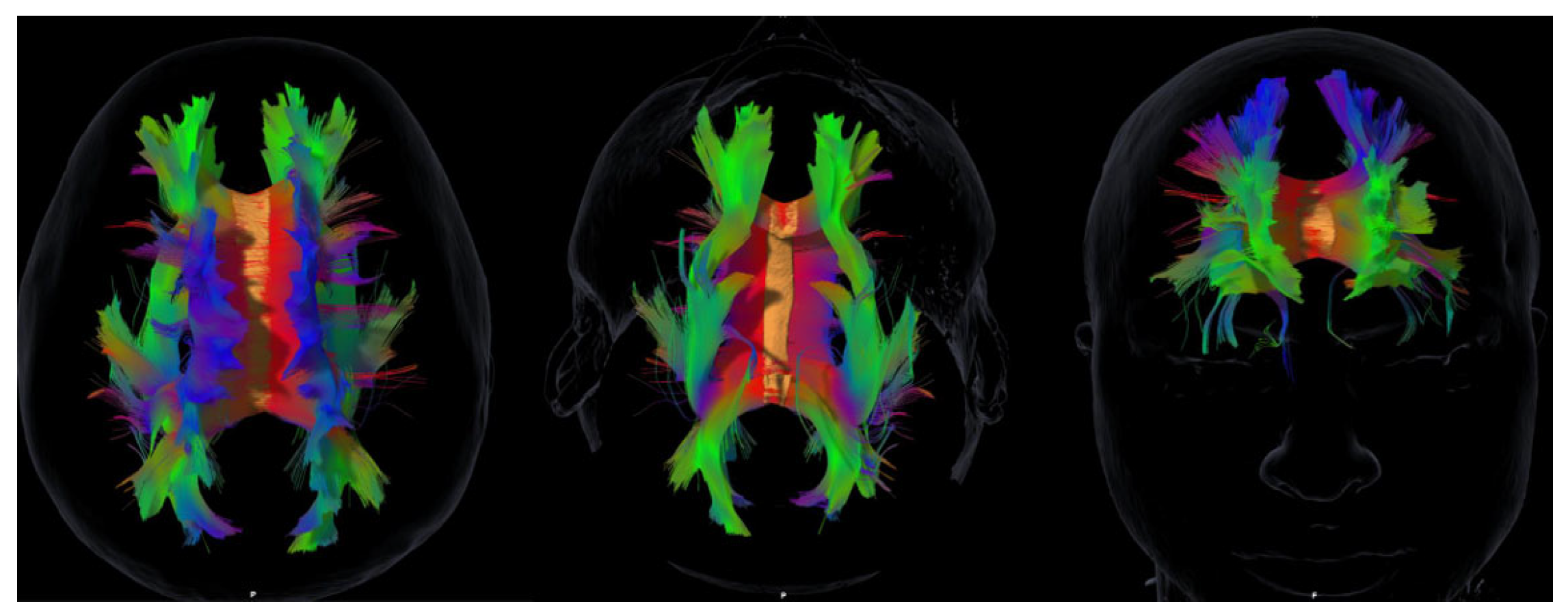
2.3. MRI Acquisition and Tractography

MRI data were acquired using a clinical scanner with diffusion-weighted imaging sequences suitable for tractography. Standard pre-processing (motion and eddy-current correction) was applied, and major association tracts (SLF, ILF, UF, IFOF, cingulum) were reconstructed using deterministic fiber-tracking algorithms (Brainlab Elements, v2.0). Volumetric estimates (mm3) were obtained by summing voxels traversed by streamlines. All reconstructions were performed by an experienced neuroradiologist in collaboration with neurosurgeons, blinded to neuropsychological results. Fiber-tracking principles followed standard recommendations [25].
The following association tracts were reconstructed: superior longitudinal fasciculus (SLF), inferior longitudinal fasciculus (ILF), uncinate fasciculus (UF), inferior fronto-occipital fasciculus (IFOF), and cingulum. The superior fronto-occipital fasciculus (SFOF) was also reconstructed. However, given the ongoing controversy about its existence in humans [26], SFOF volumes are reported for transparency but not emphasized in the main discussion or interpretation of results. All reconstructions were performed by an experienced neuroradiologist in collaboration with neurosurgeons, blinded to neuropsychological data. Representative tractography reconstructions are shown in Figure 1, images are illustrative and do not provide diagnostic information at the individual level.

2.4. Statistical Analyses

Descriptive statistics were used for demographic and clinical data. Normality of distributions was examined with the Shapiro–Wilk test. Given the small sample size and the exploratory nature of this pilot study, associations between tract volumes and neuropsychological outcomes were examined using Spearman’s rank correlation (two-tailed, p < 0.05). No correction for multiple comparisons was applied, as the aim was to identify potential tract–behavior relationships to guide future hypothesis-driven studies with larger samples. All analyses were conducted in R (v.4.3.2) and Python (Scikit-learn, NetworkX, Matplotlib, Pandas).
Exploratory k-means clustering (k = 3) and simple network analysis were also conducted on the correlation matrix; nodes with |ρ| > 0.70 were considered hubs for descriptive purposes.

2.5. Ethics

The study was approved by the Institutional Ethics Committee (Ref: CEIm 2022-206-1). Written informed consent was obtained from all participants, in line with the Declaration of Helsinki.

3. Results

3.1. Participant Characteristics

Ten ABI patients (6 women, 4 men; mean age = 51.1 years, range = 29–63) were included. All presented with cognitive sequelae sufficient to warrant evaluation. Descriptive demographic and clinical data are presented in Table 1.

3.2. Neuropsychological Outcomes

All participants scored below the MoCA cutoff (<25), confirming global cognitive impairment [19]. Mean scores indicated consistent difficulties in attention and working memory (Digit Span forward = 5.4 ± 1.6; backward = 3.6 ± 0.7). Processing speed and flexibility (TMT A and B) were markedly heterogeneous, with several participants performing in the pathological range. RCFT scores reflected variable visuoconstructive and planning abilities.
Regarding emotional symptoms, mean HADS scores suggested mild-to-moderate anxiety (10.6 ± 4.3) and depression (8.9 ± 5.2). Social cognition, assessed via TECA, revealed reduced empathy across subdomains, particularly empathic distress and empathic joy.

3.3. White Matter Tract Volumes

Diffusion-based tractography successfully reconstructed all major association tracts (SLF, ILF, UF, IFOF, cingulum).

3.4. Exploratory Tract–Behavior Associations

Spearman correlations revealed several significant associations between tract volumes and neuropsychological outcomes (Table 2):
  • SLF (left): negatively correlated with MoCA (ρ = –0.64, p = 0.046) and RCFT (ρ = –0.66, p = 0.039); positively correlated with slower TMT performance (ρ = 0.86, p = 0.001).
  • UF (left): negatively correlated with Digit Span forward (ρ = –0.66, p = 0.038).
  • UF (right): positively correlated with Digit Span backward (ρ = 0.68, p = 0.029).
  • ILF (left): negatively correlated with total empathy scores (ρ = –0.66, p = 0.039).
  • Cingulum (left): negatively correlated with empathic distress (ρ = –0.64, p = 0.046).
  • IFOF (right): negatively correlated with empathic joy (ρ = –0.71, p = 0.022).
  • No significant associations were observed for HADS scores.
  • Age was negatively correlated with left cingulum volume (ρ = –0.73, p = 0.016).

3.5. Cluster and network analyses

Cluster analysis (k = 3) identified subgroups with distinct profiles. Network analysis highlighted the left SLF and bilateral UF as structural hubs (|ρ| > 0.7):
  • Cluster 1: marked cognitive impairment with preserved empathy.
  • Cluster 2: mixed cognitive and affective deficits.
  • Cluster 3: milder deficits but reduced empathy across domains.

4. Discussion

This exploratory pilot study examined associations between white matter tract integrity and cognitive, emotional, and social outcomes in patients with acquired brain injury (ABI). The results revealed paradoxical structure–function associations, consistent with compensatory or maladaptive mechanisms of reorganization, and highlighted the role of specific tracts in supporting empathy and other socio-emotional functions.

4.1. Structure–Function Paradox and Network Reorganization

One of the most striking findings was the negative association between tract volumes and performance in cognitive tasks, particularly for the left superior longitudinal fasciculus (SLF). Larger SLF volumes correlated with poorer global cognition (MoCA), reduced visuospatial organization (RCFT), and slower executive performance (TMT). These paradoxical associations challenge the assumption that larger structural volumes necessarily imply better function. Instead, they may reflect inefficient or maladaptive reorganization, where volumetric increases occur without corresponding microstructural efficiency [18].
Similar findings have been reported in post-stroke and aging populations, where volumetric hypertrophy coexists with functional decline [3]. Such evidence reinforces the perspective that recovery is determined less by absolute tract size and more by network integration and microstructural quality [4,5].

4.2. Lateralization and Tract-Specific Contributions

The study also revealed hemispheric asymmetries. The left UF correlated negatively with immediate memory (Digit Span forward), whereas the right UF correlated positively with working memory (Digit Span backward). These findings align with lateralization models, suggesting that left tracts support verbal–executive functions while right tracts contribute more strongly to socio-emotional and memory regulation.
These results are consistent with tractography studies linking the UF and cingulum to memory and emotion, and the SLF to executive control [12,13]. Such asymmetries highlight the need for individualized rehabilitation, as patients may present with lateralized vulnerabilities requiring targeted interventions.

4.3. Social Cognition and Empathy as Novel Contributions

A distinctive contribution of this study is the inclusion of empathy. Few tractography studies in ABI have directly examined social cognition, despite its critical role in quality of life and reintegration. Our results revealed consistent associations between empathy (TECA scores) and ILF, UF, and cingulum volumes.
The negative correlations observed suggest that volumetric remodeling may represent compensatory mechanisms that sustain socio-emotional performance through alternative pathways. This aligns with prior evidence implicating fronto-limbic and temporo-occipital networks in empathy and socio-emotional processing [16,17]. By demonstrating that empathy can be structurally anchored in ABI, this study expands the scope of neurorehabilitation beyond executive and motor domains.

4.4. Clinical Implications

Clinically, these findings underscore the potential of tractography to complement neuropsychological assessment. Two patients with comparable test scores may differ substantially in tract integrity, which could explain divergent rehabilitation trajectories. Identifying such tract-specific profiles may help clinicians tailor interventions: for example, prioritizing attentional and executive training in patients with SLF involvement, or incorporating emotional regulation therapies when UF or cingulum asymmetries are evident.
This perspective supports the emerging paradigm of precision neurorehabilitation, in which interventions are individualized based on residual structural and functional connectivity rather than group averages [4]. Novel experimental approaches in TBI rehabilitation further underscore the need for individualized, multimodal strategies that extend beyond cognitive training alone [29].

4.5. Future Directions

While exploratory, this study lays the foundation for future multimodal investigations. Larger longitudinal cohorts should confirm whether the associations reported here persist over time and predict recovery. Integrating tractography with advanced microstructural metrics (fixel-based analysis, NODDI), functional modalities (resting-state fMRI, EEG), and connectome-based modeling will be critical to capture dynamic reorganization [15].
Artificial intelligence methods are increasingly being applied to multimodal neuroimaging and may enable predictive models that combine structural, functional, and clinical variables to forecast rehabilitation outcomes [6,7,8]. Incorporating empathy and social cognition into such models could help optimize not only functional but also social reintegration.

4.6. Limitations

Several limitations must be acknowledged. First, the small sample size constrains statistical power and generalizability. Nevertheless, sample sizes in tractography studies of ABI often range between 8 and 15 patients due to the logistical complexity and costs of acquisition [27,28]. Accordingly, this work should be interpreted as hypothesis-generating rather than confirmatory. Second, the absence of a control group limits between-group comparisons; however, the study was intentionally designed to characterize intra-individual variability, which is highly relevant for personalized neurorehabilitation. Third, multiple correlations were tested without statistical correction, increasing the risk of false positives. This decision was deliberate, given the exploratory design, and future larger-scale studies will be required to confirm these preliminary associations. Finally, volumetric indices provide indirect estimates of tract integrity; combining tractography with advanced microstructural (e.g., NODDI, fixel-based analysis) and functional methods will be essential in subsequent work.
Finally, although SFOF volumes were reconstructed and reported in the results tables, their interpretation should be considered tentative. The existence of the SFOF in humans remains debated, with some studies supporting its identification and others suggesting that it may reflect spurious reconstruction in tractography [26]. Given this controversy and the small sample size, SFOF data are provided for transparency but were not emphasized in the discussion.

5. Conclusions

This exploratory pilot study suggests that associations between white matter tract volumes and cognitive, affective, and empathy-related outcomes in acquired brain injury may be more complex than traditionally assumed. Preliminary findings point to the involvement of association tracts such as the ILF, UF, and cingulum in empathy and emotional regulation, extending the discussion of rehabilitation targets beyond conventional cognitive domains. These results should be interpreted with caution, yet they support the feasibility of integrating tractography with neuropsychological assessment and provide hypotheses for future research with larger samples to validate and expand these observations.

Author Contributions

Conceptualization, R.B.G. and W.P.C.; methodology, R.B.G. and W.P.C.; formal analysis, R.B.G.; investigation, R.B.G. and E.F.D.V.; resources, C.S.P., S.B.A. and J.M.M.; data curation, R.B.G. and E.F.D.V.; writing—original draft preparation, R.B.G.; writing—review and editing, R.B.G. and W.P.C.; supervision, W.P.C. and J.L.H.F.; visualization, C.S.P., S.B.A. and J.M.M.; project administration, R.B.G. All authors have read and agreed to the published version of the manuscript.

Funding

This research received no external funding.

Institutional Review Board Statement

The study was conducted in accordance with the Declaration of Helsinki and approved by the Institutional Ethics Committee of CEIm Las Palmas (protocol code 2022-206-1).

Informed Consent Statement

Informed consent was obtained from all subjects involved in the study.

Data Availability Statement

The data presented in this study are available on reasonable request from the corresponding author. The data are not publicly available due to privacy restrictions.

Acknowledgments

We thank Francisco Rodríguez and Abidan Cerdeña for their generous and voluntary collaboration

Conflicts of Interest

The authors declare no conflict of interest.

Abbreviations

The following abbreviations are used in this manuscript:
ABI Acquired brain injury
Anx Anxiety
Dep Depression
DST Digit Span Test
ED Empathic distress
EJ Empathic joy
EU Emotional understanding
Fw Forward (Digit Span)
HADS Hospital Anxiety and Depression Scale
ILF Inferior longitudinal fasciculus
IFOF Inferior fronto-occipital fasciculus
MoCA Montreal Cognitive Assessment
PC Percentile score
PT Perspective taking
RCFT Rey–Osterrieth Complex Figure Test
Rv Reverse (Digit Span)
SD Standard deviation
SFOF Superior fronto-occipital fasciculus
SLF Superior longitudinal fasciculus
TECA Cognitive and Affective Empathy Test
TIPO Total drawing performance-adjusted percentile score
TMT Trail Making Test
UF Uncinate fasciculus
WAIS Wechsler Adult Intelligence Scale
WM White matter
ρ Spearman’s correlation coefficient

References

  1. Pulido ML. Neurocognitive outcomes in acquired brain injury. Rev Neurol. 2023;76(2):87–95.
  2. Njomboro P, Deb S, Humphreys GW. Social cognition deficits in traumatic brain injury: a clinical perspective. Brain Inj. 2014;28(9):1146–51. [CrossRef]
  3. Filley CM, Fields RD. White matter and cognition: making the connection. J Neurophysiol. 2016;116(5):2093–104. [CrossRef]
  4. Forkel SJ, Thiebaut de Schotten M, Kawadler JM, Dell’Acqua F, Danek A, Catani M. Disconnection and dysfunction in the human brain: from lesions to networks. Brain. 2021;144(1):3–20. [CrossRef]
  5. Chin R, Chang SWC, Holmes AJ. Beyond cortex: the evolution of the human brain. Psychol Rev. 2023;130(2):285–307. [CrossRef]
  6. Armstrong E. Precision connectomics: mapping and predicting brain networks in health and disease. Trends Neurosci. 2024;47(2):123–36. [CrossRef]
  7. Cai W, Chen T, Ryali S, Kochalka J, Li CS, Menon V. AI-driven tractography pipelines for massively parallel reconstruction. Neuroinformatics. 2024;22(3):445–60. [CrossRef]
  8. Orenuga O, Ogunyemi O, Bamisile O, Koshy S. Traumatic brain injury and artificial intelligence: shaping the future of neurorehabilitation—A review. Life. 2025;15(3):424. [CrossRef]
  9. Newcombe VF, Correia MM, Ledig C, Abate MG, Outtrim JG, Chatfield D, et al. Dynamic changes in white matter abnormalities correlate with late improvement and deterioration following traumatic brain injury: a diffusion tensor imaging study. Neurorehabil Neural Repair. 2016;30(1):49–62. [CrossRef]
  10. He X, Xu W, Zhang Q, Wang J, Zhang M. Cingulum bundle microstructure predicts affective regulation after brain injury. Hum Brain Mapp. 2021;42(18):6045–58. [CrossRef]
  11. Voelbel GT, Genova HM, Chiaravalloti ND, Hoptman MJ. Diffusion tensor imaging of traumatic brain injury review: implications for neurorehabilitation. NeuroRehabilitation. 2012;31(3):281–93. [CrossRef]
  12. Conner AK, Briggs RG, Rahimi M, Sali G, Baker CM, Burks JD, et al. A connectomic atlas of the human superior longitudinal fasciculus. Oper Neurosurg (Hagerstown). 2018;15(Suppl 1):S407–14. [CrossRef]
  13. Von der Heide RJ, Skipper LM, Klobusicky E, Olson IR. Dissecting the uncinate fasciculus: disorders, controversies and a hypothesis. Brain. 2013;136(6):1692–707. [CrossRef]
  14. Comes-Fayos J, García-García I, Peña-Casanova J. Role of association tracts in socio-emotional integration. Rev Neurol. 2018;67(7):263–72.
  15. Takahashi M, Iwamoto K, Fukatsu H, Naganawa S, Iidaka T, Ozaki N. White matter microstructure of the cingulum and cerebellar peduncle is related to sustained attention and working memory. Neurosci Lett. 2010;477(2):72–6. [CrossRef]
  16. Catani M, Dell’Acqua F, Thiebaut de Schotten M. A revised limbic system model for memory, emotion and behaviour. Neurosci Biobehav Rev. 2020;103:19–31. [CrossRef]
  17. Krick S, Koob JL, Latarnik S, Volz LJ, Fink GR, Grefkes C, et al. Neuroanatomy of post-stroke depression: association between symptom clusters and lesion location. Brain Commun. 2023;5(5):fcad275. [CrossRef]
  18. Bartolomeo P, Thiebaut de Schotten M. Let thy left brain know what thy right brain doeth: inter-hemispheric compensation of functional deficits after brain damage. Neuropsychologia. 2016;93(Pt B):407–12. [CrossRef]
  19. Nasreddine ZS, Phillips NA, Bédirian V, Charbonneau S, Whitehead V, Collin I, et al. The Montreal Cognitive Assessment (MoCA): a brief screening tool for mild cognitive impairment. J Am Geriatr Soc. 2005;53(4):695–9. [CrossRef]
  20. Reitan RM. Validity of the Trail Making Test as an indicator of organic brain damage. Percept Mot Skills. 1958;8:271–6. [CrossRef]
  21. Wechsler D. WAIS-III: Wechsler Adult Intelligence Scale. 3rd ed. San Antonio: Psychological Corporation; 1997.
  22. Rey A. L’examen clinique en psychologie. Paris: Presses Universitaires de France; 1964.
  23. Zigmond AS, Snaith RP. The Hospital Anxiety and Depression Scale. Acta Psychiatr Scand. 1983;67(6):361–70. [CrossRef]
  24. López-Pérez B, Fernández-Pinto I, Abad FJ. TECA: Test de Empatía Cognitiva y Afectiva. Madrid: TEA Ediciones; 2008.
  25. Mori S, van Zijl PC. Fiber tracking: principles and strategies—a technical review. NMR Biomed. 2002;15(7–8):468–80. [CrossRef]
  26. Forkel SJ, Friedrich P, Thiebaut de Schotten M, Howells H. Is the “superior fronto-occipital fasciculus” really there in humans? Reproducibility, reliability and controversies in diffusion tractography. Neuroimage Clin. 2020;27:102192. [CrossRef]
  27. Ressel V, O’Gorman Tuura R, Scheer I, van Hedel HJ. Diffusion tensor imaging predicts motor outcome in children with acquired brain injury. Brain Imaging Behav. 2017;11(5):1373–84. [CrossRef]
  28. Lennartsson F, Holmström L, Eliasson AC, Flodmark O, Forssberg H, Tournier JD, et al. Advanced fiber tracking in early acquired brain injury causing cerebral palsy. AJNR Am J Neuroradiol. 2015;36(1):181–7. [CrossRef]
  29. Pilipović K. Traumatic brain injury: novel experimental approaches. Life. 2025;15(6):884. [CrossRef]
Figure 1. Representative tractography reconstructions in a patient with acquired brain injury. The images illustrate whole-brain association fibers, from which tract volumes were quantified for subsequent analyses.
Preprints 178183 g001
Table 1. Patient characteristics, test scores, and volumes of association tracts.
N=10
Mean age (range) 51.1 (29-63)
Proportion of women 60%
Social Cognition, Cognitive Functions, And Emotional Outcomes
Montreal Cognitive Assessment test. Mean score (SD) 21.9 (3.03)
Digit Span Test. Mean score (SD) Forward 5.4 (1.6)
Reverse 3.6 (0.7)
Trail Making Test. Mean time, seconds (SD) Part A 51.3 (28.6)
Part B 147.5 (173.3)
Rey Complex Figure Test. Mean score (SD) Type 42.0 (26.5)
Percentile (PC) 60.0 (25.8)
Hospital Anxiety and Depression Scale. Mean score (SD) Depression 8.9 (5.2)
Anxiety 10.6 (4.3)
Empathy Test. Mean score (SD) Total 38.8 (32.4)
Perspective taking 33.5 (27.7)
Emotional understanding 30.1 (24.4)
Empathic distress 43.1 (31.4)
Empathic joy 33.7 (25.6)
Volumes of Association Tracts Mean volume mm3 (SD)
Fronto-occipital Right superior 501.8 (100.9)
Left superior 518.8 (89.7)
Right inferior 525.5 (60.8)
Left inferior 524.5 (66.2)
Uncinate fasciculus Right 392.6 (130.6)
Left 400.8 (142.3)
Longitudinal fasciculus Right superior 425.2 (111.9)
Left superior 418.2 (85.7)
Right inferior 515.2 (78.4)
Left inferior 496.4 (76.9)
Cingulum Right 461.7 (71.8)
Left 495.8 (88.7)
Abbreviations: SD, standard deviation.
Table 2. Spearman’s correlation coefficient between test scores and volumes of association tracts.
Tract/Test MoCA DST TMT RCFT HADS TECA
Fw Rv Part A Part B TIPO PC Dep Anx Total PT EU ED EJ
Right superior fronto-occipital -0.13 (0.724) 0.06 (0.862) 0.09 (0.811) -0.18 (0.629) -0.16 (0.663) -0.09 (0.797) 0.11 (0.772) -0.37 (0.287) -0.1 (0.776) -0.08 (0.827) -0.23 (0.521) -0.14 (0.699) -0.61 (0.064) 0.00 (1.00)
Left superior fronto-occipital -0.28 (0.431) 0.02 (0.958) 0.24 (0.502) 0.30 (0.399) 0.46 (0.179) -0.38 (0.285) -0.29 (0.413) -0.12 (0.748) -0.09 (0.815) -0.40 (0.254) -0.38 (0.275) -0.07 (0.854) -0.32 (0.370) -0.26 (0.476)
Right inferior fronto-occipital 0.10 (0.787) -0.53 (0.112) 0.19 (0.589) 0.27 (0.457) 0.05 (0.894) 0.09 (0.796) 0.32 (0.371) 0.09 (0.813) 0.03 (0.940) -0.20 (0.586) 0.14 (0.693) -0.37 (0.293) -0.06 (0.866) -0.71 (0.022)
Left inferior fronto-occipital 0.23 (0.528) -0.39 (0.268) -0.07 (0.846) 0.44 (0.208) 0.29 (0.422) -0.19 (0.603) 0.13 (0.719) -0.06 (0.866) -0.26 (0.463) -0.33 (0.358) 0.30 (0.397) -0.30 (0.400) -0.03 (0.940) -0.54 (0.105)
Right uncinate fasciculus 0.43 (0.217) -0.15 (0.688) 0.68 (0.029) -0.35 (0.321) -0.38 (0.275) 0.03 (0.932) 0.29 (0.413) -0.03 (0.933) 0.05 (0.894) 0.01 (0.987) 0.29 (0.413) -0.24 (0.508) -0.24 (0.507) -0.69 (0.028)
Left uncinate fasciculus 0.36 (0.306) -0.66 (0.038) 0.60 (0.069) -0.19 (0.592) -0.25 (0.487) 0.34 (0.331) 0.55 (0.097) 0.30 (0.399) 0.16 (0.662) -0.15 (0.672) 0.20 (0.578) -0.48 (0.165) -0.28 (0.441) -0.66 (0.037)
Right superior longitudinal fasciculus -0.31 (0.390) -0.10 (0.781) -0.18 (0.617) 0.58 (0.078) 0.18 (0.614) -0.47 (0.172) -0.28 (0.434) -0.01 (0.987) 0.26 (0.464) 0.07 (0.853) 0.07 (0.841) 0.16 (0.662) 0.03 (0.933) -0.24 (0.498)
Left superior longitudinal fasciculus -0.64 (0.046) -0.03 (0.944) -0.44 (0.205) 0.86 (0.001) 0.72 (0.020) -0.55 (0.099) -0.66 (0.039) 0.15 (0.670) 0.19 (0.599) 0.06 (0.865) -0.15 (0.679) 0.41 (0.238) 0.29 (0.412) 0.13 (0.711)
Right inferior longitudinal fasciculus 0.57 (0.082) 0.49 (0.147) 0.44 (0.201) -0.49 (0.147) -0.32 (0.374) -0.34 (0.331) -0.4 (0.247) 0.47 (0.168) 0.47 (0.171) 0.08 (0.827) 0.22 (0.544) 0.23 (0.531) 0.49 (0.151) 0.23 (0.521)
Left inferior longitudinal fasciculus 0.39 (0.263) 0.15 (0.688) 0.42 (0.224) 0.13 (0.731) 0.34 (0.336) -0.22 (0.544) -0.16 (0.668) 0.06 (0.880) -0.22 (0.542) -0.66 (0.039) -0.06 (0.868) -0.28 (0.432) 0.29 (0.410) -0.58 (0.077)
Right cingulum 0.02 (0.946) -0.48 (0.162) 0.66 (0.039) -0.14 (0.703) 0.06 (0.873) 0.38 (0.282) 0.18 (0.616) 0.60 (0.067) 0.34 (0.331) -0.06 (0.865) -0.11 (0.756) -0.16 (0.666) 0.03 (0.926) -0.35 (0.328)
Left cingulum -0.23 (0.523) -0.28 (0.429) 0.46 (0.177) 0.03 (0.938) -0.10 (0.776) 0.09 (0.796) 0.11 (0.771) 0.15 (0.671) 0.24 (0.513) 0.16 (0.659) 0.07 (0.847) 0.24 (0.507) -0.64 (0.046) -0.09 (0.815)
Abbreviations: Spearman’s correlation coefficients (ρ) between tract volumes and neuropsychological outcomes. Bold values indicate statistically significant correlations (p < 0.05). Abbreviations: Anx, anxiety; Dep, depression; DST, Digit Span test; ED, empathic distress; EJ, empathic joy; EU, emotional understanding; Fw, forward; HADS, Hospital Anxiety and Depression Scale; MoCA, Montreal Cognitive Assessment; PC, percentile score; PT, perspective taking; RCFT, Rey Complex Figure Test; Rv, reverse; TECA, Cognitive and Affective Empathy Test; TIPO, total drawing performance-adjusted percentile score; TMT, Trail Making Test.
Disclaimer/Publisher’s Note: The statements, opinions and data contained in all publications are solely those of the individual author(s) and contributor(s) and not of MDPI and/or the editor(s). MDPI and/or the editor(s) disclaim responsibility for any injury to people or property resulting from any ideas, methods, instructions or products referred to in the content.
Copyright: This open access article is published under a Creative Commons CC BY 4.0 license, which permit the free download, distribution, and reuse, provided that the author and preprint are cited in any reuse.
Prerpints.org logo

Preprints.org is a free preprint server supported by MDPI in Basel, Switzerland.

Subscribe

Disclaimer

Terms of Use

Privacy Policy

Privacy Settings

© 2025 MDPI (Basel, Switzerland) unless otherwise stated