
Submitted:
21 August 2025
Posted:
25 August 2025
You are already at the latest version
Abstract
The many uses of biochar extend to microbial enhancement in fermentation processes because it acts as a catalyst and a support medium. This study explores how varying biochar properties specifically concentration, temperature, and particle size affect the growth of hyper-ammonia-producing bacteria (HAB), using commercially sourced pine wood-derived biochar. Fermentation experiments were conducted using enriched rumen fluid under controlled conditions to monitor bacterial growth via optical density (OD600) over a 48-hour period. Our results showed that microbial proliferation was significantly influenced by all three parameters. The optimal growth was observed at 0.15% biochar concentration, a temperature of 45°C, and a particle size of 250 µm. While lower concentrations and smaller particles promoted microbial adhesion and colonization, higher levels appeared to hinder growth, likely due to surface saturation and reduced pore accessibility. SEM imaging supported these findings by revealing structural changes on the biochar surface at different concentrations, highlighting its role in microbial distribution. Regression analysis confirmed a strong correlation between biochar parameters and microbial activity, although no single variable stood out statistically due to likely multicollinearity and sample limitations. These findings suggest tuning biochar properties for optimal microbial processing, taking into consideration environmental factors and using additional imaging techniques to better understand HAB- biochar interactions.
Keywords:
biochar
; microbial fermentation
; hyper-ammonia-producing bacteria
; biochar particle size
; temperature optimization
; microbial growth kinetics
1. Introduction
Microbial fermentation plays a vital role in a wide range of industries, from producing foods and beverages to creating pharmaceuticals, biofuels and biochemicals [1]. It is also an essential part of environmental and waste management, supporting processes like anaerobic digestion for converting waste into energy and composting for recycling organic materials. Despite its broad application, microbial induced reactions are inherently slow, unlike chemical reactions. For instance, fermentation of complex feedstocks like lignocellulosic biomass, which doesn’t break down easily without proper pretreatment, is slow [2]. Fermentation rates are limited by microbial physiology and growth kinetics. Fermentative microbes are sensitive to medium compositions which can boost or hinder their proliferation [3]. Creating a supportive environment for microbes can help improve the fermentation process.
One effective approach is immobilizing or attaching the cells to a solid support matrix. [4] explain that immobilizing microbial cells helps them stay stable and more concentrated, which can lead to better yields and faster fermentation. This claim is supported by [5], who found that supported cultures generally offer better stress tolerance, reusability, and catalytic performance than free-floating cells. Hence, the need to provide microbes with a supportive environment to enable them to adapt easily to changing conditions leading to a more efficient fermentation process. However, not all materials may be suitable, sustainable and applicable for large-scale use. Some common materials like synthetic resins, polymer gels, or activated carbon are often expensive and require significant resources to produce. To keep fermentation cost-effective and eco-friendly, the support materials need to be affordable, widely available, and ideally made from renewable resources. Recent efforts have therefore turned to natural, low-cost support materials [6]. Biochar, produced by pyrolyzing biomass, is becoming a popular, affordable, and sustainable support material. By transforming agricultural residues into a valuable product, it promotes recycling and supports a circular economy. Additionally, biochar’s effects on microbial communities are generally positive, as studies have shown it doesn’t harm microbes. Although microbes will react differently to biochar depending on the condition [7], the proliferation of hyper ammonia producing bacteria in the presence of biochar is still a mystery.
Biochar’s porous structure, large surface area, and nutrient-rich particles offer a great environment for microbes to attach and thrive. It also contains functional groups and minerals like carbon, hydrogen, oxygen, and nitrogen, as well as key ratios like H/C and O/C [8,9]. The negatively charged surface of biochar helps attract nutrient ions and beneficial microbes [10]. Because biochar can create small, protective spaces that help microbes resist stress, it has proven to be an effective carrier for the bacterial strain N33 [11]. These traits make it an ideal environment for fermentative microbes, acting like a supportive scaffold that keeps them clustered and active. Biochar stands out as an affordable, eco-friendly material that’s easy to produce in large quantities and features a porous structure ideal for microbial growth [6]. Amending anaerobic microbial consortia with biochar led to high levels of biohydrogen yields from organic waste by encouraging biofilm formation and enriching beneficial bacteria [12]. Similarly, using biochar in ethanolic fermentation allowed microbes to handle normally inhibitive by-products, leading to significantly higher ethanol production under stress [13].
Toward the development of biofertilizer, our research group has previously developed bio-ammonia production via fermentation using hyper ammonia-producing bacteria (HAB) [14]. Inclusion of biochar in the bio-ammonia fermentation system would enhance biofertilizer mineral composition and potentially serve as support for HAB proliferation. However, not much is known about how biochar could affect HAB proliferation during fermentation, thereby evincing the importance of pursuing this research. This study aims to explore how biochar concentration, temperature, and particle size affect the growth of hyper-ammonia-producing bacteria (HAB) during fermentation, with the goal of identifying conditions that best support microbial proliferation and using SEM imaging to visually explore how biochar structure affects bacterial attachment and colonization.
2. Materials and Methods
Figure 1.
Process Flowchart for Biochar-Based Microbial Fermentation Study.
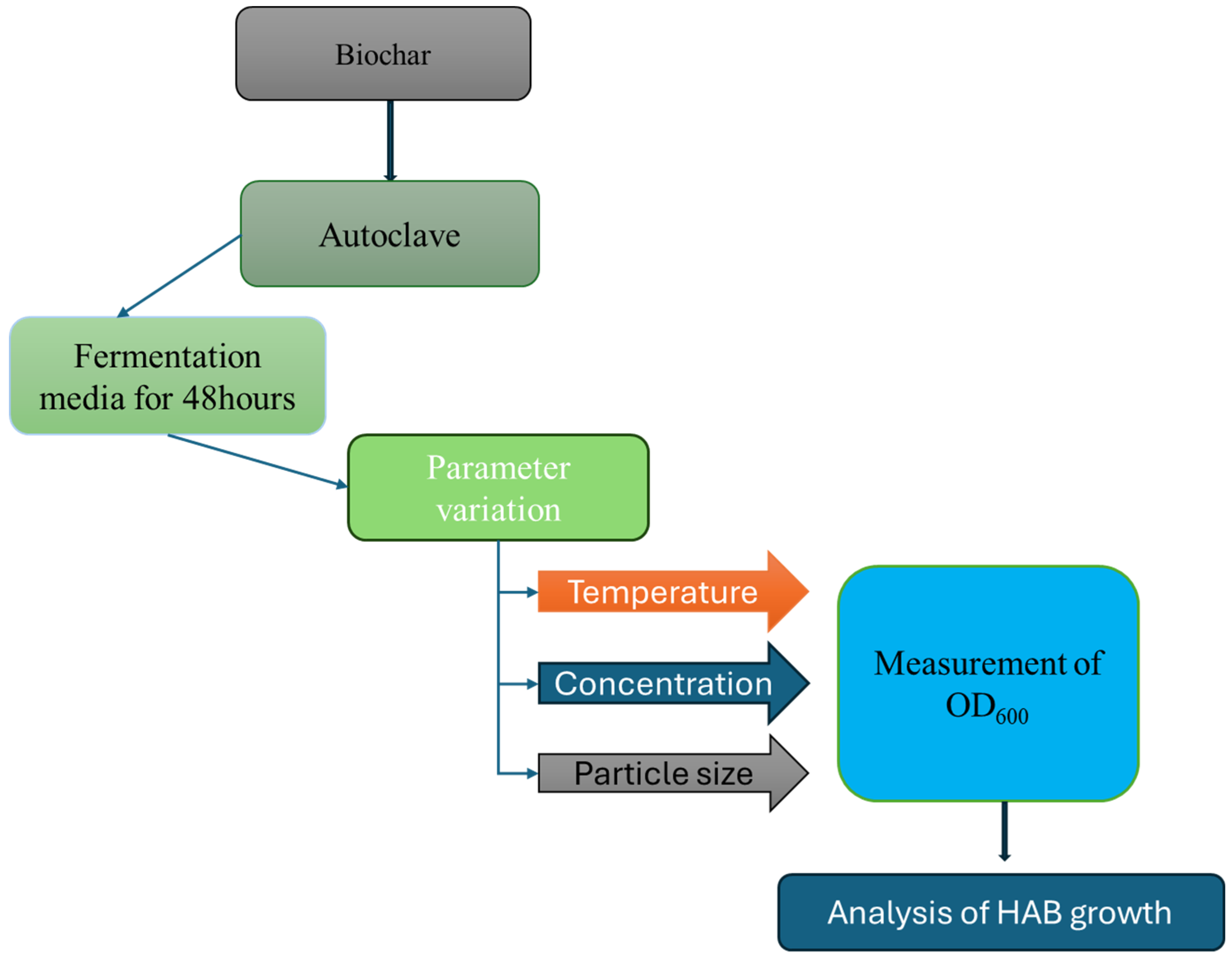
2.1. Media and Fermentation Conditions
The study utilized commercial sourced biochar derived from pine wood, which was sieved to obtain the desired particle sizes and sterilized at 121 °C to eliminate any remaining microbes. Fresh rumen fluid was collected from fistulated dairy cows at the North Dakota State University (NDSU) Animal Nutrition and Physiology Center (ANPC) and stored in 50 mL centrifuge tubes. Samples were kept in an incubation chamber at 39 °C until use.
To cultivate and isolate hyper-ammonia-producing bacteria (HAB), an enriched medium was prepared following the formulation described by [14]. The medium per liter consisted of: Na₂SO₄ (0.480 g), NaCl (0.10 g), CaCl₂·2H₂O (0.064 g), Na₂CO₃ (4.0 g), K₂HPO₄ (0.292 g), KH₂PO₄ (0.292 g), yeast extract (0.50 g), and of casamino acids (15 g). The pH was adjusted to 6.5 using HCl, and the medium was sterilized by autoclaving at 121 °C.
Prior to inoculation, the rumen fluid was allowed to settle, and the supernatant was diluted 1:100 to achieve an optical density (OD) of approximately 0.07. This diluted fluid was then added to the sterile medium and incubated at 39 °C with shaking at 130 rpm for 12 hours. The resulting culture was subsequently used as the inoculum for all experiments.
2.2. Experimental Set Up
The fermentation experiments were conducted using a Thermo Scientific MaxQ™ 4000 shaker, which ensured consistent agitation and precise temperature control. Each treatment was prepared in 250 mL sterile Erlenmeyer flasks containing 40 mL of autoclaved growth medium, inoculated with 0.4 mL of prepared HAB culture. Sterilized pine wood-derived biochar was added according to the specific variable under investigation concentration, temperature, or particle size. The flasks were securely clamped onto the shaker platform and incubated for 48 hours under controlled conditions, with the temperatures ranging from 30°C to 50°C and a shaking speed maintained at130 rpm. These conditions promoted uniform mixing, prevented particle settling, and enhanced interaction between the biochar and microbes. Samples were collected every six hours for optical density measurements at 600 nm (OD600) and filtered using Whatman qualitative filter paper to separate the biochar particles.
2.2. Biochar Concentration Experiments
The effect of varying biochar concentrations on the growth rates of HAB was evaluated using six different biochar loadings: 0%, 0.05%, 0.1%, 0.15%, 0.2%, and 0.25% (w/v). This range aligns with previous findings suggesting that low to moderate biochar levels can enhance microbial activity, while higher concentrations such as those above 2% reported by [15], may reduce digestibility and hinder microbial function. Optical density (OD600) was measured at 6-hours intervals over a 48-hour incubation period using a spectrophotometer set at 600 nm. The growth medium was inoculated HAB and incubated at 37 °C on a shaker set to 120 rpm.
A biochar particle size of 850 μm was selected to balance microbial accessibility, as supported by He et al. (2020), who found that particles <1 mm enhance microbial colonization and sustained methanogenesis. To maintain consistency across treatments, 0.1 g of biochar was added to 40 ml of media, ensuring a representative biochar-to-liquid ratio, while 0.4 ml of HAB inoculum was used to provide a uniform microbial load for tracking growth patterns. All treatments were prepared in duplicate for reliability.
Bacteria growth phases were identified based on OD trends. Samples were collected and filtered through qualitative Whatman filter paper, and the optimal biochar concentration for bacterial growth was determined from the OD measurements.
2.3. Temperature Experiments
Experiments were conducted to evaluate the effect of temperature on the growth of HAB in the presence of biochar at five distinct temperatures: 30°C, 35°C, 40°C, 45°C, and 50°C. The study included two treatment groups: the first comprised biochar and medium only (BM), while the second included biochar, medium, and HAB (BMH). Each treatment was prepared in duplicate using 2g of 850 µm biochar/800ml of media. For each experimental unit, 40ml of media containing 0.1 g of biochar and 0.4ml of HAB inoculant (for BMH only) was used. Samples were collected every 6-hour interval for 48 hours. OD was measured at each time point, and all samples were filtered using Whatman qualitative filter paper to remove biochar particles. A two-way ANOVA was conducted to assess the significance of differences in bacterial growth across treatments and temperatures.
2.4. Biochar Particle Size Experiments
The impact of biochar particle size on microbial interactions was evaluated using three biochar particles sizes: 250 µm, 425 µm, and 1.40 mm. Experiments were conducted in duplicate using growth media inoculated with HAB. Each treatment consisted of 40 mL of media, 0.1 g of biochar of the specified particle size, and 0.4 mL of HAB inoculum. Samples were collected every six hours over a 48-hours period and filtered using Whatman qualitative filter paper to remove biochar particles. Microbial interaction levels were quantified by measuring OD values, and statistical analyses were performed to determine significant differences among particle sizes, thereby identifying the size that best supports microbial growth and activity.
2.5. Statistical Analysis
Two-way ANOVA was used to assess the effects of time and experimental variables specifically biochar concentration, temperature, and particle size on microbial growth. Statistical significance was determined at a p-value threshold of < 0.05. All analyses were performed using R (version 4.3.0). Additionally, regression analysis was performed to evaluate the relationship between each experimental factor (biochar concentration, temperature, and particle size) and microbial growth, as measured by optical density (OD). This approach enabled us to identify optimal conditions for enhancing the growth of HAB.
2.6. Scanning Electron Microscope Analysis (SEM)
To prepare the samples, biochar was applied to adhesive carbon tabs that were subsequently mounted on cylindrical aluminum holders. To ensure optimal imaging conditions, the excess biochar was carefully removed using a nitrogen gas stream, which aids in preventing contamination and ensuring a clear view of the surface features. Before imaging, a conductive gold coating was applied to the samples using a Cressington 108auto sputter coater (Ted Pella Inc., Redding CA, USA). Coating will help to enhance the conductivity of the biochar samples, by improving the quality of the SEM images and reducing charging effects during electron beam exposure. Images were obtained using a JEOL JSM-6490LV scanning electron microscope (JEOL USA, Peabody MA, USA) at an accelerating voltage of 15 kV.
3. Results
3.1. Temperature Effect
Temperature is a key factor influencing microbial growth and interactions within their communities [16]. Different microbial species respond uniquely to temperature changes, which can lead to variations in behavior and community structure [17]. Some microbes thrive at elevated temperatures, exhibiting accelerated growth, while others experience thermal stress that impairs survival and metabolic function [18,19].
Biochar helps microbes stay active under high temperatures by protecting their enzymes and supporting normal metabolic function, even during heat stress [20]. Its hydrophobic properties also facilitate interactions at the gas-liquid interface in static systems potentially stabilizing the thermal characteristics of the surrounding medium [21]. Furthermore, biochar enhances enzyme stability at higher temperatures, boosting microbial performance and making it a valuable tool for optimizing microbial processes [22]
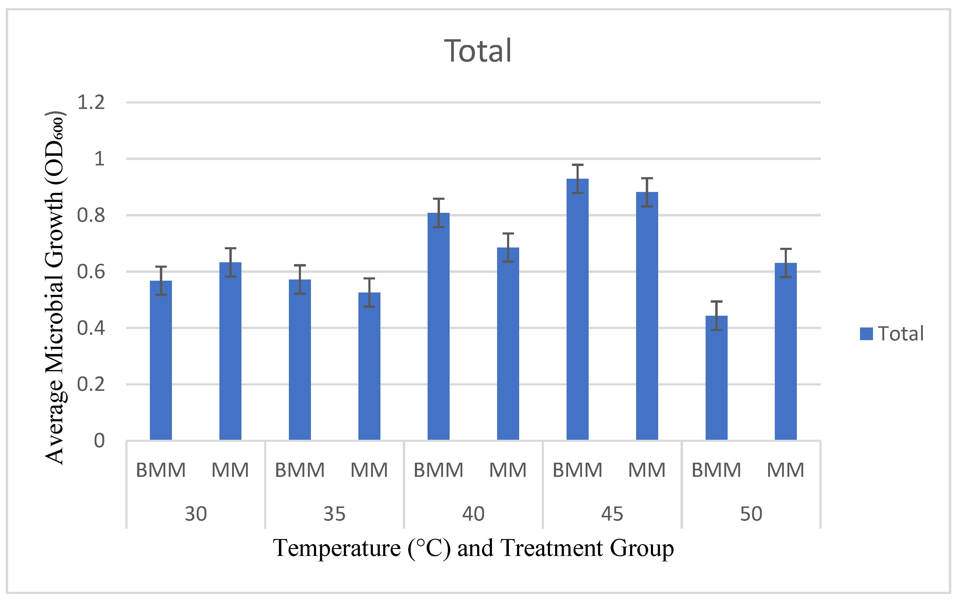
Achieving optimal microbial growth requires careful temperature regulation. In our study, we examined the growth patterns of HAB in the presence of biochar across a temperature range of 30°C to 50°C, over a 48-hours period. Treatments that included biochar, medium, and HAB (BMH) consistently showed higher microbial growth than those containing only biochar and medium (BM), especially at higher temperatures. The highest microbial activity was observed in BMH treatments at 45°C, with OD values occurring at 48-hour mark. This supports previous findings which suggest that increased temperatures accelerate microbial biomass turnover, potentially favoring fast-growing microbial species [23].
Conversely, Microbial activity was notably reduced at lower temperatures, with OD₆₀₀ values of 0.567 at 30 °C and 0.572 at 35 °C. The minimal difference between these values suggests that HAB growth was similarly limited under both conditions, likely due to temperature-induced constraints on enzyme efficiency and metabolic function. Reduced molecular movement at these temperatures leads to diminished enzyme activity due to insufficient kinetic energy for effective substrate binding and catalysis. This is particularly evident in mesophilic microbes, which typically exhibit optimal enzyme activity around 37°C. As the temperatures drop below this threshold, metabolic rates decline, resulting in slower microbial growth and reduced metabolic efficiency [24]. This trend is consistent with findings by [25], who reported changes in microbial community structure and metabolic efficiency at suboptimal temperatures.
Interestingly, temperatures above 45°C had a detrimental effect on microbial activity. This decline is likely due to enzyme denaturation, which disrupts essential metabolic pathways. These observations highlight the importance of maintaining an optimal temperature around 45°C for maximizing the synergistic effects of biochar and promoting robust microbial growth. It can be concluded that although biochar significantly enhances microbial activity, its effectiveness is closely tied to environmental factors, particularly temperature.
Figure 2.
Average growth rate for biochar, HAB and media at different temperatures.
3.2. Particle Sizes
Biochar particle size is a crucial parameter influencing its efficiency and microbial activity in both soil systems and fermentation media. Recent studies, such as [26], highlight that microbial colonization, diversity, and activity are strongly influenced by biochar particle size, with smaller particles boosting microbial growth thanks to their greater surface area and porosity. These characteristics offer ample sites for microbial attachment and create favorable microenvironments that support microbial growth [27,28,29]. Studies exploring the impact of biochar particle size on microbial activity and gas production consistently emphasize the advantages of finer particles. [30] demonstrated that smaller biochar particles significantly improved forage digestibility and gas output compared to larger sizes. Supporting this, [31] found that while most particle sizes enhanced methane yields during fermentation, biochar particles in the 1–3 cm range were less effective, likely due to their tendency to float and limit physical contact with microbial communities. These trends are further explained by [32], who noted that fine biochar particles provide a higher surface area-to-volume ratio, increasing the availability of reactive sites for microbial colonization and facilitating electron transfer critical in syntrophic microbial interactions. On the other hand, large biochar particles can still benefit fermentation by acting as stable scaffolds for microbial communities, as [33] showed they support the accumulation of methanogenic archaea and secondary fermenters in anaerobic digesters.
To optimize biochar particle size for HAB fermentation, we used different sizes of biochar to see how each one affected the growth of hyper-ammonia-producing bacteria. The treatments included 250 µm, 425 µm and 1.40 mm biochar particle sizes and were applied for 6, 24, 30, 42 and 48 hours, respectively. In the first stage of the experiment (6–24 h), the levels of microbial interaction were low for all the treatments, which is in accordance with the lag phase that is seen when bacteria are adjusting to a new environment [34]. It was expected that microbial interaction levels would increase as bacteria continued to adapt to the biochar environment.
At the end of 48hours, microbial interaction levels increased significantly, particularly with 250 and 425 µm biochar particles. These findings corroborate prior research emphasizing the contribution of smaller particles to microbial colonization and activity via increased surface area and porosity [28].
On the other hand, the 1.40 mm biochar particles had a slower rate of microbial interaction throughout the experiment. Their low surface area and pore volume probably suppressed microbial colonization and activity, and thus were less effective in supporting HAB. The finding agrees with the previous research that has shown that larger biochar particles are less effective in microbial environments [35]. These findings suggest the need to use the right size of biochar particles to enhance microbial activity.
3.3. Biochar Concentration
Biochar concentration plays a critical role in microbial-driven processes such as fermentation, where its application can enhance nutrient retention and provides surfaces for microbial immobilization. Although biochar offers notable advantages, [36] emphasized that there is no universally optimal biochar concentration. Instead, it must be tailored to specific application goals and environmental contexts. This underscores the importance of optimizing biochar dosage to support the growth of hyper-ammonia-producing bacteria (HAB) without introducing adverse effects.
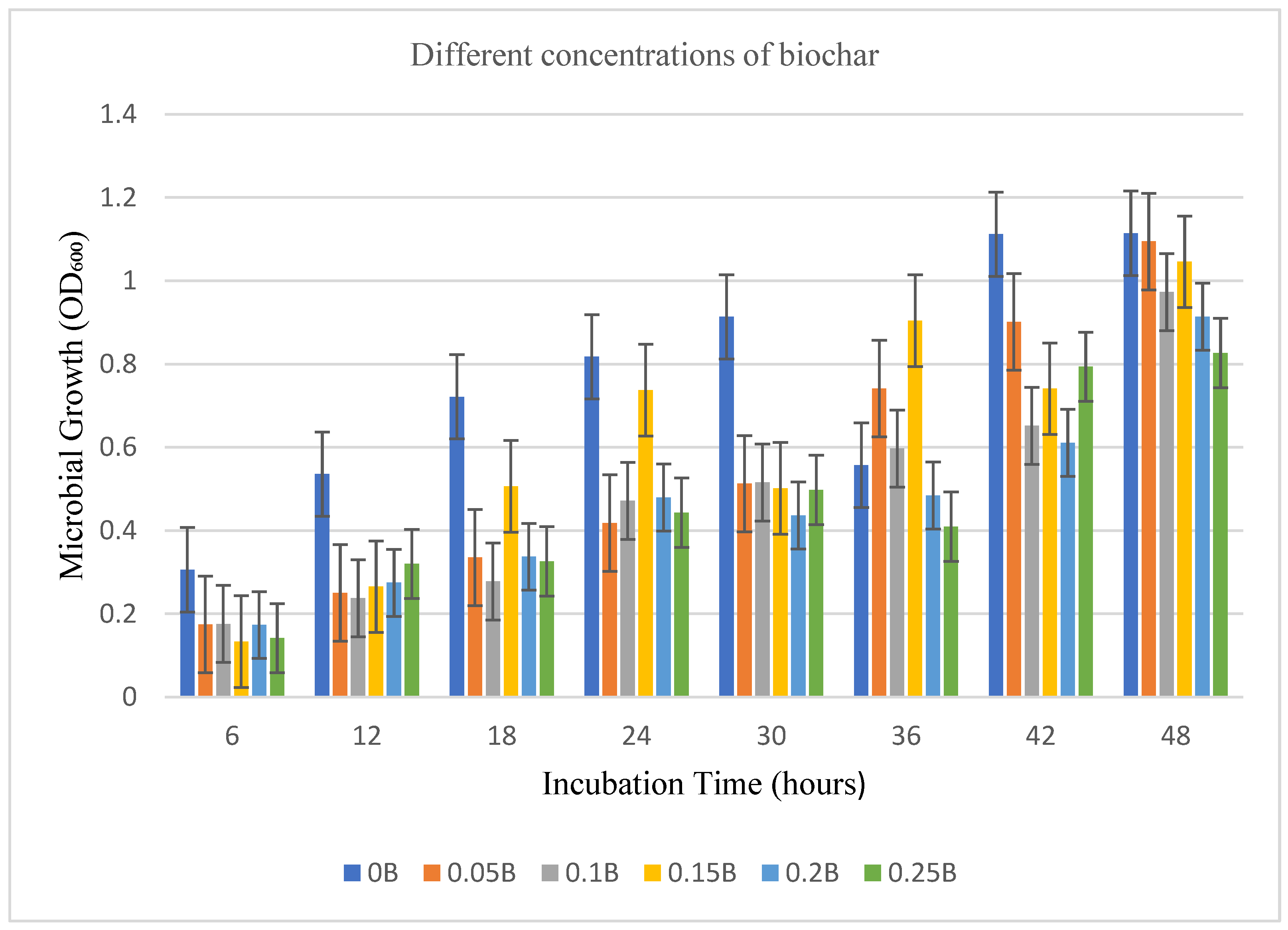
In this study, we evaluated the impact of various biochar concentrations (ranging from 0% to 0.25% w/v) on the growth kinetics of free-living HAB (Figure 3). The results show that smaller biochar particles support more robust HAB proliferation, likely due to greater surface area and porosity. Compared to the control (0% biochar), the addition of biochar generally reduced the concentration of free HAB in solution, presumably due to microbial entrapment or adhesion to biochar surfaces. This observation aligns with previous findings that biochar offers a porous and structurally favorable substrate for bacterial attachment and immobilization [37].
During the initial phases of incubation (6–18 hours), microbial growth was observed across all treatments, although no consistent trend emerged across biochar concentrations. This variability suggests that early microbial activity may be influenced by additional factors such as microbial adaptation, competition, or bioavailability of nutrients within the substrate.
Between 24 and 30 hours, particularly in the control (0% biochar) and the 0.15% biochar treatment, which exhibited the highest growth rates. Growth was reduced at elevated concentrations (2.0% and 2.5%), which shows inhibiting effects, likely due to overpopulation or depleting resources [38]. A similar inhibitory effect was reported by [39,40] that observed that high biochar concentrations inhibited enzyme activity and microbial growth due to substrate saturation and nutrient immobilization.
At the end of the 48-hour incubation period, optical density values across treatments aligned, with the control and 0.15% biochar treatments showing slightly elevated values. Interestingly, increasing biochar concentrations show a reduction in free microbial populations possibly due to increasing adhesion HAB with biochar concentration. Based on this observation we propose the following 2 stages namely (i) Adaptation and colonization stage: HAB progressively colonized the biochar surface, possibly utilizing its structure to their advantage, and (ii) Detachment and redistribution stage: more HAB become free into the surrounding solution thereby contributed to the increased free microbial numbers. These insights suggest that optimal microbial proliferation and overall process efficiency depends on finely tuned biochar concentrations. This necessitates further studies employing advanced nano-imaging techniques (e.g., SEM, TEM, or confocal microscopy) to directly observe microbial adhesion, colonization patterns, and biofilm formation on biochar surfaces.
Figure 4.
Different biochar concentrations at a 6 hourly incubation period.
The results of the two-way ANOVA revealed that time (hours) has a highly significant effect on microbial growth outcomes, as measured by optical density (F (1, 56) =782.970F, p < 2×10-16). Additionally, the experimental condition (encompassing variations in biochar concentration, temperature, and particle size) also had a significant effect (F (11, 56) = 8.427, p = 1.65×10⁻⁸). Importantly there was a statistically significant interaction between time and condition (F (11,56) =3.958, p =0.000285). This interaction suggests that the effect of time on microbial growth is not uniform across all experimental conditions; rather, it varies depending on the specific treatment applied. Such a result implies that both time and condition contribute to microbial growth in both dependent and independent ways.
The regression analysis assessed the impacts of biochar concentration, temperature, particle size, and their interactions on microbial growth, quantified as optical density (OD). The model exhibited a robust fit, accounting for 99.31% of the variation in OD (R² = 0.9931) with an adjusted R² = 0.9758. None of the individual predictors or interaction terms exhibited statistical significance (p > 0.05), presumably attributable to the limited sample size or multicollinearity among the variables. Although the model displays a variability in OD, the absence of substantial impacts indicates that more comprehensive data or more studies with larger sample sizes are required to validate the relationships among biochar concentration, temperature, and particle size.
3.3. Biochar, Microbe and Composite Morphology
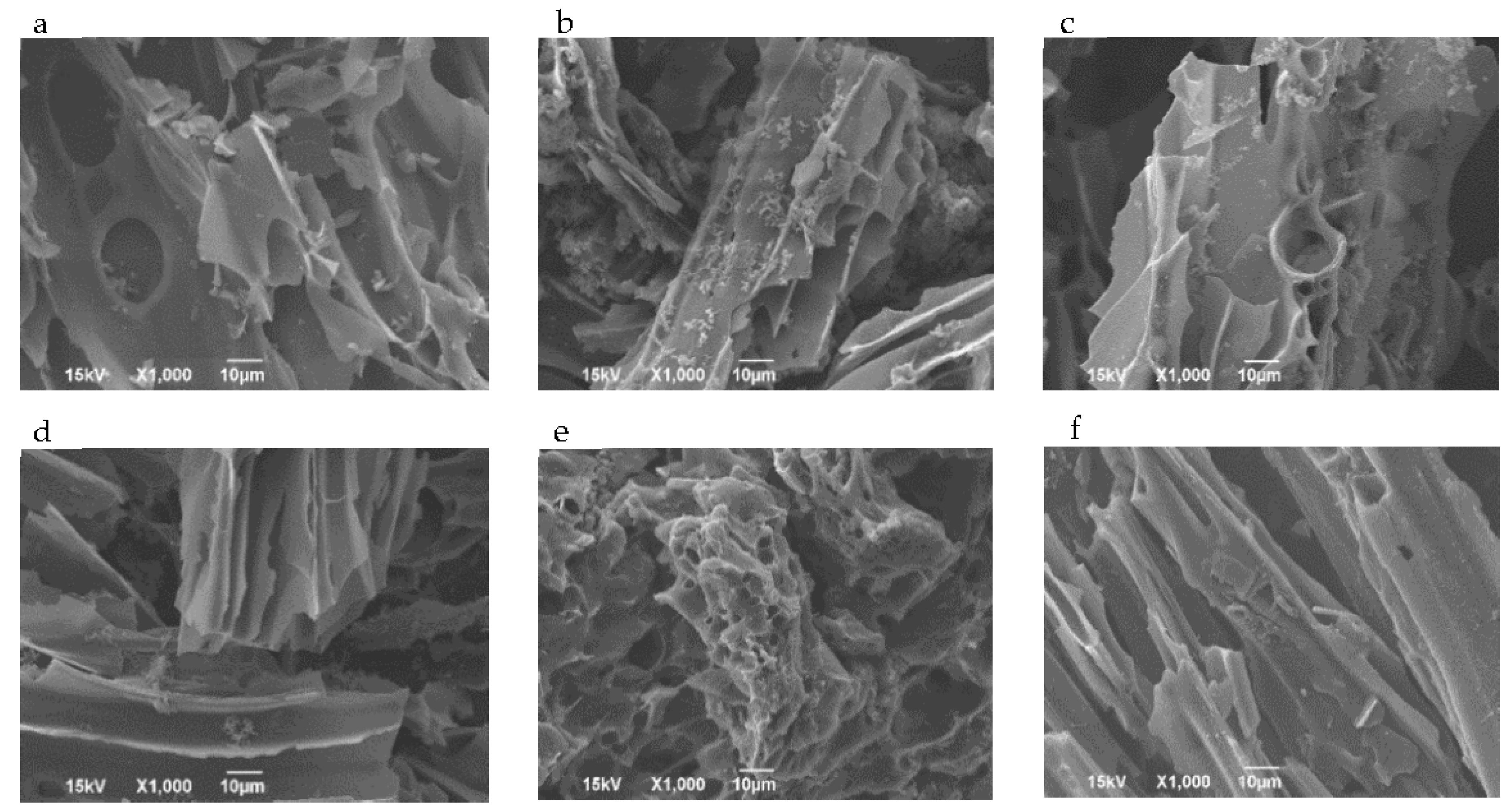
The scanning electron microscopy (SEM) images of biochar inoculated with hyper ammonia producing bacteria (HAB) at varying concentrations, specifically from 0.5 to 0.25, were analyzed to assess the structural changes. The results (Figure 5) show clear differences in structure across various biochar concentrations, emphasizing how the amount of biochar affects microbial growth and its ability to absorb substances.
The SEM images showed an uneven textured surface and a presence of pores, due to biochar’s interaction with the HABs, facilitating adsorption and microbial colonization. Studies by Tao et al. (2019) support this finding, showing that biochar’s porous surface created a favorable environment for Bacillus subtilis bacteria. Similarly, [41] found that surface texture played a role in the interaction of biochar and E. coli bacteria, with smoother surfaces attracting a greater number of bacteria. Furthermore, microbial treatment has been shown to enhance biochar's surface properties. [42] reported that biochar treated with microbes exhibited increased surface area and porosity compared to untreated ones, which improved their adsorption capabilities. Our results show that lower concentrations of biochar (0.5% - 0.15%), possess a highly porous and well-distributed structure with minimal aggregation, indicating that open pores and accessibility, providing an ideal environment for the HABs to grow. In contrast, as the concentration increases (0.20% - 0.25%), there is an appearance of a distorted surface, more compact and with evidence of pore blockage and particle aggregation. Such structural alterations may impact the biochar’s adsorption ability and may limit microbial colonization. Thus, to effectively apply biochar, there is a need to understand the amount of biochar needed to create a balance between biochar and microbial interaction.
4. Conclusions
The study confirms that biochar has the potential to support microbial fermentation, especially hyper-ammonia-producing bacteria (HAB). We discovered that there is a relationship between biochar concentration, temperature variations, and particle size that influences microbial proliferation. The results suggest that biochar can provide a conducive environment for bacterial attachment and nutrient retention. Optimal microbial proliferation was observed at a biochar concentration of 0.15%, a temperature of 45°C, and a particle size of 250 µm. However, higher concentrations and biochar particle size displayed inhibitory effects, which could be because of surface saturation and limited microbial access. The varying temperature used revealed the influence of thermal conditions on microbial dynamics. Although biochar enhanced microbial tolerance to heat variations, higher growth rates were observed around 45°C, with increasing temperature showing a reduction in microbial growth suggesting an enzyme denaturalization state. The biochar particle size for this experiment shows that surface area, porosity in microbial adhesion and proliferation were influenced by particle size. Smaller particle sizes (250–425 µm) provided favorable microbial colonization sites while larger particles (>1.40 mm) were less effective, likely due to reduced available surface area and pore connectivity. We carried out a regression analysis and it shows that there is a strong correlation between biochar properties and microbial growth, with the model explaining 99.31% of OD variability. However, there is an absence of statistical significance in individual variables suggesting the complexity of HAB-biochar interactions. The SEM analysis showed a microstructural view of how biochar surface morphology changes with increasing concentration and its impact on bacterial distribution. Hence the need for tuning the biochar to suit specific application. Further research can be done to evaluate additional environmental factors, and advanced imaging techniques to elucidate the mechanisms that influence biochar-microbial interactions.
References
- Siddiqui SA, Erol Z, Rugji J, Taşçı F, Kahraman HA, Toppi V, et al. An overview of fermentation in the food industry - looking back from a new perspective. Bioresour Bioprocess 2023;10:85. [CrossRef]
- Kotarska K, Dziemianowicz W, Świerczyńska A. The Effect of Detoxification of Lignocellulosic Biomass for Enhanced Methane Production. Energies 2021;14:5650. [CrossRef]
- Huang Y, Wang Y, Shang N, Li P. Microbial Fermentation Processes of Lactic Acid: Challenges, Solutions, and Future Prospects. Foods 2023;12:2311. [CrossRef]
- Berillo D, Malika T, Baimakhanova BB, Sadanov AK, Berezin VE, Trenozhnikova LP, et al. An Overview of Microorganisms Immobilized in a Gel Structure for the Production of Precursors, Antibiotics, and Valuable Products. Gels 2024;10:646. [CrossRef]
- Najim AA, Radeef AY, al-Doori I, Jabbar ZH. Immobilization: the promising technique to protect and increase the efficiency of microorganisms to remove contaminants. J Chem Technol Biotechnol 2024;99:1707–33. [CrossRef]
- Manikandan SK, Pallavi P, Shetty K, Bhattacharjee D, Giannakoudakis DA, Katsoyiannis IA, et al. Effective Usage of Biochar and Microorganisms for the Removal of Heavy Metal Ions and Pesticides. Molecules 2023;28:719. [CrossRef]
- Benchaar C, Hassanat F, Côrtes C. Assessment of the Effects of Commercial or Locally Engineered Biochars Produced from Different Biomass Sources and Differing in Their Physical and Chemical Properties on Rumen Fermentation and Methane Production In Vitro. Animals 2023;13:3280. [CrossRef]
- Huff MD, Marshall S, Saeed HA, Lee JW. Surface oxygenation of biochar through ozonization for dramatically enhancing cation exchange capacity. Bioresour Bioprocess 2018;5:18. [CrossRef]
- Wijitkosum S, Jiwnok P. Elemental Composition of Biochar Obtained from Agricultural Waste for Soil Amendment and Carbon Sequestration. Appl Sci 2019;9:3980. [CrossRef]
- Kayoumu M, Wang H, Duan G. Interactions between microbial extracellular polymeric substances and biochar, and their potential applications: a review. Biochar 2025;7:62. [CrossRef]
- Jiang Z, Li Q, Peng F, Yu J. Biochar Loaded with a Bacterial Strain N33 Facilitates Pecan Seedling Growth and Shapes Rhizosphere Microbial Community. Plants 2024;13:1226. [CrossRef]
- Lu J-H, Chen C, Huang C, Lee D-J. Glucose fermentation with biochar-amended consortium: microbial consortium shift. Bioengineered 2020;11:272–80. [CrossRef]
- Wang W, Dai L, Wu B, Qi B, Huang T, Hu G, et al. Biochar-mediated enhanced ethanol fermentation (BMEEF) in Zymomonas mobilis under furfural and acetic acid stress. Biotechnol Biofuels 2020;13:28. [CrossRef]
- Adeniyi A, Bello I, Mukaila T, Sarker N, Hammed A. Trends in Biological Ammonia Production 2023. [CrossRef]
- O’Reilly GC, Huo Y, Meale SJ, Chaves AV. Dose response of biochar and wood vinegar on in vitro batch culture ruminal fermentation using contrasting feed substrates. Transl Anim Sci 2021;5:txab107. [CrossRef]
- Burman E, Bengtsson-Palme J. Microbial Community Interactions Are Sensitive to Small Changes in Temperature. Front Microbiol 2021;12. [CrossRef]
- Fu Z, Chen Y, Lu Y, Wang Y, Chen J, Zhao Y, et al. KMnO4 modified biochar derived from swine manure for tetracycline removal. Water Pract Technol 2022;17:2422–34. [CrossRef]
- Chang X, Wang S, Luo C, Zhang Z, Duan J, Zhu X, et al. Responses of soil microbial respiration to thermal stress in alpine steppe on the Tibetan plateau. Eur J Soil Sci 2012;63:325–31. [CrossRef]
- Ye J-S, Bradford MA, Maestre FT, Li F-M, García-Palacios P. Compensatory Thermal Adaptation of Soil Microbial Respiration Rates in Global Croplands. Glob Biogeochem Cycles 2020;34:e2019GB006507. [CrossRef]
- Shan S, Coleman MD. Biochar influences nitrogen availability in Andisols of north Idaho forests. SN Appl Sci 2020;2:362. [CrossRef]
- Prévoteau A, Ronsse F, Cid I, Boeckx P, Rabaey K. The electron donating capacity of biochar is dramatically underestimated. Sci Rep 2016;6:32870. [CrossRef]
- Bednik M, Medyńska-Juraszek A, Ćwieląg-Piasecka I, Dudek M. Enzyme Activity and Dissolved Organic Carbon Content in Soils Amended with Different Types of Biochar and Exogenous Organic Matter. Sustainability 2023;15:15396. [CrossRef]
- Zheng Q, Hu Y, Zhang S, Noll L, Böckle T, Richter A, et al. Growth explains microbial carbon use efficiency across soils differing in land use and geology. Soil Biol Biochem 2019;128:45–55. [CrossRef]
- Isaksen GV, Åqvist J, Brandsdal BO. Enzyme surface rigidity tunes the temperature dependence of catalytic rates. Proc Natl Acad Sci 2016;113:7822–7. [CrossRef]
- Lv Y, Tang C, Liu X, Zhang M, Chen B, Hu X, et al. Optimization of Environmental Conditions for Microbial Stabilization of Uranium Tailings, and the Microbial Community Response. Front Microbiol 2021;12. [CrossRef]
- Ding J, Zhen F, Kong X, Hu Y, Zhang Y, Gong L. Effect of Biochar in Modulating Anaerobic Digestion Performance and Microbial Structure Community of Different Inoculum Sources. Fermentation 2024;10:151. [CrossRef]
- Ahmad W, Nepal J, Zou Z, Munsif F, Khan A, Ahmad I, et al. Biochar particle size coupled with biofertilizer enhances soil carbon-nitrogen microbial pools and CO2 sequestration in lentil. Front Environ Sci 2023;11. [CrossRef]
- Gu Y, Zhang H, Liang X, Fu R, Li M, Chen C. Effect of different biochar particle sizes together with bio-organic fertilizer on rhizosphere soil microecological environment on saline–alkali land. Front Environ Sci 2022;10. [CrossRef]
- Tang E, Liao W, Thomas SC. Optimizing Biochar Particle Size for Plant Growth and Mitigation of Soil Salinization. Agronomy 2023;13:1394. [CrossRef]
- Tahery S, Parra MC, Munroe P, Mitchell DRG, Meale SJ, Joseph S. Developing an activated biochar-mineral supplement for reducing methane formation in anaerobic fermentation. Biochar 2025;7:26. [CrossRef]
- Zhang L, Lim EY, Loh K-C, Ok YS, Lee JTE, Shen Y, et al. Biochar enhanced thermophilic anaerobic digestion of food waste: Focusing on biochar particle size, microbial community analysis and pilot-scale application. Energy Convers Manag 2020;209:112654. [CrossRef]
- Wu S-L, Wei W, Xu Q, Huang X, Sun J, Dai X, et al. Revealing the Mechanism of Biochar Enhancing the Production of Medium Chain Fatty Acids from Waste Activated Sludge Alkaline Fermentation Liquor. ACS EST Water 2021;1:1014–24. [CrossRef]
- Heitkamp K, Latorre-Pérez A, Nefigmann S, Gimeno-Valero H, Vilanova C, Jahmad E, et al. Monitoring of seven industrial anaerobic digesters supplied with biochar. Biotechnol Biofuels 2021;14:185. [CrossRef]
- Tang E, Liao W, Thomas SC. Optimizing Biochar Particle Size for Plant Growth and Mitigation of Soil Salinization. Agronomy 2023;13:1394. [CrossRef]
- Abd-Elhamied AS, El-Shiekha AM. Effect of Biochar Source, Particle Size and Application Rates on Soil Properties and Maize Yield (Zea mays L.) under Sandy Soil Conditions. J Soil Sci Agric Eng 2021;12:71–80. [CrossRef]
- Waqas M, Nizami AS, Aburiazaiza AS, Barakat MA, Ismail IMI, Rashid MI. Optimization of food waste compost with the use of biochar. J Environ Manage 2018;216:70–81. [CrossRef]
- Schommer VA, Nazari MT, Melara F, Braun JCA, Rempel A, dos Santos LF, et al. Techniques and mechanisms of bacteria immobilization on biochar for further environmental and agricultural applications. Microbiol Res 2024;278:127534. [CrossRef]
- Krichen E, Harmand J, Torrijos M, Godon JJ, Bernet N, Rapaport A. High biomass density promotes density-dependent microbial growth rate. Biochem Eng J 2018;130:66–75. [CrossRef]
- Frenkel O, Jaiswal AK, Elad Y, Lew B, Kammann C, Graber ER. The effect of biochar on plant diseases: what should we learn while designing biochar substrates? J Environ Eng Landsc Manag 2017;25:105–13. [CrossRef]
- Li K, Yang B, Wang H, Xu X, Gao Y, Zhu Y. Dual effects of biochar and hyperaccumulator Solanum nigrum L. on the remediation of Cd-contaminated soil. PeerJ 2019;7:e6631. [CrossRef]
- Hill RA, Hunt J, Sanders E, Tran M, Burk GA, Mlsna TE, et al. Effect of Biochar on Microbial Growth: A Metabolomics and Bacteriological Investigation in E. coli. Environ Sci Technol 2019;53:2635–46. [CrossRef]
- Zhang B, Li R, Zheng Y, Chen S, Su Y, Zhou W, et al. Biochar Composite with Enhanced Performance Prepared Through Microbial Modification for Water Pollutant Removal. Int J Mol Sci 2024;25:11732. [CrossRef]
Figure 3.
Trend for biochar and HAB at different particle sizes.
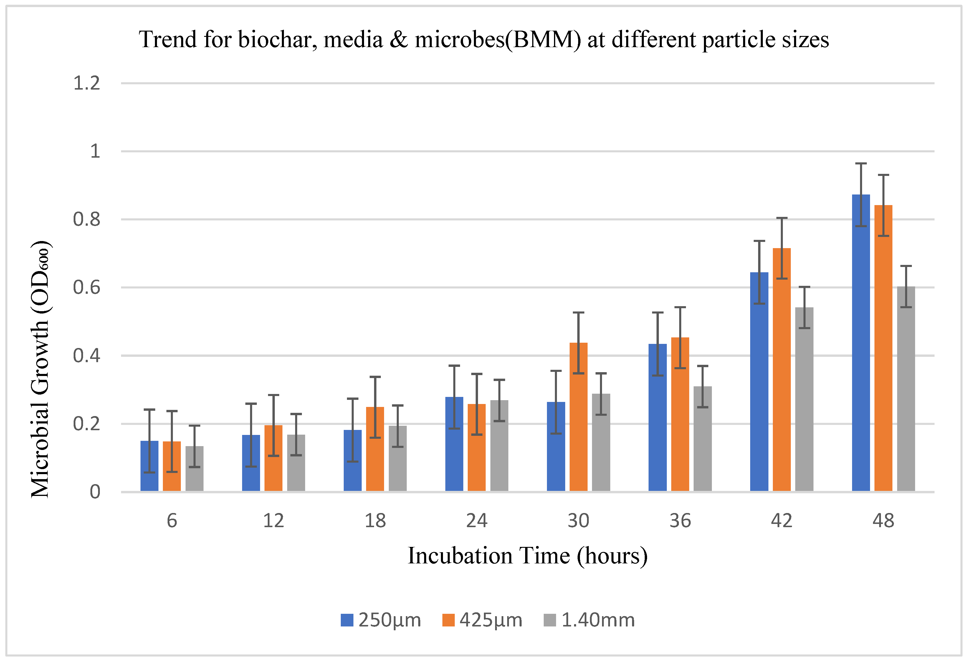
Figure 5.
Scanning electron microscopy (SEM) images of biochar inoculated with hyper-ammonia-producing bacteria (HAB) at varying concentrations. The pore structure biochar (A) Raw biochar (B).0.05% biochar with HABs (C). 0.10% biochar with HABs (D). 0.15% biochar with HABs (E). 0.20% biochar with HABs (F). 0.25% biochar with HABs.
Disclaimer/Publisher’s Note: The statements, opinions and data contained in all publications are solely those of the individual author(s) and contributor(s) and not of MDPI and/or the editor(s). MDPI and/or the editor(s) disclaim responsibility for any injury to people or property resulting from any ideas, methods, instructions or products referred to in the content. |
© 2025 by the authors. Licensee MDPI, Basel, Switzerland. This article is an open access article distributed under the terms and conditions of the Creative Commons Attribution (CC BY) license (http://creativecommons.org/licenses/by/4.0/).
Copyright: This open access article is published under a Creative Commons CC BY 4.0 license, which permit the free download, distribution, and reuse, provided that the author and preprint are cited in any reuse.


