
Submitted:
22 July 2025
Posted:
23 July 2025
You are already at the latest version
Abstract
The study investigates the antifungal potential of 1,5-diarylidene-4-piperidones. These compounds were synthesized via Claisen-Schmidt condensation, yielding 26 derivatives with varied electronic and hydrophobic properties. Their antifungal efficacy against Cryptococcus neoformans was evaluated using minimum inhibitory concentration (MIC) assay, revealing that piperidone with tetrabutoxy groups exhibited potent activity (MIC of 7.8 µM), surpassing standard antifungals like fluconazole. The selectivity index (SI) values confirmed their safety profile across various human cell lines. Mechanistic insights via docking studies suggest that these compounds inhibit ergosterol biosynthesis by targeting sterol 14-demethylase, with E binding score of 8.2 close to that of fluconazole. The compounds also demonstrated broad-spectrum activity against other pathogenic yeasts and fungi, including Candida and Aspergillus species, with some efficacy against fluconazole-resistant strains. These findings underscore the potential of 1,5-diarylidene-4-piperidones as promising antifungal candidates with a favorable safety profile.
Keywords:
antifungal
; cryptococcus neoformans
; piperidones
; curcumins
1. Introduction
The infectious agent Cryptococcus neoformans is a capsulated yeast present in soils and organic debris transmitted by air. This invasive fungal infection with C. neoformans is a serious cause of morbidity in HIV/AIDS immunocompromised patients. The most clinical form of infection is meningoencephalitis or pneumonia which is fatal when it is not treated even in immunocompetent patients.[1] In these cases, the standard treatment is amphotericin B combined with flucytosine.[2] However, both agents are toxic and laboratory monitoring of the patients is needed. Moreover, intravenous administration of amphotericin B is required limiting therefore the access of patients from resource-constrained countries. [3,4] In infections with no meningoencephalitis, fluconazole has been a primary antifungal agent in managing cryptococcosis, however, the excess use of azole drugs has caused the drug-resistance. Indeed, the ability of Cryptococcus to alter its genomic architecture in the face of antifungal stress primarily occurs through a phenomenon referred to as heteroresistance, necessitating the search and identification of novel antifungal drugs directed toward C. neoformans.
Numerous reports have demonstrated that curcumins derived from piperidones have a wide range of biological activities such as anti-cancer,[5] anti-bacterial anti-inflammatory and anti-fungal.[6,7,8] α,β-Unsaturated carbonyl compounds are known to be Michael acceptors particularly to thiol nucleophiles,[9] this faculty has been nicely exploited to design curcuma derivatives as potential anti-fungal drugs including 1,5-diarylidene-4-piperidones.[10,11,12] This assumption prompted us to design new piperidone library to investigate their potential anti-fungal activities against C. neoformans. The synthesis of curcumin derived drug is simple and based on Claisen-Schmidt reaction in which ketone reacts with two molecules of benzaldehyde analogues in the presence of sodium hydroxide to reach benzylidene structures. Motivated by the simplicity and the potential of such molecules, we aimed to target series of benzylidene compounds since there is a need for the development of novel hits to face the drug-resistance.[13]
2. Results
2.1. Synthesis of Piperidones
To target structure-activity relationship (SAR), preparation of several analogues of 1,5-diarylidene-4-piperidones (Scheme 1) was planned by varying their electronic and/or hydrophobic properties (Figure 1). The Claisen-Schmidt condensation of piperidones and aldehyde derivatives gave rise to twenty-six compounds in one-step procedure with, in most cases, no need for column chromatography since a precipitation in ethanol is conclusive to obtain pure products with yields ranging from 48 to 86%.[14] We introduced a structural diversity through the modification of alkoxy groups, N-alkyl functions and the lateral moieties. Except 3h and 3iso-h (Figure 1), all compounds were isolated as intensely colored solids and characterized by 1H NMR, 13C NMR and Mass spectroscopy.
2.1. Antifungal Activities and Toxicity of Synthesized Piperidones
The antifungal activities of the twenty-three piperidone compounds were first determined against C. neoformans using minimum inhibitory concentration (MIC) assay and are reported in Table 1. The antifungal potency was found to be modest for the monosubstituted aryl series 1a-1f as well as for compound 2j, with MIC ranging from 125 to 250 μM, regardless of the substitution (R1= OMe, Me, CN, NO2, Cl, F, CF3). Disubstituted methoxy and allyloxy groups (2a and 2g) afforded even worse results. However, the dibutoxy compound 2h exhibited a high ability to inhibit the growth of C. neoformans (MIC of 31.2 μM). Compound 2i, derived from piperonal, showed weak activity with a MIC of 250 μM. Piperidone 2l, possessing a quinoline scaffold analogous to those reported by Shingate,[15] was tested but showed lower efficiency (MIC = 250 μM). Since butoxy groups appeared to exhibit the best activity against C. neoformans, we decided to synthesize and evaluate the compound 2m which contains six butoxy groups enhancing therefore the hydrophobic ability of the piperidone. Compound 2m displayed a good MIC of 62.5 µM, but it was less efficient than 2h. Bicyclic piperidone 2n was found to be inactive against C. neoformans. We then varied the nature of N-alkyl substitution. The results indicated that cyclopropyl, Boc, mesyl and Cbz groups were detrimental to the activity. Benzyl amines (e.g. butenafine) are known to be antifungal agents [16] with activity on the cell membrane through squalene epoxidase bounding.[17] We decided therefore to synthesize compound 3h that significantly improved activity, presenting a MIC as low as 7.8 μM. To confirm this effect, we compared the activity of 2k and 3j by switching from ethyl alkyl to benzyl group on the nitrogen atom. Again, the results showed that benzyl substitution (compound 3j) was benefit presenting a lower MIC (62.5 μM) compared to the ethyl-substituted piperidone 2k (MIC = 250 μM). Testing compound 2o, which contains one butoxy group on each aromatic moiety, again proved detrimental, with a MIC of 125 μM (vs 7.8 μM for 3h) highlighting the importance of the presence two butoxy groups on each aromatic moiety. A similar trend was observed when changing the position of the alkoxy group from ortho to meta (3iso-h being less active than 3h, with a MIC of 62.5 and 7.8 µM, respectively). The comparison between compounds 3a (MIC >250 µM) with methoxy instead of butoxy groups and 3h strongly suggests that the hydrophobic nature and the length of the alkyl chains are crucial for the activity of these piperidone derivatives. The longer butyl chains in 3h likely facilitate stronger hydrophobic interactions with a specific region (hydrophobic pocket) on the target protein, leading to enhanced biological activity. In addition, to underscore the significance of the piperidone core, we synthesized and evaluated 1,5-diarylidene cyclohexanone analogues 8-10h which demonstrated no activity with MIC > 250 µM. These results suggest that the amphiphilic character of piperidones- due to the presence of four alkyl chains and nitrogen atom- plays an important role. Interestingly, a control test revealed that compound 3h was more active than fluconazole (MIC of 25 µM), an antifungal recommended to treat C. neoformans infection,[18] particularly in low-income and middle-income countries.[19]
To better evaluate the therapeutic potential of the active compounds, their toxicity was then measured using various human cells: i.e. A498 (kidney), BEAS-2B (lung), Caco-2 (intestine), HaCaT (skin) and HepG2 (liver) cells (Figure 2). For comparison, the toxicity of fluconazole was also determined (Figure 3). The CC50 values, i.e. the concentrations causing 50% reduction of the cell viability, were determined from the graphs and are given in Table 1.
The CC50 values of compounds range from 18.3 to > 1000 µM depending on the compound and of the cell model tested. Compounds possessing mono-substituted aryl groups in para-position 1b, 1c, 1e, 1f, and 2j afforded the highest toxicity (CC50 of 18.3 to 313.9 µM). Introducing substituted alkoxy group, like in 1a and 2o, was beneficial giving lower toxicity (CC50 of 64.9 to >1000 µM). In addition to higher anti-fungal activity, disubstituted butoxy groups 2h revealed low toxicity (CC50 of 140 to 696.6 µM). When the aryl groups were connected to acetal, a detrimental effect was observed on toxicity of compound 2l (CC50 of.30.0 to 89.5 µM). However, incorporating a third butoxy group in 2m drug, afforded even a lower toxicity (CC50 of 663 to >1000 µM). We then evaluated the effect of the substitution on the nitrogen atom by introducing alkyl groups. Benzyl group 3h revealed a small elevation of toxicity when it is compared to 2h (CC50 of 42.4 to 628.6 µM) whereas compound with cyclopropyl group 4h showed the lowest toxicity regarding all the cell lines tested (>1000 µM).
For comparison, Fluconazole gave CC50 ranging from 841.2 to >1000 µM, respectively (Figure 3 and Table 1). In order to further identify the most promising candidates, the Selectivity Indexes (SI) of each compound (i.e. the ratio of CC50 to MIC values) were calculated (Table 1).
These investigations revealed that 2h, 2m, 3iso-h, and 3h have the best safety/activity profile with very interesting SI up to 80.5. Importantly, 3h gave the highest SI values ranging from 5.4 to 80.5 depending on the human cell model considered. Although less active than 3h (with MIC values on C. neoformans of 7.8 and 62.5 µM, respectively), 2m showed to be the less toxic among the active compounds, with CC50 ranging from 663.7 to > 1000 µM and SI ranging from 10.6 to > 16. For comparison, Fluconazole gave SI ranging from 33.6 to >40, close to the ones obtained with the safest compounds of this study.
The spectrum of antifungal activity of the more active compounds (i.e. 2h, 2m, 3iso-h, and 3h) was further screened using various fungal strains (yeasts and filamentous species) infecting humans or plants (Table 2 and Table 3). Regarding human pathogenic yeasts (Table 2), 2h, 2m, 3iso-h and 3h gave MIC values similar to the ones obtained on C. neoformans when tested against Candida auris and C. glabrata (MIC of 7.8 to 31.2 µM) but were found less active against C. albicans and C. tropicalis (MIC of 62.5 to 250 µM). Regarding filamentous fungi (Table 3), for the human pathogen Aspergillus fumigatus and the plant pathogen A. flavus, 3h was the only active compound (MIC of 31.2 µM), 2h, 2m, and 3iso-h being found less or not active (MIC of 125 to > 250 µM). 2h, 2m, 3iso-h and 3h were also found less or not active against the plant pathogens Colletotrichum graminicola, Fusarium graminearum, and Penicillium verrucosum (MIC of 125 to > 250 µM) except for 3h giving an MIC of 62.5 µM on P.verrucosum. Regarding the plant pathogens Magnaporthe oryzae and Microdochium bolleyi and the human pathogen Trichophyton rubrum, 2h, 2m, 3iso-h and 3h displayed good activities (MIC of 7.8 to 62.5 µM) except for 2m on M. bolleyi (MIC >250µM) and 3iso-h on T. rubrum (MIC of 125 µM), 3h giving the lowest MIC on those strains (i.e. 7.8 µM). Interestingly, it must be noted that yeasts and filamentous fungi found resistant to Fluconazole (MIC of 250 to >1000 µM) were still sensitive to 2h, 2m, 3iso-h and 3h. Thus, again, 3h gave the best antifungal activity with the lowest MIC values for each strain.
Importantly, the twenty-three piperidone compounds were tested against Gram-positive and Gram-negative bacteria belonging to the ESKAPE group and were found inactive with MIC > 250 µM for all compounds, except 2k giving an MIC of 250 µM on the Gram-negative bacteria A. baumannii and E cloacae and 2l and 3j giving an MIC of 250 µM on the Gram-positive bacteria S. aureus. This demonstrates that the compounds possess an antimicrobial activity selectively directed against yeasts and fungi that can be explained by the specific targeting of fungal enzyme(s).
2.3. Molecular Modeling
It has been demonstrated that similar compounds are able to interact and inhibit cytochrome P450 14α-sterol 14-demethylase (CYP51), an enzyme involved in the ergosterol biosynthesis in fungal.[15] Docking analysis was therefore performed to evaluate the affinity of best piperidones molecules with sterol 14-demethylase using its crystal structure (PDB code: 3KHM). Except for piperidone 2m which contains three butoxy groups, the results indicated that compounds 2h, 3h, 3iso-h effectively interacted in the active site of CYP51 complex. The docking binding energy for 3h score was found -8,2 kcal/mol similar to that found with Fluconazole (binding energy = -8,1 kcal/mol) which supports the fact that 3h may act through inhibition of ergosterol synthesis as previously shown for other piperidone compounds described in the literature.[11,15] In this molecular docking study, all docked compounds (2m, 2h, 3h, 3iso-h) formed interactions with Hem300 and hydrophobic interactions with numerous amino-acids residues (Table 4 and Figure 5-8) ). These hydrophobic interactions with butoxy chains may explain the difference in activity between 3h (MIC=7.8 µM) and its methyl analogue 3a (MIC > 250 µM). In addition, piperidones 2h and 3h revealed T-shaped interactions with Tyr103 whereas 3iso-h showed T-shaped interactions with His294 and Phe290.
Figure 4.
Best binding mode of Fluconazole into the active site of CYP51. Docking score: -8.1 kcal/mol.
Figure 4.
Best binding mode of Fluconazole into the active site of CYP51. Docking score: -8.1 kcal/mol.
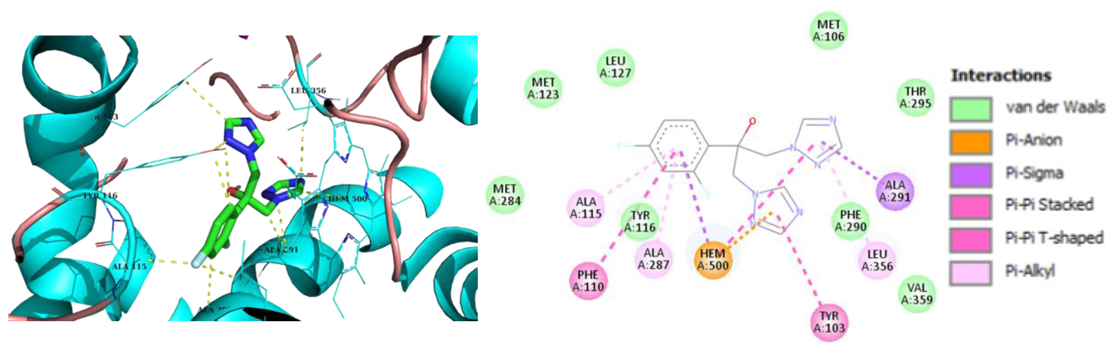
Figure 5.
Best binding mode of 2h into the active site of CYP51. Docking score: -7.8 kcal/mol.

Figure 6.
Best binding mode of 3h into the active site of CYP51. Docking score: -8.2 kcal/mol.
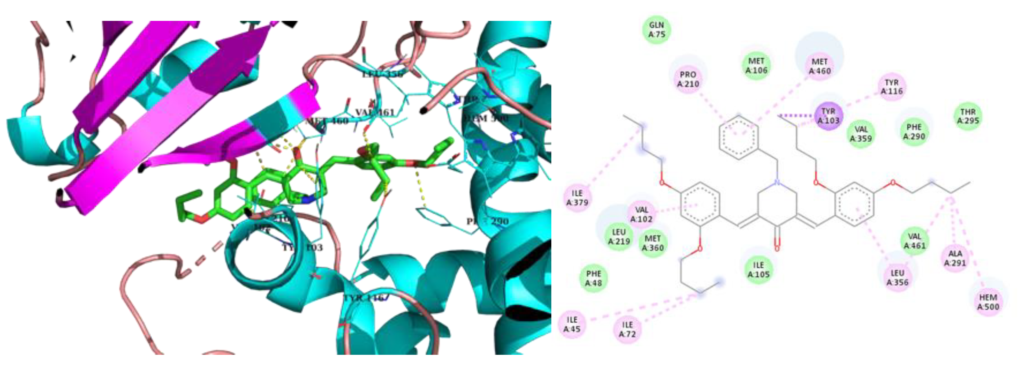
Figure 7.
Best binding mode of 2m into the active site of CYP5. Docking score: -7.2 kcal/mol.

Figure 8.
Best binding mode of 3iso-h into the active site of CYP51. Docking score: -8.8 kcal/mol.
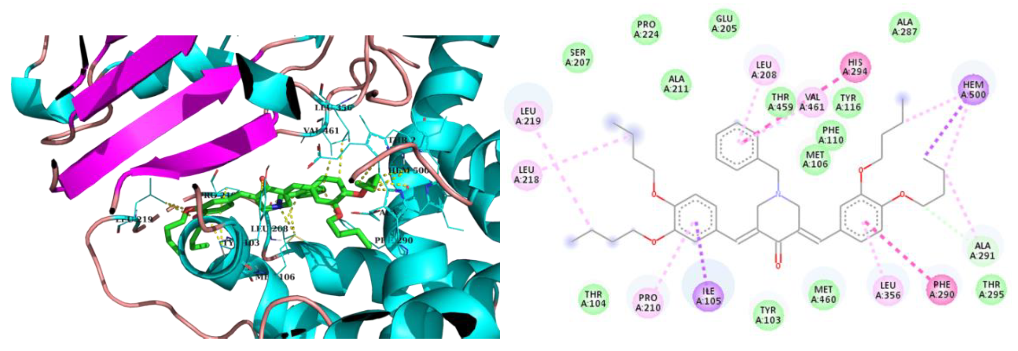
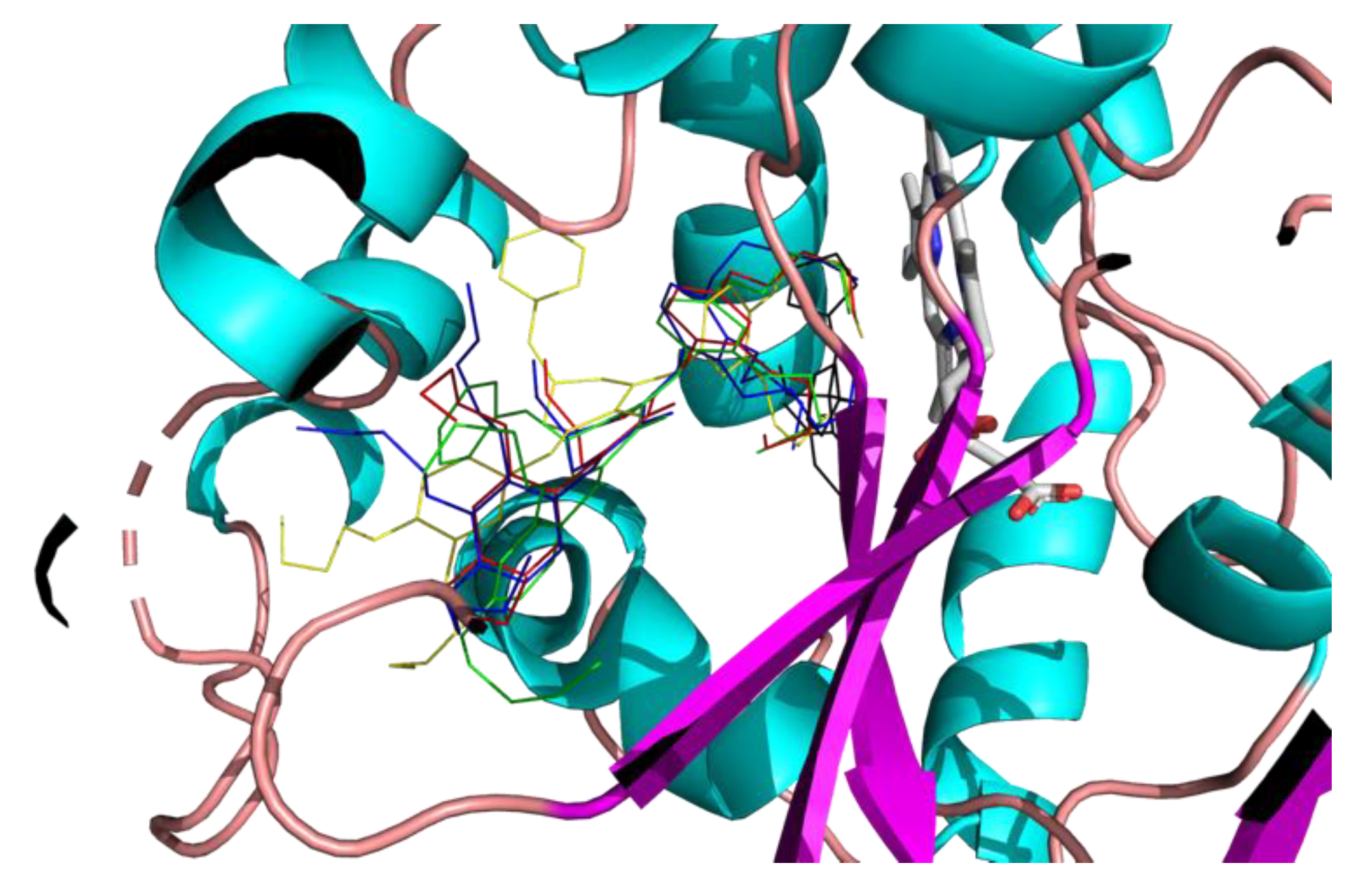
Figure 9.
Best binding mode of 2h(red), 3h(green), 2m(blue), 3iso-h(yellow) and Fluconazole (black) into the active site of CYP51.
Figure 9.
Best binding mode of 2h(red), 3h(green), 2m(blue), 3iso-h(yellow) and Fluconazole (black) into the active site of CYP51.
3. Discussion
C. neoformans is an important human pathogen but, other fungi should be considered when developing antifungal drugs. Candida sp infect mainly immunocompromised patients but also immunocompetent ones leading every year to more than 1.5 millions bloodstream infection or invasive candidiasis and around 900,000 deaths.[20] In addition to C. albicans, among the various Candida sp infecting humans, C. auris, C. glabrata, and C. tropicalis have attracted more attention due to their increasing prevalence and their ability to develop resistance to antifungal drugs used in medicine, including fluconazole.[21,22,23,24,25] Similarly, Aspergillus fumigatus is responsible for infection mainly in immunocompromised patients and is estimated to be responsible for over 600,000 deaths annually.[26] Although not infecting humans, fungi infecting plants and crops are also important to consider when developing antifungal since they cause massive losses (for example, Fusarium graminearum only causes every year losses of around 28 million metric tons of wheat grain valued at $5.6 billion[27] and/or produce mycotoxins harmful for humans and animals).[28] When testing the spectrum of antifungal activity of the best piperidone compounds, data showed that in addition to possess good activity against C. neoformans, 2h, 2m, 3iso-h, and 3h demonstrated also antifungal activity against important humans and plants pathogens, such as C. albicans, C. auris, C. glabrata, A. fumigatus, and T. rubrum but also A. flavus, M. oryzae, M. bolleyi, and P. verrucosum. In all case, 3h was identified as the most active piperidone compound with MIC ranging from 7.8 to 62.5 µM. Importantly, fungi resistant to Fluconazole were found still sensitive to 2h, 2m, 3h, and/or 3iso-h. This observation is not in contradiction with the docking data indicating that these molecules target ergosterol biosynthesis like Fluconazole. Indeed, the resistance to Fluconazole is not due to mutation in its target but classically involves a reduction of its entry and/or an increase in its efflux in resistant strains.[29] The fact that strains resistant to Fluconazole remain sensitive to 2h, 2m, 3h, and/or 3iso-h is of major importance due to the growing incidence of Fluconazole resistance in fungal strains infecting humans including Candida species and A. fumigatus.[30,31]
4. Materials and Methods
4.1. Chemistry
All reagents and solvents were purchased from Aldrich, FluoroChem or TCI and used as received without further purification. Mass spectroscopy was performed by the Spectropole of Aix-Marseille University. ESI mass spectral analyses were recorded with a 3200 QTRAP (Applied Biosystems SCIEX) mass spectrometer. The HRMS mass spectral analysis was performed with a QStar Elite (Applied Biosystems SCIEX) mass spectrometer. Elemental analyses were recorded with a Thermo Finnigan EA 1112 elemental analysis apparatus driven by the Eager 300 software. 1H and 13C NMR spectra were determined at room temperature in 5 mm o.d. tubes on a Bruker Avance 400 spectrometer or a Bruker Avance 300 spectrometer of the Spectropole: 1H (400 MHz), 1H (300 MHz), 13C (100 MHz) and 13C (75 MHz). The 1H chemical shifts were referenced to the solvent peak CDCl3 (7.26 ppm) or DMSO_d6 (2.50 ppm) and the 13C chemical shifts were referenced to the solvent peak CDCl3 (77.16 ppm) or DMSO_d6 (39.52 ppm). All compounds were prepared with analytical purity up to accepted standards for new organic compounds (>98%) which was checked by high field NMR analysis.
N-alkylpiperidin-4-one (10 mmol) was dissolved in 20 mL of ethanol, benzaldehyde derivative (20 mmol) and 40% of NaOH (2 equiv.) were then added at 0°C. The solution was stirred at room temperature overnight. The yellow precipitate was filtrated off, washed with cold ethanol and dried under vacuum (48-86% yield). Spectral data, structure, and NMR spectra of the compounds are given in the supplementary materials.
4.2. Antimicrobial Activity
The antifungal effect of compounds was measured following the reference methods for yeasts (NCCLS M27-A) and molds (M38-P) as previously described. [32] Reference yeast strains tested were Candida albicans (DSM 10697), C. auris (DSM 21092), C. glabrata (DSM 11226), C. tropicalis (DSM 9419), and Cryptococcus neoformans (DSM11959). Filamentous fungi tested were either human pathogens (i.e. A. fumigatus (DSM 819) and Trichophyton rubrum (DSM16111)) or plant pathogens (i.e. Aspergillus flavus (DSM 1959), Colletotrichum graminicola (DSM 63127), Fusarium graminearum (DSM 1095), Magnaporthe oryzae (gift from Richard O’Connell UMR Bioger, Paris Saclay, (DOI: 10.1094/Phyto-82-421)), Microdochium bolleyi (DSM 62073), and Penicillium verrucosum (DSM 12639)). Yeast suspensions were prepared by resuspending colonies collected from PDA plates in sterile NaCl 0.9% solution. Yeasts were then diluted at 1–2 × 10³ yeasts/mL in RMPI media supplemented with glucose (1,8%) buffered with MOPS (final concentration of 0.165 mol/L (pH 7.0)). For filamentous fungi, conidi were collected from mycelium grown on PDA plates using sterile solution of NaCl 0.9% supplemented with Tween at 0.1%. After counting under microscope, dilution at 2–3 × 10⁴ conidi/mL were also prepared in MOPS-buffered RMPI media supplemented with glucose. Diluted yeast or fungi were then exposed to increasing concentrations of compounds (1/2 serial dilution) in 96-wells plates, pure DMSO (maximal concentration of 1%) being used as negative control. Plates were incubated at 35 ◦C for 24-48 h for yeasts and filamentous fungi infecting humans (i.e. A. fumigatus and T. rubrum) or 25°C for 48-72 h for the other filmantous fungi from the environment or infecting plants. Minimum Inhibiting Concentrations (MIC) were determined as the lowest concentrations of compounds totally inhibiting the growth of the fungi. The antibacterial effect of the compounds was measured through liquid MIC determination using Gram-negative (Acinetobacter baumannii (DSM 30007), Enterobacter cloacae (DSM 30054), Klebsiella pneumoniae (DSM 26371), and Pseudomonas aeruginosa (ATCC 9027) and Gram-positive (Enterococcus faecalis (DSM 2570), Enterococcus faecium (DSM 20477), and Staphylococcus aureus (ATCC 6538) bacteria from the ESKAPE group following the National Committee of Clinical Laboratory Standards (NCCLS, 1997) procedure as previously described. [33] In all case, MIC were determined in n=2-3.
4.3. Cytotoxicity Studies
The toxicity of the compounds was tested using human cells (i.e. A498 (human kidney cell line), BEAS-2B (normal human airway epithelial cells), Caco-2 (human intestinal cell line), HaCaT (normal human skin cells), and HepG2 (human liver cell line)) as previously described [34]. Cells were maintained at 37°C in a 5% CO2 incubator in Dulbecco's modified essential medium (DMEM) supplemented with 10% fetal calf serum (FCS), 1% L-glutamine and 1% antibiotics (all from Invitrogen). Cells grown on 75 cm2 flasks were detached using trypsin-EDTA solution (Thermofisher), counted using Mallasez’s chamber, and seeded into 96-well cell culture plates (Greiner Bio-one) at approximately 104 cells per well. After 24-48h, when the cells reached 80-90 % confluence, wells were aspirated and increasing concentrations of compounds (from 0 to 1000 µM, ½ dilution) were added to the cells, DMSO (maximal concentration of 1%) being used as negative control. After 48 h incubation at 37°C in a 5% CO2 incubator, wells were aspirated, and the cell viability was measured by adding 100 µL of resazurin solution at 0.03 mg/mL final in phosphate buffer saline with calcium and magnesium chloride (PBS++). After 1 h incubation at 37°C, the fluorescence intensity of the wells was measured using a microplate reader (SynerMix, Biotek, Ex 530 nm/Em 590 nm). The fluorescence values were normalized by the negative controls (DMSO treated cells) and expressed as the percentage of cell viability. The cytotoxic concentrations 50, (i.e. CC50) of compounds corresponding to the concentrations causing a reduction of 50% of the cell viability were calculated using GraphPad® Prism 8 software (n=3).
4.4. Molecular Docking
All the ligands were fully optimized with the Gaussian16 Rev. A03 package at the HF/6-31G(d) level of theory. The atomic charges were computed at the HF/6-31G(d) level of theory with the RESP scheme. For docking studies, X-ray crystal structure of sterol 14alpha-demethylase (CYP51) from Trypanosoma cruzi in complex with inhibitor fluconazole was obtained from the Protein Data Bank (PDB), (PDB code: 3KHM). The enzyme was prepared by removing fluconazole, then adding hydrogen atoms and atomic charges with AutoDock Tools software. The molecular docking studies were carried out with the AutoDock 4.2.6 software. The docking box set at 70 × 70 × 70 Å and was centered at x = 2.270 Å, y = -23.537 Å and z = 16.924 Å (center of fluconazole). The Lamarckian Genetic Algorithm with default parameters was applied for simulation. The number of docking runs was 20. The pose drawings and interaction calculations were performed with ProteinPlus and BIOVIA Discovery Studio. The 3D active sites were drawn with PyMOL. Details of molecular docking analysis are given in the supplementary materials.
5. Conclusions
This study highlights the potential of 1,5-diarylidene-4-piperidones as effective antifungal agents, particularly against Cryptococcus neoformans and other clinically significant fungi. Among the synthesized compounds, 3h emerged as the most promising candidate, displaying superior antifungal potency. Its ability to inhibit fluconazole-resistant strains further underscores its therapeutic value. Docking studies suggest that these compounds act by targeting sterol 14α-demethylase, disrupting ergosterol biosynthesis in fungal cells. Additionally, their broad-spectrum antifungal activity and low cytotoxicity support their potential application in both medical and agricultural settings. Further in vivo studies and clinical evaluations are necessary to fully explore their therapeutic potential and to address the growing need for new antifungal drugs to combat resistant fungal pathogens.
Supplementary Materials
The following supporting information can be downloaded at the website of this paper posted on Preprints.org, Supplementary File S1: Spectral data, NMR spectra and Molecular Docking. References [x-x] are cited in the supplementary materials
Author Contributions
Conceptualization, M.N., M.M., and D.S.; methodology, M.N., M.M., E.C.D., and D.S.; validation, M.N., M.M. and D.S.; formal analysis, E.C.D., H.R., A.G., L.D., Y.C., F.D., D.S. M.M., and M.N.; investigation, E.C.D., H.R., A.G., L.D., Y.C., F.D., D.S. M.M., and M.N.; writing—original draft preparation, M.M., M.N., and D.S.; writing—review and editing, M.M. and M.N.; supervision, M.M. and M.N.; project administration, M.M. and M.N. All authors have read and agreed to the published version of the manuscript.
Funding
This research received no external funding.
Data Availability Statement
The original contributions presented in this study are included in the article/supplementary material. Further inquiries can be directed to the corresponding authors.
Acknowledgments
We thank Aix-Marseille Université and CNRS for financial support. This work was supported by the computing facilities of the CRCMM,“Centre Régional de Compétences en Modélisation Moléculaire de Marseille”
Conflicts of Interest
The authors declare no conflicts of interest.
References
- Perfect, J.R. Efficiently Killing a Sugar-Coated Yeast. N. Engl. J. Med. 2013, 368, 1354–1356. [Google Scholar] [CrossRef]
- Day, J.N.; Chau, T.T.H.; Wolbers, M.; Mai, P.P.; Dung, N.T.; Mai, N.H.; Phu, N.H.; Nghia, H.D.; Phong, N.D.; Thai, C.Q.; et al. Combination Antifungal Therapy for Cryptococcal Meningitis. N. Engl. J. Med. 2013, 368, 1291–1302. [Google Scholar] [CrossRef]
- Hartland, K.; Pu, J.; Palmer, M.; Dandapani, S.; Moquist, P.N.; Munoz, B.; DiDone, L.; Schreiber, S.L.; Krysan, D.J. High-Throughput Screen in Cryptococcus Neoformans Identifies a Novel Molecular Scaffold That Inhibits Cell Wall Integrity Pathway Signaling. ACS Infect. Dis. 2016, 2, 93–102. [Google Scholar] [CrossRef]
- Beattie, S.R.; Schnicker, N.J.; Murante, T.; Kettimuthu, K.; Williams, N.S.; Gakhar, L.; Krysan, D.J. Benzothiourea Derivatives Target the Secretory Pathway of the Human Fungal Pathogen Cryptococcus Neoformans. ACS Infect. Dis. 2020, 6, 529–539. [Google Scholar] [CrossRef] [PubMed]
- Pati, H.N.; Das, U.; Das, S.; Bandy, B.; De Clercq, E.; Balzarini, J.; Kawase, M.; Sakagami, H.; Quail, J.W.; Stables, J.P.; et al. The Cytotoxic Properties and Preferential Toxicity to Tumour Cells Displayed by Some 2,4-Bis(Benzylidene)-8-Methyl-8-Azabicyclo[3.2.1] Octan-3-Ones and 3,5-Bis(Benzylidene)-1-Methyl-4-Piperidones. Eur. J. Med. Chem. 2009, 44, 54–62. [Google Scholar] [CrossRef]
- Prasad, S.; Gupta, S.C.; Tyagi, A.K.; Aggarwal, B.B. Curcumin, a Component of Golden Spice: From Bedside to Bench and Back. Biotechnol. Adv. 2014, 32, 1053–1064. [Google Scholar] [CrossRef] [PubMed]
- Bazzaro, M.; Linder, S. Dienone Compounds: Targets and Pharmacological Responses. J. Med. Chem. 2020, 63, 15075–15093. [Google Scholar] [CrossRef] [PubMed]
- Noureddin, S.A.; El-Shishtawy, R.M.; Al-Footy, K.O. Curcumin Analogues and Their Hybrid Molecules as Multifunctional Drugs. Eur. J. Med. Chem. 2019, 182, 111631. [Google Scholar] [CrossRef]
- Dao, T.T.; Sehgal, P.; Tung, T.T.; Møller, J.V.; Nielsen, J.; Palmgren, M.; Christensen, S.B.; Fuglsang, A.T. Demethoxycurcumin Is A Potent Inhibitor of P-Type ATPases from Diverse Kingdoms of Life. PLOS ONE 2016, 11, e0163260. [Google Scholar] [CrossRef]
- Lawson, S.; Arumugam, N.; Almansour, A.I.; Suresh Kumar, R.; Thangamani, S. Dispiropyrrolidine Tethered Piperidone Heterocyclic Hybrids with Broad-Spectrum Antifungal Activity against Candida Albicans and Cryptococcus Neoformans. Bioorganic Chem. 2020, 100, 103865. [Google Scholar] [CrossRef]
- Nagargoje, A.A.; Akolkar, S.V.; Subhedar, D.D.; Shaikh, M.H.; Sangshetti, J.N.; Khedkar, V.M.; Shingate, B.B. Propargylated Monocarbonyl Curcumin Analogues: Synthesis, Bioevaluation and Molecular Docking Study. Med. Chem. Res. 2020, 29, 1902–1913. [Google Scholar] [CrossRef]
- Synthesis and Evaluation of Novel Antifungal Agents Targeted to the Plasma Membrane H[+]-ATPase. Br. J. Pharm. 2018, 2. [CrossRef]
- Howard, K.C.; Dennis, E.K.; Watt, D.S.; Garneau-Tsodikova, S. A Comprehensive Overview of the Medicinal Chemistry of Antifungal Drugs: Perspectives and Promise. Chem. Soc. Rev. 2020, 49, 2426–2480. [Google Scholar] [CrossRef] [PubMed]
- Pigot, C.; Noirbent, G.; Bui, T.-T.; Péralta, S.; Gigmes, D.; Nechab, M.; Dumur, F. Push-Pull Chromophores Based on the Naphthalene Scaffold: Potential Candidates for Optoelectronic Applications. Materials 2019, 12, 1342. [Google Scholar] [CrossRef]
- Nagargoje, A.A.; Akolkar, S.V.; Siddiqui, M.M.; Subhedar, D.D.; Sangshetti, J.N.; Khedkar, V.M.; Shingate, B.B. Quinoline Based Monocarbonyl Curcumin Analogs as Potential Antifungal and Antioxidant Agents: Synthesis, Bioevaluation and Molecular Docking Study. Chem. Biodivers. 2020, 17, e1900624. [Google Scholar] [CrossRef]
- Fioravanti, R.; Biava, M.; Porretta, G.; Landolfi, C.; Simonetti, N.; Villa, A.; Conte, E.; Porta-Puglia, A. Research on Antibacterial and Antifungal Agents. XI. Synthesis and Antimicrobial Activity of N-Heteroaryl Benzylamines and Their Schiff Bases. Eur. J. Med. Chem. 1995, 30, 123–132. [Google Scholar] [CrossRef]
- Hammoudi Halat, D.; Younes, S.; Mourad, N.; Rahal, M. Allylamines, Benzylamines, and Fungal Cell Permeability: A Review of Mechanistic Effects and Usefulness against Fungal Pathogens. Membranes 2022, 12, 1171. [Google Scholar] [CrossRef]
- Perfect, J.R.; Dismukes, W.E.; Dromer, F.; Goldman, D.L.; Graybill, J.R.; Hamill, R.J.; Harrison, T.S.; Larsen, R.A.; Lortholary, O.; Nguyen, M.-H.; et al. Clinical Practice Guidelines for the Management of Cryptococcal Disease: 2010 Update by the Infectious Diseases Society of America. Clin. Infect. Dis. 2010, 50, 291–322. [Google Scholar] [CrossRef]
- Loyse, A.; Burry, J.; Cohn, J.; Ford, N.; Chiller, T.; Ribeiro, I.; Koulla-Shiro, S.; Mghamba, J.; Ramadhani, A.; Nyirenda, R.; et al. Leave No One behind: Response to New Evidence and Guidelines for the Management of Cryptococcal Meningitis in Low-Income and Middle-Income Countries. Lancet Infect. Dis. 2019, 19, e143–e147. [Google Scholar] [CrossRef]
- Denning, D.W. Global Incidence and Mortality of Severe Fungal Disease. Lancet Infect. Dis. 2024, 24, e428–e438. [Google Scholar] [CrossRef]
- Long, B.; Lacy, A.J.; Koyfman, A.; Liang, S.Y. Candida Auris: A Focused Review for Emergency Clinicians. Am. J. Emerg. Med. 2024, 84, 162–167. [Google Scholar] [CrossRef]
- Abe, M.; Kinjo, Y.; Koshikawa, T.; Miyazaki, Y. Basic Research on Candida Species: Disease Mechanism, Virulence, and Relationship with Environmental Factors. Med. Mycol. J. 2024, 65, 67–74. [Google Scholar] [CrossRef] [PubMed]
- Dawoud, A.M.; Saied, S.A.; Torayah, M.M.; Ramadan, A.E.; Elaskary, S.A. Antifungal Susceptibility and Virulence Determinants Profile of Candida Species Isolated from Patients with Candidemia. Sci. Rep. 2024, 14, 11597. [Google Scholar] [CrossRef] [PubMed]
- Beardsley, J.; Kim, H.Y.; Dao, A.; Kidd, S.; Alastruey-Izquierdo, A.; Sorrell, T.C.; Tacconelli, E.; Chakrabarti, A.; Harrison, T.S.; Bongomin, F.; et al. Candida Glabrata ( Nakaseomyces Glabrata ): A Systematic Review of Clinical and Microbiological Data from 2011 to 2021 to Inform the World Health Organization Fungal Priority Pathogens List. Med. Mycol. 2024, 62, myae041. [Google Scholar] [CrossRef] [PubMed]
- Keighley, C.; Kim, H.Y.; Kidd, S.; Chen, S.C.-A.; Alastruey, A.; Dao, A.; Bongomin, F.; Chiller, T.; Wahyuningsih, R.; Forastiero, A.; et al. Candida Tropicalis —A Systematic Review to Inform the World Health Organization of a Fungal Priority Pathogens List. Med. Mycol. 2024, 62, myae040. [Google Scholar] [CrossRef]
- Dhingra, S.; Cramer, R.A. Regulation of Sterol Biosynthesis in the Human Fungal Pathogen Aspergillus Fumigatus: Opportunities for Therapeutic Development. Front. Microbiol. 2017, 8. [Google Scholar] [CrossRef]
- Liu, S.; Giacoletto, N.; Schmitt, M.; Nechab, M.; Graff, B.; Morlet-Savary, F.; Xiao, P.; Dumur, F.; Lalevée, J. Effect of Decarboxylation on the Photoinitiation Behavior of Nitrocarbazole-Based Oxime Esters. Macromolecules 2022, 55, 2475–2485. [Google Scholar] [CrossRef]
- Deligeorgakis, C.; Magro, C.; Skendi, A.; Gebrehiwot, H.H.; Valdramidis, V.; Papageorgiou, M. Fungal and Toxin Contaminants in Cereal Grains and Flours: Systematic Review and Meta-Analysis. Foods 2023, 12, 4328. [Google Scholar] [CrossRef]
- Maebashi, K.; Niimi, M.; Kudoh, M.; Fischer, F.J.; Makimura, K.; Niimi, K.; Piper, R.J.; Uchida, K.; Arisawa, M.; Cannon, R.D.; et al. Mechanisms of Fluconazole Resistance in Candida Albicans Isolates from Japanese AIDS Patients. J. Antimicrob. Chemother. 2001, 47, 527–536. [Google Scholar] [CrossRef]
- Janowski, M.; Demchuk, O.M.; Wujec, M. Fluconazole Analogs and Derivatives: An Overview of Synthesis, Chemical Transformations, and Biological Activity. Molecules 2024, 29, 2855. [Google Scholar] [CrossRef]
- Wiederhold, N. Antifungal Resistance: Current Trends and Future Strategies to Combat. Infect. Drug Resist. 2017, Volume 10, 249–259. [Google Scholar] [CrossRef]
- E. ; Perrier, J.; Maresca, M. Comparative Structure–Activity Analysis of the Antimicrobial Activity, Cytotoxicity, and Mechanism of Action of the Fungal Cyclohexadepsipeptides Enniatins and Beauvericin. Toxins 2019, 11, 514. [Google Scholar] [CrossRef]
- Benkhaled, B.T.; Hadiouch, S.; Olleik, H.; Perrier, J.; Ysacco, C.; Guillaneuf, Y.; Gigmes, D.; Maresca, M.; Lefay, C. Elaboration of Antimicrobial Polymeric Materials by Dispersion of Well-Defined Amphiphilic Methacrylic SG1-Based Copolymers. Polym. Chem. 2018, 9, 3127–3141. [Google Scholar] [CrossRef]
- Olleik, H.; Yacoub, T.; Hoffer, L.; Gnansounou, S.M.; Benhaiem-Henry, K.; Nicoletti, C.; Mekhalfi, M.; Pique, V.; Perrier, J.; Hijazi, A.; et al. Synthesis and Evaluation of the Antibacterial Activities of 13-Substituted Berberine Derivatives. Antibiotics 2020, 9, 381. [Google Scholar] [CrossRef]
Scheme 1.
Chemical syntheses of 1,5-diarylidene-4-piperidinones.
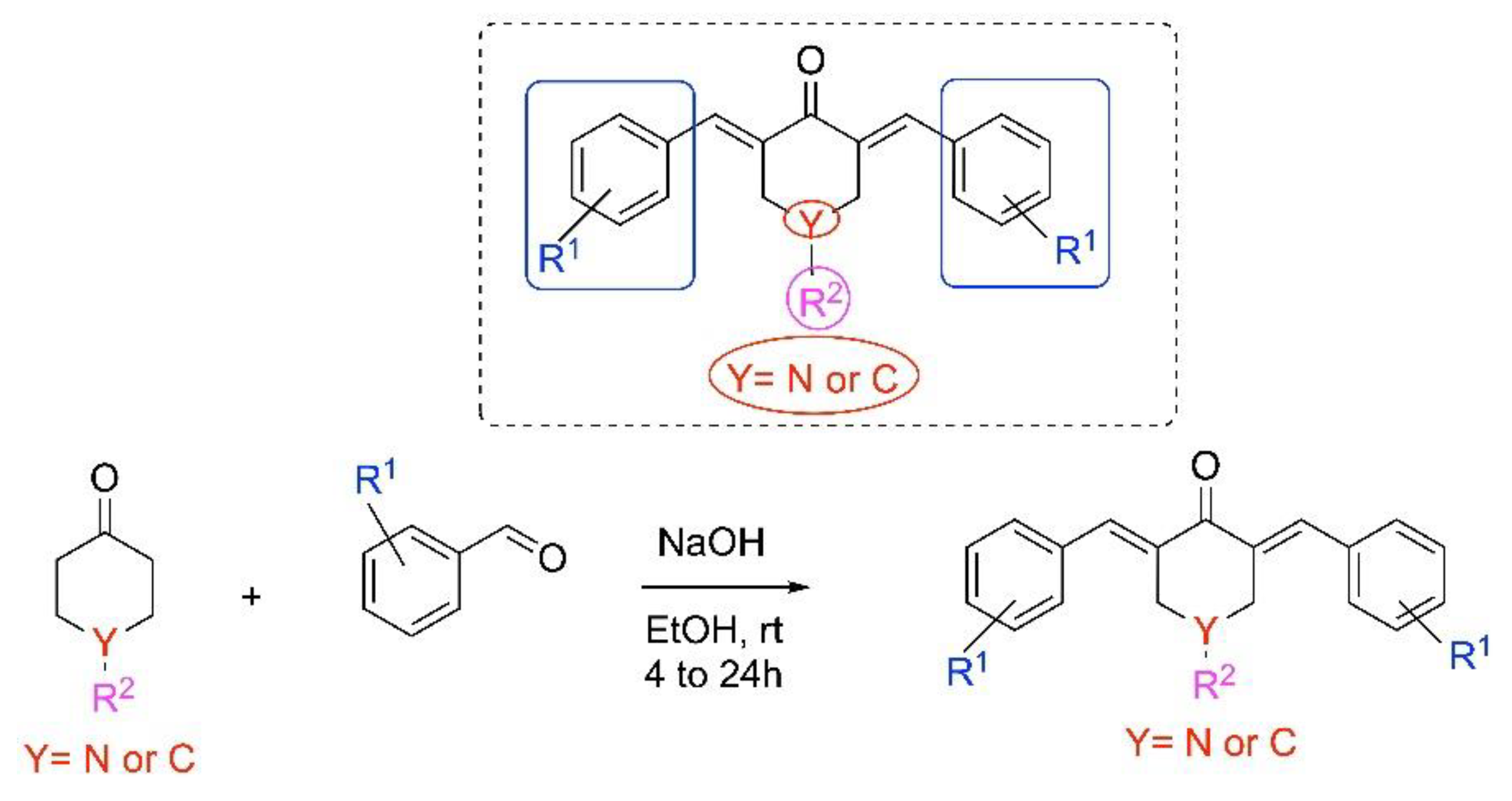
Figure 1.
Chemical structures of tested 1,5-diarylidene-4-piperidinones.
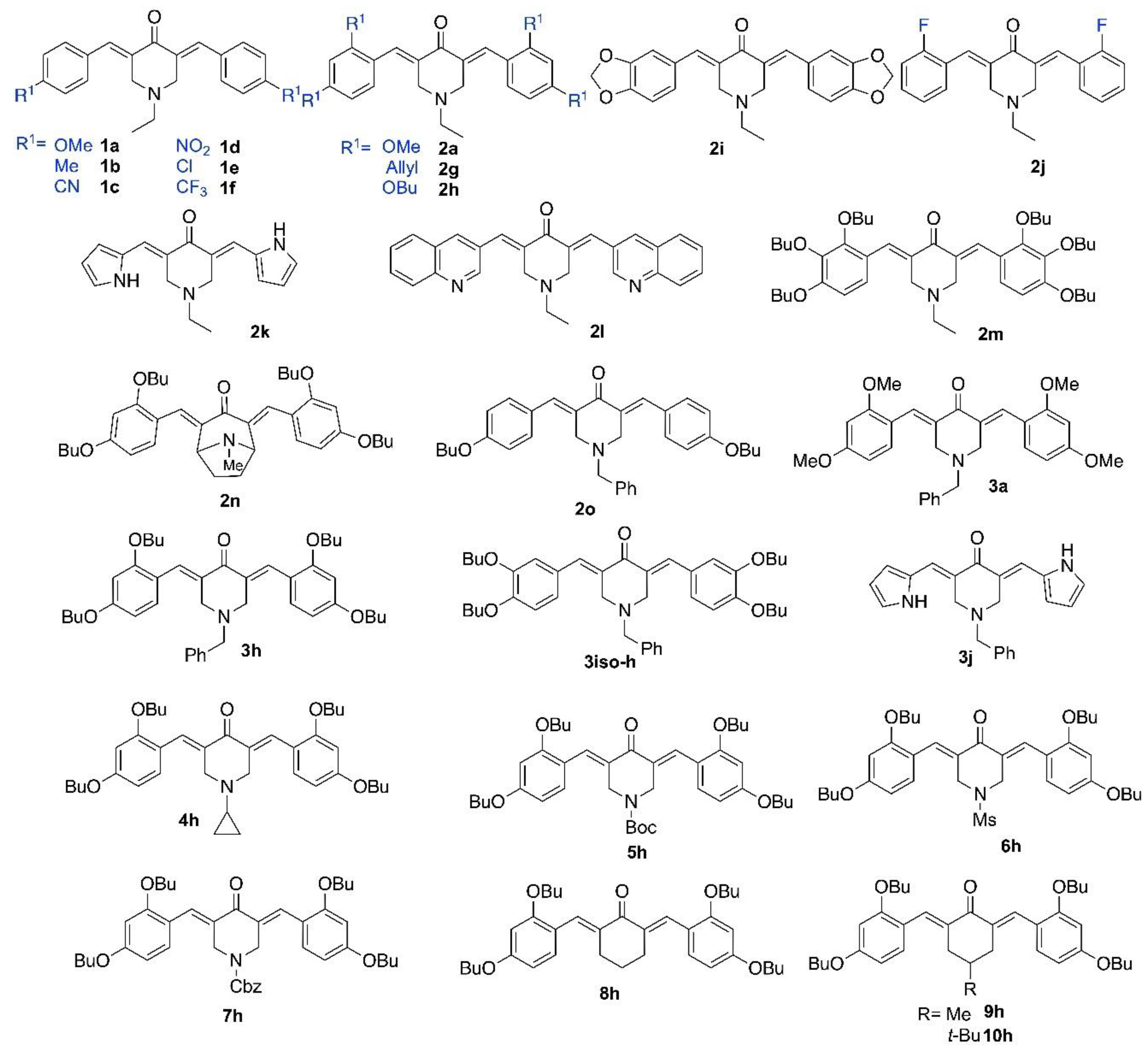
Figure 2.
Toxicity of piperidone compounds against human cells. Human cells were exposed to increasing concentrations of piperidone compounds active against C. neoformans (1a, 1b, 1c, 1d, 1e, 1f, 2h, 2i, 2j, 2m, 2o, 3h, 3iso-h, 3j, and 4h) for 48h before measurement of the cell viability using resazurin assay. A-C: A498 cells (kidney), D-F: BEAS-2B cells (lung), G-I: Caco-2 cells (intestine), J-L: HaCaT cells (skin), and M-O: HepG2 cell (liver). Curves were fitted using GraphPad® Prism 8 software (means +/- SD, n=3).

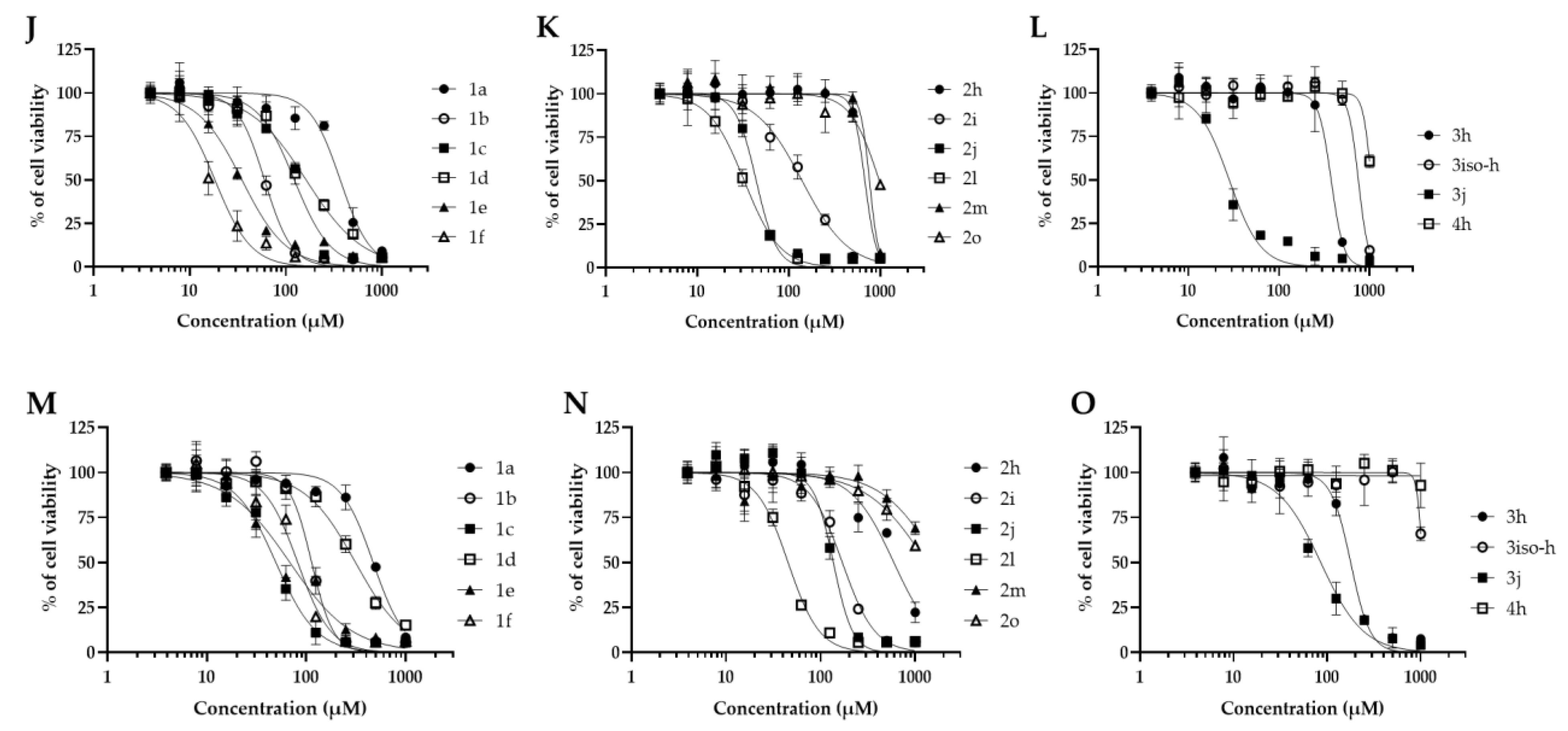
Figure 3.
Toxicity of Fluconazole against human cells. Human cells were exposed to increasing concentrations of Fluconazole for 48h before measurement of the cell viability using resazurin assay. Curves were fitted using GraphPad® Prism 8 software (means +/- SD, n=3).
Figure 3.
Toxicity of Fluconazole against human cells. Human cells were exposed to increasing concentrations of Fluconazole for 48h before measurement of the cell viability using resazurin assay. Curves were fitted using GraphPad® Prism 8 software (means +/- SD, n=3).
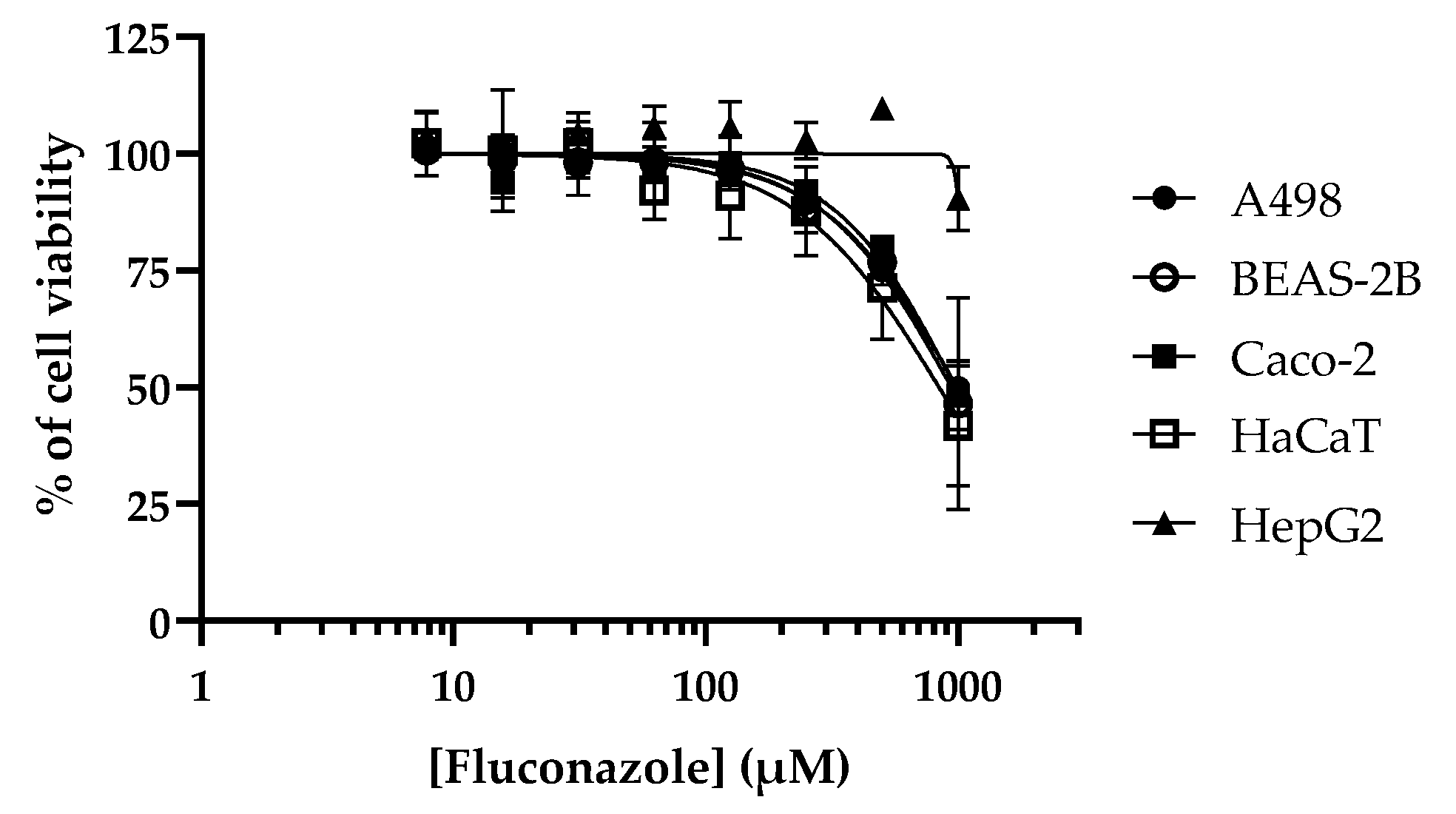

Table 1.
Anti-C. neoformans activity and cytotoxicity of piperidone compounds. The MIC values (in µM) of piperidone compounds and fluconazole were measured on C. neoformans as explained in Materials and Methods section. Their Cytotoxic Concentration 50 (CC50) (in µM) were calculated from Figure 1 and Figure 2 after 48 h incubation of human cells with increasing concentrations of molecules (n=3). Selectivity Indexes (SI) were calculated by dividing CC50 by MIC values. Molecules with MIC superior or equal to 250 µM were not tested in toxicity assay and are indicated as “NT”.
| Compound | MIC (μM) | CC50 (μM) | SI | ||||
| A498 | BEAS-2B | Caco-2 | HaCaT | HepG2 | Min-Max | ||
| 1a | 125 | 80.4 | 64.9 | 313.7 | 368.6 | 471.9 | 0.5-3.7 |
| 1b | 250 | 175.6 | 87.9 | 86.2 | 60.0 | 113.5 | 0.2-0.7 |
| 1c | 250 | 313.9 | 191.8 | 70.3 | 122.7 | 49.5 | 0.1-1.2 |
| 1d | 250 | 486.2 | 363.5 | 95.4 | 162.9 | 313.6 | 0.3-1.9 |
| 1e | 250 | 31.3 | 23.7 | 98.6 | 34.9 | 68.5 | 0.09-0.4 |
| 1f | 250 | 30.0 | 30.9 | 47.0 | 18.3 | 82.1 | 0.07-0.3 |
| 2a | >250 | NT | NT | NT | NT | NT | NT |
| 2g | >250 | NT | NT | NT | NT | NT | NT |
| 2h | 31.2 | 203.7 | 140.2 | 696.6 | 680.3 | 595.6 | 4.5-22.3 |
| 2i | 250 | 266.6 | 241.2 | 152.0 | 140.0 | 169.6 | 0.5-1.0 |
| 2j | 250 | 177.3 | 139.0 | 68.3 | 44.2 | 136.4 | 0.1-0.7 |
| 2k | >250 | NT | NT | NT | NT | NT | ND |
| 2l | 250 | 89.5 | 84.5 | 30.0 | 32.6 | 45.3 | 0.1-0.3 |
| 2m | 62.5 | 829.4 | 663.7 | 686.9 | 764.3 | >1000 | 10.6->16 |
| 2n | >250 | NT | NT | NT | NT | NT | NT |
| 2o | 125 | 297.4 | 230.5 | 454.7 | 979.3 | >1000 | 1.8->8 |
| 3a | >250 | NT | NT | NT | NT | NT | NT |
| 3h | 7.8 | 76.4 | 42.4 | 628.6 | 377.3 | 178.3 | 5.4-80.5 |
| 3iso-h | 62.5 | 122.4 | 106.9 | >1000 | 753.8 | >1000 | 1.7->16 |
| 3j | 62.5 | 34.5 | 82.9 | 26.1 | 28.2 | 85.6 | 0.4-1.3 |
| 4h | 250 | >1000 | >1000 | >1000 | >1000 | >1000 | >4 |
| 5h | >250 | NT | NT | NT | NT | NT | NT |
| 6h | >250 | NT | NT | NT | NT | NT | NT |
| 7h | >250 | NT | NT | NT | NT | NT | NT |
| 8h | >250 | NT | NT | NT | NT | NT | NT |
| 9h | >250 | NT | NT | NT | NT | NT | NT |
| 10h | >250 | NT | NT | NT | NT | NT | NT |
| Fluconazole | 25 | 993.6 | 942.5 | 977.0 | 841.2 | >1000 | 33.6->40 |
Table 2.
Activity of piperidone compounds against pathogenic yeasts. The MIC values (in µM) of piperidone compounds 2h, 2m, 3h, and 3iso-h were measured using various yeast models as explained in Materials and Methods section. Fluconazole was used as antifungal control.
Table 2.
Activity of piperidone compounds against pathogenic yeasts. The MIC values (in µM) of piperidone compounds 2h, 2m, 3h, and 3iso-h were measured using various yeast models as explained in Materials and Methods section. Fluconazole was used as antifungal control.
| Compound | C. albicans | C. auris | C. glabrata | C. tropicalis | C. neoformans |
| 2h | 250 | 31.2 | 31.2 | 250 | 31.2 |
| 2m | 125 | 31.2 | 15.6 | 250 | 62.5 |
| 3h | 62.5 | 7.8 | 7.8 | 125 | 7.8 |
| 3iso-h | 250 | 31.2 | 62.5 | 125 | 62.5 |
| Fluconazole | 6.25 | 500 | 250 | 1000 | 25 |
Table 3.
Activity of piperidone compounds against pathogenic filamentous fungi. The MIC values (in µM) of piperidone compounds 2h, 2m, 3h, and 3iso-h were measured using various filamentous fungi models (human pathogens or plant pathogens) as explained in Materials and Methods section. Fluconazole was used as antifungal control.
| Compound | A. flavus | A.fumigatus | C. graminicola | F.graminearum | M.oryzae | M. bolleyi | P.verrucosum | T. rubrum |
| 2h | >250 | 125 | >250 | >250 | 15.6 | 62.5 | 125 | 62.5 |
| 2m | >250 | 250 | >250 | >250 | 62.5 | >250 | >250 | 31.2 |
| 3h | 31.2 | 31.2 | 250 | 125 | 7.8 | 31.2 | 62.5 | 7.8 |
| 3iso-h | >250 | 125 | 125 | >250 | 15.6 | 62.5 | 125 | 125 |
| Fluconazole | >1000 | >1000 | 50 | >1000 | 12.5 | >1000 | >1000 | >1000 |
Table 4.
Interaction parameters. The interaction analysis of the sterol 14 α-demethylase protein on the basis of molecular docking studies with piperidones 2h, 3h, 2m, 3iso-h.
Table 4.
Interaction parameters. The interaction analysis of the sterol 14 α-demethylase protein on the basis of molecular docking studies with piperidones 2h, 3h, 2m, 3iso-h.
| Compound | Binding energy kcal/mol |
Hydrophobic interactions with amino acids | Aromatic interactions |
| Fluconazole | -8.1 | Phe110, Tyr116, Leu127, Ala287, Ala291, Leu356 | T-Shaped (Tyr103), Hem500 |
| 2h | -7.8 | Tyr103, Met106, Tyr116, Leu219, Phe290, Ala291, Leu356, Met360, Met460 | Hem500 T-Shaped (Tyr103), |
| 3h | -8.2 | Val102, Tyr103, Ile105, Pro210, Phe290, Ala291, Leu356, Met360, Met460 | Hem500 T-Shaped (Tyr103), |
| 2m | -7.2 | Ile72, Tyr103, Phe110, Pro210, Leu219, Phe290, Thr295, Leu356, Met360, Met460, Val461 | Hem500 |
| 3iso-h | -8.8 | Ile105, Met106, Leu208, Pro210, Leu219, Phe290, Ala291, Leu356, Thr459, Val461 | Hem500 T-Shaped (His294, Phe290), |
Disclaimer/Publisher’s Note: The statements, opinions and data contained in all publications are solely those of the individual author(s) and contributor(s) and not of MDPI and/or the editor(s). MDPI and/or the editor(s) disclaim responsibility for any injury to people or property resulting from any ideas, methods, instructions or products referred to in the content. |
© 2025 by the authors. Licensee MDPI, Basel, Switzerland. This article is an open access article distributed under the terms and conditions of the Creative Commons Attribution (CC BY) license (http://creativecommons.org/licenses/by/4.0/).
Copyright: This open access article is published under a Creative Commons CC BY 4.0 license, which permit the free download, distribution, and reuse, provided that the author and preprint are cited in any reuse.


