Submitted:
14 May 2025
Posted:
14 May 2025
You are already at the latest version
Abstract
Philodendron ‘White Knight’ is a popular climbing evergreen plant typically propagated through stem cuttings. However, this method is slow and inefficient, making it challenging to meet the rising market demand. In vitro culture could enhance the multiplication rate of this cultivar. However, research on its in vitro propagation is limited. Therefore, the objectives of the present study were to investigate the impact of silver nanoparticles (Ag NPs) on the surface sterilization of Philodendron ‘White Knight’ petioles, to study the influence of plant growth regulators on adventitious shoot regeneration, shoot multiplication, and root induction. Aseptic petiole explants (100%) were obtained after treatment with 40 mg L-1 Ag NPs for 60 minutes. The highest rate of adventitious shoot induction (52.6%) and the maximum shoot number (13.9 shoots per petiole) were achieved on Murashige and Skoog shoot multiplication B (MS-B) medium with 20 µM of 2-isopentenyl adenine (2-IP) and 5.0 µM of naphthalene acetic acid (NAA). The highest number of shoots (34 shoots per shoot tip) with a length of 5.1 cm was obtained on MS-B medium with 5.0 µM 2-IP and 2.5 µM NAA. The highest root induction (100%), the maximum root number (8.2 roots per shoot), and the greatest plantlet height (9.1 cm) were achieved on half-strength Murashige and Skoog medium containing indole-3-butyric acid (10.0 μM). The rooted plantlets of Philodendron ‘White Knight’ were transplanted in a transplanted into a substrate composed of 10% peat moss, 50% orchid stone, and 40% coconut husk chips, and acclimatized in a greenhouse environment, achieving a survival rate of 100%. This micropropagation protocol can be used for the commercial propagation of Philodendron ‘White Knight’ and related species.
Keywords:
auxin
; cytokinin
; in vitro propagation
; petiole
; silver nanoparticles
1. Introduction
The genus Philodendron Schott, belonging to the Araceae family, comprises 625 species native to South America, Central America, the Caribbean, and parts of Colombia, French Guiana, and Guyana [1]. Many Philodendron species are popular indoor plants due to their attractive foliage and low maintenance requirements, with several cultivars (cv.) emerging in recent decades [2]. Philodendron cultivars with orange, red, and yellow foliage, either variegated or non-variegated, tend to attract more attention and command higher market value, especially the variegated varieties (Chen et al. 2002, Oboni and Hossain 2025). Although they are typically propagated through stem cuttings and seeds, these methods do not meet market demand due to the lack of healthy plant materials and the lengthy propagation process [2,3,4]. In contrast, in vitro propagation techniques offer excellent alternatives for mass propagating Philodendron species and cultivars, using less plant material and allowing for year-round production with reduced space and a shorter time to satisfy market demand [2].
Micropropagation is a powerful in vitro plant culture technique widely used for the commercial propagation of various ornamental plants [5], including Philodendron billietiae Croat [6], Philodendron bipinnatifidum Schott ex Endl. [7], Philodendron erubescens cv. Pink Princess [4], Philodendron pertusum Kunth & C.D.Bouché [8], Philodendron selloum Koch [9], Philodendron tuxtlanum G.S.Bunting [10], Philodendron cv. Birkin [11], Imperial Green, Imperial Red, and Imperial Rainbow [2], as well as Blue Mistic, Painted Lady, Pink Prince, Pluto, Royal Queen, and Green Emperor [3]. The authors have utilized various explants, including axillary buds, axillary shoots, leaf laminae, nodes, petioles, shoot tips, and stem nodal segments, cultured on media containing a range of plant growth regulators such as 2,4-dichlorophenoxyacetic acid (2,4-D), 2-isopentenyl adenine (2IP), benzyladenine (BA)/6-benzylaminopurine (BAP), indole-3-butyric acid (IBA), kinetin (KN), naphthalene acetic acid (NAA), and thidiazuron (TDZ), either alone or in combination, to achieve adventitious shoot regeneration or axillary shoot multiplication. However, shoot induction depends on explant type, culture media composition, genotype, and plant growth regulators [2]. Therefore, the development of a micropropagation protocol for the mass production of a new cultivar of Philodendron is necessary.
Micropropagation involves multiple stages, including the establishment of aseptic culture, shoot multiplication, shoot elongation, rooting, and acclimatization [5]. Although all stages are equally important, the initial establishment of in vitro plant culture is a critical stage, and contamination poses a significant challenge. Success depends on effectively disinfecting explant surfaces while preserving their viability [12]. Philodendron explants were often surface disinfected with 70-95% ethyl alcohol (EtOH), 0.16-5.0% sodium hypochlorite (NaOCl), and 0.05-0.1% mercuric chloride (HgCl2) [3,5,8,11,13,14]. Nanoparticles (NPs) effectively combat various bacteria, fungi, and viruses that can contaminate plant explants [15]. Their small size allows them to penetrate microbial cell walls easily, making them more efficient than traditional disinfectants. NPs can be used in lower concentrations, reducing toxicity risks to plant tissues while remaining effective. Furthermore, they are generally more environmentally friendly than HgCl2, making them a promising alternative for disinfecting plant tissue culture explants [16,17]. Silver (Ag) NPs are environmentally friendly chemicals that have been used to eliminate microorganisms in plant tissue culture [14] because of their powerful antimicrobial properties. Recently, Lara-Ascencio et al. [17] demonstrated that the Ag NPs treatment reduces the presence of contaminants in the Philodendron xanadu Croat explants. Philodendron ‘White Knight’ is a climbing evergreen plant native to South America. It is one of the popular Philodendron cultivars due to its variegated foliage and is used as both an indoor and garden plant. It is commonly multiplied through stem cutting; however, vegetative propagation is slow and requires effective alternative methods for multiplying this important cultivar to meet market demand. So far, no reports have documented the in vitro propagation of Philodendron ‘White Knight’. Thus, this study aimed to develop an efficient micropropagation protocol for the mass production of Philodendron ‘White Knight’ through adventitious shoot regeneration using petiole explants. The objectives of the present study were to evaluate the effect of Ag NPs on the surface disinfection of petiole explants and to investigate the effect of plant growth regulators (PGRs) on adventitious shoot regeneration, shoot multiplication, and rooting of Philodendron ‘White Knight’.
2. Materials and Methods
2.1. Plant Materials and Surface Disinfection
Petioles were collected from fully developed leaves of three-year-old Philodendron ‘White Knight’ shoots cultivated in a polyhouse. The cut ends of the petioles were sealed, thoroughly cleaned under running tap water for 20 minutes, and then washed with distilled water (DH2O) containing 0.15% (w/v) polyvinylpyrrolidone (PVP). Petioles were surface disinfected in laminar airflow by treating them with 70% (v/v) EtOH for 30 seconds; rinsed three times in sterile DH2O containing 0.15% PVP (SDH2O-PVP) and 1.0% (v/v) NaOCl solution for 12 minutes; and washed five times with SDH2O-PVP. To study the effect of Ag NPs (< 100 nm particle size, 99.5% purity, Sigma-Aldrich) on surface disinfection, the NaOCl-treated petioles were immersed in 10, 20, 40, or 80 mg L-1 Ag NPs for 30, 60, or 90 minutes and then washed five times with SDH2O-PVP. The sealed ends were discarded, and the petioles were cut transversely into 0.4-0.6 mm segments and then cultured on Murashige and Skoog shoot multiplication B (MS-B) medium (Duchefa Biochemie, The Netherlands) containing 8.0 µM BA. All media contained 0.6 g L⁻¹ activated charcoal (AC), 30 g L⁻¹ sucrose, 0.8 g L⁻¹ agar, and the pH of the media was adjusted to 5.70-5.80 before autoclaving at 123 ºC for 23 minutes. For each treatment, 15 petiole explants with three replicates were used, and the experiment was repeated twice. The cultures were kept at 23 ± 1°C under a 12-hour photoperiod with a photosynthetic photon flux density (PPFD) of 18-23 μMol s-1 m-2. The rates of contamination and survival of Philodendron ‘White Knight’ explants were recorded after 21 days of incubation. Explants that turned black were deemed dead, while those that remained purple or increased in size were considered alive.
2.2. Effect of PGRs on Adventitious Shoot Regeneration
To study the impact of PGRs on adventitious shoot induction, the explants were prepared from petioles treated with 40 mg L-1 Ag NPs for 60 minutes and cultured on MS-B medium containing 5, 10, 20, or 30 µM of BA or 2-IP combined with 2.5 or 5.0 µM of NAA or IBA. The medium MS-B, devoid of PGRs, served as a control. For each treatment, 20 petiole explants with three replicates were used, and the experiment was repeated twice. The cultures were kept at 23 ± 1°C under a 12-hour photoperiod with a PPFD of 35-40 μMol s-1 m-2. The rate of shoot formation and the number of shoots per Philodendron ‘White Knight’ petiole were recorded after 13 weeks of incubation.
2.3. Effect of Combinations of 2-IP and NAA on Shoot Multiplication and Shoot Elongation
The adventitious shoot buds were transferred to PGR-free MS-B medium and subcultured at four-week intervals. After three subcultures, the in vitro developed shoots were utilized to obtain shoot tips explants. For shoot multiplication, shoot tips were cultured on the MS-B medium containing 2.5, 5.0, or 10.0 µM 2-IP combined with 2.5 µM NAA. The medium MS-B, devoid of 2-IP and NAA, served as a control. For each treatment, 30 shoot buds with three replicates were used, and the experiment was repeated twice. The cultures were kept at 23 ± 1°C under a 14-hour photoperiod with a PPFD of 45-50 μMol s-1 m-2. The number of shoots per Philodendron ‘White Knight’ shoot bud, and shoot length (mm) were recorded after 13 weeks of incubation.
2.4. Effect of IBA and NAA on Rooting
Well-developed shoots separated from the shoot clusters were transferred to half-strength Murashige and Skoog (MS) [18] medium containing 0, 2.5, 5.0, 10.0, or 20.0 µM NAA or IBA to induce roots. For each treatment, 30 shoots with three replicates were used, and the experiment was repeated twice. The cultures were kept at 23 ± 1°C under a 14-hour photoperiod with a PPFD of 60-65 μMol s-1 m-2. The rate of root induction and the number of roots per Philodendron ‘White Knight’ shoot, were recorded after 8 weeks of incubation.
2.5. Acclimatization
The well-developed 300 Philodendron ‘White Knight’ plantlets were separated from the rooting medium, thoroughly washed to remove any traces of it, and then transplanted into trays containing a mixture of peat moss (10%), orchid stone (50%), and coconut husk chips (40%). They were subsequently acclimatized in a greenhouse at a temperature of 20-25 °C and a relative humidity of 95-100% for 2 weeks, which was gradually reduced to 60%. The plants were fertigated with ¼ MS nutrients, and the survival of the plantlets was recorded after 6 weeks of cultivation.
2.6. Statistical Analysis
The data were subjected to analysis of variance (ANOVA) tests, and means were distinguished by Duncan’s multiple range test (DMRT) at p ≤ 0.05. The statistical analyses were performed using SAS Release 9.4.
3. Results and Discussion
3.1. Impact of Ag NPs on the Surface Disinfection of Philodendron ‘White Knight’ Petiole Explants
The petiole explants of Philodendron ‘White Knight’ treated with EtOH and NaOCl (control) yielded 25.9% aseptic explants, while 74.1% of the petioles were contaminated with bacteria and fungi; however, the survival rate of Philodendron ‘White Knight’ explants remained unaffected. The percentage of aseptic cultures increased when the petioles were treated with Ag NPs compared to the control (Table 1). The positive effect of Ag NPs on the surface disinfection of explants has also been documented in Ardisia mamillata [12], Begonia tuberous [19], Limonium sinuatum ‘White’ [20], Monstera deliciosa ‘Thai Constellation’ [21], Ocimum basilicum ‘Italian Large Leaf’, Salvia farinecae, and Thymus vulgaris [22]. Increasing the concentration of Ag NPs and soaking period reduced explant contamination; however, higher concentrations and a longer soaking period also resulted in a decrease in the explant survival rate (Table 1). Similar results have been observed in A. mamillata [12], Kaempferia parviflora [23], M. deliciosa ‘Thai Constellation’ [21], O. basilicum ‘Italian Large Leaf’, S. farinecae, and T. vulgaris [22]. In this study, soaking Philodendron ‘White Knight’ explants in 10 mg L-1 silver nanoparticles for 30, 60, and 90 minutes resulted in 29.3%, 40.1%, and 51.4% of aseptic explants, respectively. Additionally, the survival rate (100%) of explants remained unaffected. Among the various Ag NPs treatments, 40 mg L-1 Ag NPs for 60 minutes was found to be the most effective, resulting in 100% aseptic explants with a 100% survival rate. In contrast, the survival rate of explants decreased when treated with 40 mg L-1 Ag NPs for 90 minutes (Table 1).
3.2. Influence of PGRs on Adventitious Shoot Regeneration from Petiole Explants of Philodendron ‘White Knight’
3.2.1. Combination of 2-IP and Auxins (NAA and IBA)
Direct adventitious shoot regeneration is an effective technique that enhances efficiency and sustainability in commercial plant propagation [24] and is essential for Agrobacterium-mediated genetic transformation [25]. It has been reported that the petiole segments of Philodendron scandens [26] and Philodendron ‘Imperial Green’ [2] do not produce shoots. In this study, the petiole explants of Philodendron ‘White Knight’ did not yield adventitious shoots on the MS-B medium lacking PGRs or containing 5.0 µM 2-IP plus auxins, and they died after 13 weeks of culture. However, adventitious shoot buds developed from the explants within 8 weeks of culture when the MS-B medium was fortified with higher concentrations of 2-IP and auxins (Figure 1A and B). It has been revealed that supplementing both auxin and cytokinin can induce adventitious shoot regeneration in several members of the Araceae family, including Aglonema ‘Lady Valentine’ [27], Anthurium andraeanum [28], Epipremnum aureum [29], Spathiphyllum wallsii ‘Domino’ [30], and Zamioculcas zamiifolia [31]. When the MS-B medium was fortified with 10-30 µM of 2-IP and 2.5 µM of NAA, 15-38.9% of Philodendron ‘White Knight’ petioles developed 2.8-7.0 adventitious shoots per explant (Figure). The highest adventitious shoot induction (52.6%) and the maximum shoot production (13.9 shoots per explant) were achieved on MS-B medium with 20 µM of 2-IP and 5.0 µM of NAA (Table 2). This result surpassed previous findings, in which only 2.8% of petioles from Philodendron Imperial Red and 11.1% from Imperial Rainbow produced shoots [2]. When 2-IP was combined with IBA, 14.5-34.9% of Philodendron ‘White Knight’ petioles developed 1.9-6.3 adventitious shoots per explant (Table 2). The optimal shoot production (34.9% explants induced 6.3 shoots per petiole) was obtained on MS-B medium containing 20 µM of 2-IP and 5.0 µM of IBA.
3.2.2. Combination of BA and Auxins (NAA and IBA)
When BA (10-30 µM) was combined with NAA (2.5 or 5.0 µM), 8.3-38.8% of petioles produced 1.2-4.8 shoots per explant. The maximum adventitious shoot induction (38.8%) and the highest number of shoots (4.8 shoots per petiole) were achieved on MS-B medium containing 20 µM BA and 2.5 µM NAA. However, the shoot production potential of Philodendron ‘White Knight’ petioles decreased when the medium MS-B was fortified with BA and 5.0 µM NAA (Table 3). The presence of BA and IBA in the MS-B medium enhanced shoot production, with the best adventitious shoot induction at 46.1% and the maximum number of shoots at 7.1 shoots per petiole achieved on MS-B medium with 20 µM BA and 5.0 µM IBA (Table 3, Figure 1C). The results (Table 2 and Table 3) indicated that adventitious shoot production from petiole explants of Philodendron ‘White Knight’ was influenced by the type of cytokinin and auxin. The petioles of Philodendron ‘White Knight’ showed the best response to MS-B medium containing 2-IP and NAA. The presence of 2-IP and NAA in the culture medium has been shown to effectively promote optimal shoot formation from petioles of Bixa orellana [32].
3.3. Combination of 2-IP and NAA on Philodendron ‘White Knight’ Shoot Multiplication
Chen et al. [2] utilized in vitro regenerated shoots of Philodendron cultivars as the explant (node) source to examine the impact of cytokinins (BA, KN, and TDZ) on shoot multiplication. Similarly, the shoot multiplication of Philodendron cultivars or species has been achieved using in vitro derived nodes, protocorm-like bodies, and shoot tips on culture media containing BA alone or in combination with auxins [4,6,7,33]. These studies have demonstrated that BA/BAP, whether used alone or in combination with auxins, is more effective for shoot multiplication than other cytokinin treatments. In this study, the results indicated that a combination of 2-IP and NAA produced a higher number of shoots. Therefore, the impact of various concentrations of 2-IP combined with 2.5 µM NAA was examined on multiple shoot induction from in vitro derived shoot tips of Philodendron ‘White Knight’. The shoot tips of Philodendron ‘White Knight’ cultured on MS-B medium devoid of 2-IP and NAA produced multiple shoots (4.9 shoots per shoot tip) with a length of 6.4 cm (Table 4). In contrast, Alawaadh et al. [7] reported that shoot tip explants of Philodendron bipinnatifidum developed only a single shoot on PGR-free media. On the other hand, P. erubescens ‘Pink Princess’ protocorm-like bodies developed 4.4 shoots on PGR-free medium [4]. The addition of 2-IP and NAA to MS-B medium significantly increased the number of shoots per Philodendron ‘White Knight’ shoot tip compared to the control, while the length of the shoot significantly decreased in a dose-dependent manner. The highest number of shoots (34 shoots per shoot tip) with a length of 5.1 cm was obtained on MS-B medium containing 5.0 µM 2-IP and 2.5 µM NAA (Table 4, Figure 2A-C). Likewise, shoot multiplication has been achieved in the presence of 2-IP and NAA in Allium sativum [34], Philodendron bipinnatifidum [7], and Philodendron goeldii [35].
3.4. Effect of IBA and NAA on Rooting
Rooting is a crucial stage of micropropagation influenced by the composition of the shoot induction medium, the rooting medium composition, the type and concentration of auxins, as well as the genotype and plant species [5]. In vitro rooting of Philodendron cultivars and species has been achieved on basal medium without [3,9] or with auxins [2,4,6,7,8,13]. Previous studies have shown that in vitro rooting of Philodendron cultivars and species is best achieved on a medium containing IBA or NAA [2,4,6,7,8,13]. Thus, this study investigated the influence of IBA and NAA on the rooting of Philodendron ‘White Knight’ shoots. In the control medium (auxin-free ½ MS), 18.9% of shoots successfully developed an average of 2.2 roots, with an average plantlet height of 3.9 cm (Table 5). The inclusion of IBA or NAA at concentrations ranging from 2.5 to 20.0 µM to the ½ MS medium significantly increased the rooting percentage (52.2-100%), the average number of roots (2.7-8.2), and the plantlet height (4.3-5.8) compared to the control. However, it is noteworthy that elevated concentrations of NAA, particularly at 20.0 µM, resulted in a marked decrease in plantlet height, which measured only 3.1 cm compared to the control. Of the various concentrations (2.5-20.0 μM) of auxins tested, 10.0 μM IBA resulted in the highest root induction (100%), the maximum root number (8.2 roots per shoot), and the greatest plantlet height (9.1 cm) (Table 5). Among the two auxins tested, IBA was found to be the best for in vitro rooting of Philodendron ‘White Knight’ shoots (Figure 2D). The beneficial effects of IBA on the rooting of Araceae members have been shown in Aglaonema ‘Lady Valentine’ [36], M. deliciosa ‘Thai Constellation’ [21], Philodendron cultivars [2], Philodendron ‘Birkin’ [11], P. erubescens ‘Pink Princess’ [4], P. pertusum [8], Philodendron xanadu [17], and Spathiphyllum wallsii ‘Domino’ [30]. In this study, most shoots that regenerated in vitro during the rooting phase displayed normal green leaves; however, a subset of shoots exhibited variegated leaves (Figure 3).
3.5. Acclimatization
Acclimatization is the final stage of micropropagation, influenced mainly by the growth substrate. Philodendron cultivars and species have been successfully acclimatized using a mixture of various substrates such as peat moss and perlite [2,7], river sand, farmyard manure, and soil [3], perlite, vermiculite, and peat moss cocopeat [4], sand, soil, and farmyard manure [8], and coir and Luffa sponge [13]. In this study, both the normal and variegated plantlets of Philodendron ‘White Knight’ were successfully transplanted into a substrate composed of 10% peat moss, 50% orchid stone, and 40% coconut husk chips and acclimatized (Figure 2E) in a greenhouse environment, achieving a survival rate of 100%.
4. Conclusions
For the first time, we have established an efficient micropropagation protocol for the commercial propagation of Philodendron ‘White Knight’. Our findings demonstrate that a combination of cytokinin and auxin is essential for inducing adventitious shoot induction and maximizing shoot multiplication. The in vitro grown shoots of Philodendron ‘White Knight’ exhibit optimal rooting when treated with IBA. In vitro regenerated normal and variegated plantlets can be utilized in breeding programs aimed at enhancing this cultivar. To ensure the genetic integrity of the acclimatized plantlets, molecular analysis will be conducted.
Author Contributions
Conceptualization, I.S.; methodology, I.S.; software, I.K.; formal analysis, I.K.; investigation, I.S.; data curation, I.K.; writing—original draft preparation, I.K.; writing—review and editing, I.S. All authors have read and agreed to the published version of the manuscript.
Funding
This research received no external funding.
Data Availability Statement
Data are contained within the article.
Acknowledgments
This article was supported by the KU Research Professor Program of Konkuk University.
Conflicts of Interest
The authors declare no conflicts of interest.
Abbreviations
The following abbreviations are used in this manuscript:
| Ag NPs | silver nanoparticles |
| MS-B | Murashige and Skoog shoot multiplication B |
| 2-IP | 2-isopentenyl adenine |
| IBA | indole-3-butyric acid |
| NAA | naphthalene acetic acid |
| BA | benzyladenine |
| PPFD | photosynthetic photon flux density |
References
- International Plant Names Index. Royal Botanic Gardens, Kew Names and Taxonomic Backbone. 2025. https://powo.science.kew.org/taxon/urn:lsid:ipni.org:names:326132-2#source-KB. Retrieved 08 May 2025.
- Chen, F.C.; Wang, C.Y.; Fang, J.Y. Micropropagation of self-heading Philodendron via direct shoot regeneration. Sci. Hortic. 2012, 141, 23–29. [Google Scholar] [CrossRef]
- Sreekumar, S.; Mukunthakumar, S.; Seeni, S. Morphogenetic response of six Philodendron cultivars in vitro. Indian J. Exp. Biol. 2001, 39, 1280–1287. [Google Scholar]
- Klanrit, P.; Kitwetcharoen, H.; Thanonkeo, P.; Thanonkeo, S. In Vitro Propagation of Philodendron erubescens ‘Pink Princess’ and Ex Vitro Acclimatization of the Plantlets. Horticulturae 2023, 9, 688. [Google Scholar] [CrossRef]
- Debergh, P.; Maene, L. A scheme for commercial propagation of ornamental plants by tissue culture. Sci. Hortic. 1981, 14, 335–345. [Google Scholar] [CrossRef]
- Maikaeo, L.; Puripunyavanich, V.; Limtiyayotin, M.; Orpong, P.; Kongpeng, C. Micropropagation and gamma irradiation mutagenesis in Philodendron billietiae. Thai J. Agric. Sci. 2024, 57, 11–19. [Google Scholar]
- Alawaadh, A.A.; Dewir, Y.H.; Alwihibi, M.S.; Aldubai, A.A.; El-Hendawy, S.; Naidoo, Y. Micropropagation of lacy tree philodendron (Philodendron bipinnatifidum Schott ex Endl.). Hortscience 2020, 55, 294–299. [Google Scholar] [CrossRef]
- Kumar, D.; Tiwari, J.P.; Singh, R. In-vitro clonal propagation of Philodendron pertusum. Ind. J. Hortic. 1998, 55, 340–343. [Google Scholar]
- Seliem, M.K.; El-Mahrouk, M.E.; El-Banna, A.N.; Hafez, Y.M.; Dewir, Y.H. Micropropagation of Philodendron selloum: Influence of copper sulfate on endophytic bacterial contamination, antioxidant enzyme activity, electrolyte leakage, and plant survival. South Afr. J. Bot. 2021, 139, 230–240. [Google Scholar] [CrossRef]
- Jambor-Benczur, E.; Marta-Riffer, A. In vitro propagation of Philodendron tuxilanum Bunting with benzylaminopurine. Acta Agron. Hung. 1990, 39, 341–348. [Google Scholar]
- Akramian, M.; Khaleghi, A.R.; Salehi-Arjmand, H. Optimization of plant growth regulators for in vitro mass propagation of Philodendron cv. Birkin through shoot tip culture. Greenh. Plant Prod. J. 2024, 1, 55–62. [Google Scholar] [CrossRef]
- Chun, S.C.; Seob, P.K.; Kang, K.W.; Sivanesan, I. Micropropagation of Ardisia mamillata Hance through axillary shoot multiplication. Propag. Ornam. Plants 2024, 24, 63–70. [Google Scholar]
- Gangopadhyay, G.; Bandyopadhyay, T.; Gangopadhyay, S.B.; Mukherjee, K.K. Luffa sponge – a unique matrix for tissue culture of Philodendron. Curr. Sci. 2004, 86, 315–319. [Google Scholar]
- Kim, D.H.; Gopal, J.; Iyyakkannu Sivanesan, I. Nanomaterials in plant tissue culture: The disclosed and undisclosed. RSC Adv. 2017, 7, 36492–36505. [Google Scholar] [CrossRef]
- Ochatt, S.; Abdollahi, M.R.; Akin, M.; Bello Bello, J.J.; Eimert, K.; Faisal, M.; Nhut, D.T. Application of nanoparticles in plant tissue cultures: minuscule size but huge effects. Plant Cell Tiss. Organ Cult. 2023, 155, 323–326. [Google Scholar] [CrossRef]
- Singh, Y.; Kumar, U.; Panigrahi, S.; Balyan, P.; Mehla, S.; Sihag, P.; Sagwal, V.; Singh, K.P.; White, J.C.; Dhankher, O.P. Nanoparticles as novel elicitors in plant tissue culture applications: Current status and future outlook. Plant Physiol. Biochem. 2023, 203, 108004. [Google Scholar] [CrossRef]
- Lara-Ascencio, M.; Andrade-Rodriguez, M.; Guillen-Sanchez, D.; Sotelo-Nava, H.; Villegas-Torres, O.G. Establishment of in vitro aseptic culture of Philodendron xanadu Croat. Rev. Cienc. Agron. 2021, 52, e20197034. [Google Scholar] [CrossRef]
- Murashige, T.; Skoog, F. A revised medium for rapid growth and bioassays with tobacco tissue cultures. Physiol. Plant. 1962, 15, 473–497. [Google Scholar] [CrossRef]
- Tung, H.T.; Van, H.T.; Bao, H.G.; Bien, L.H.; Khai, H.D.; Luan, V.Q.; Cuong, D.M.; Phong, T.H.; Nhut, D.T. Silver nanoparticles enhanced efficiency of explant surface disinfection and somatic embryogenesis in Begonia tuberous via thin cell layer culture. Vietnam J. Biotechnol. 2021, 19, 337–347. [Google Scholar] [CrossRef]
- Cuong, D.M.; Mai, N.T.; Tung, H.T.; Khai, H.D.; Luan, V.Q.; Phong, T.H.; Van The Vinh, B.; Phuong, H.T.; Van Binh, N.; Tan Nhut, D. Positive effect of silver nanoparticles in micropropagation of Limonium sinuatum (L.) Mill. ‘White’. Plant Cell Tissue Organ Cult. 2023, 22, 417–432. [Google Scholar] [CrossRef]
- Sivanesan, I.; Lee, Y.K.; Kang, K.W.; Park, H.Y. Micropropagation of Monstera delicosa Liebm. ‘Thai Constellation’. Propag. Ornam. Plants.
- Nartop, P. Effects of surface sterilisation with green synthesized silver nanoparticles on Lamiaceae seeds. IET Nanobiotechnol. 2018, 12, 663–668. [Google Scholar] [CrossRef]
- Park, H.-Y.; Kim, K.-S.; Ak, G.; Zengin, G.; Cziáky, Z.; Jekő, J.; Adaikalam, K.; Song, K.; Kim, D.-H.; Sivanesan, I. Establishment of a Rapid Micropropagation System for Kaempferia parviflora Wall. Ex Baker: Phytochemical Analysis of Leaf Extracts and Evaluation of Biological Activities. Plants 2021, 10, 698. [Google Scholar] [CrossRef]
- Yang, H.; Yang, Y.; Wang, Q.; He, J.; Liang, L.; Qiu, H.; Wang, Y.; Zou, L. Adventitious Shoot Regeneration from Leaf Explants in Sinningia Hybrida ‘Isa’s Murmur’. Plants 2022, 11, 1232. [Google Scholar] [CrossRef]
- Pérez-Caselles, C.; Faize, L.; Burgos, L.; Alburquerque, N. Improving Adventitious Shoot Regeneration and Transient Agrobacterium-Mediated Transformation of Apricot (Prunus armeniaca L.) Hypocotyl Sections. Agronomy 2021, 11, 1338. [Google Scholar] [CrossRef]
- Klimaszewska, K. Plant regeneration from petiole segments of some species in tissue culture. Acta Agrobotanica 1981, 34, 5–28. [Google Scholar] [CrossRef]
- El-Gedawey, H.I.; Hussein, S.E. Micropropagation of Aglonema ‘Lady Valentine’ by axillary shoots explants. Egyptian Academic Journal of Biological Sciences, H. Botany 2022, 13, 129–42. [Google Scholar] [CrossRef]
- Zhang, H.; Wang, G.; Qiao, Y.; Chen, C. Plant regeneration from root segments of Anthurium andraeanum and assessment of genetic fidelity of in vitro regenerates. In Vitro Cell. Dev. Biol. Plant 2021, 57, 954–964. [Google Scholar] [CrossRef]
- Qu, L.; Chen, J.; Henny, R.J.; Huang, Y.; Caldwell, R.D.; Robinson, C.A. Thidiazuron promotes adventitious shoot regeneration from pothos (Epipremnum aureum) leaf and petiole explants. Vitr. Cell Dev. Biol. Plant 2014, 50, 561–567. [Google Scholar] [CrossRef]
- Chun, S.C.; Seob, P.K.; Kang, K.W.; Sivanesan, I. In vitro propagation of Spathiphyllum wallsii Regel ‘Domino’ through adventitious shoot regeneration and somatic embryogenesis. Propag. Ornam. Plants 2024, 24, 55–62. [Google Scholar]
- Pourhassan, A.; Kaviani, B.; Kulus, D.; Miler, N.; Negahdar, N. A Complete Micropropagation Protocol for Black-Leaved Zamioculcas zamiifolia (Lodd.) Engl. ‘Dowon’. Horticulturae 2023, 9, 422. [Google Scholar] [CrossRef]
- Mohammed, A.; Chiruvella, K.K.; Namsa, N.D.; Ghanta, R.G. An efficient in vitro shoot regeneration from leaf petiolar explants and ex vitro rooting of Bixa orellana L.—A dye yielding plant. Physiol. Mol. Biol. Plants 2015, 21, 417–424. [Google Scholar] [CrossRef]
- Chiewchan, N.; Saetiew, K.; Teerarak, M. The effect of BA on inducing shoots of Philodendron erubescent ‘Pink Princes’ in vitro. International Journal of Agricultural Technology 2023, 19, 2385–2398. [Google Scholar]
- Bhojwani, S.S. In vitro propagation of garlic by shoot proliferation. Sci. Hortic. 1980, 13, 47–52. [Google Scholar] [CrossRef]
- Irawati, I. Diferensiasi Berbagai Macam Eksplan Pada Perbanyakan Philodendron Goeldii (Araceae) Secara In-vitro*[differentiation of Several Explants on In-vitro Propagation of Philodendron Goeldii (Araceae)]. Berita Biologi 2000, 5, 67669. [Google Scholar]
- Fang, J.Y.; Hsul, Y.R.; Chen, F.C. Development of an efficient micropropagation procedure for Aglaonema ‘Lady Valentine’ through adventitious shoot induction and proliferation. Plant Biotechnol. 2013, 30, 423–431. [Google Scholar] [CrossRef]
Figure 1.
Adventitious shoots developed from Philodendron ‘White Knight’ petioles cultured on MS-B medium with 2-IP and NAA (A), 2-IP and IBA (B), BA and IBA (C).
Figure 1.
Adventitious shoots developed from Philodendron ‘White Knight’ petioles cultured on MS-B medium with 2-IP and NAA (A), 2-IP and IBA (B), BA and IBA (C).
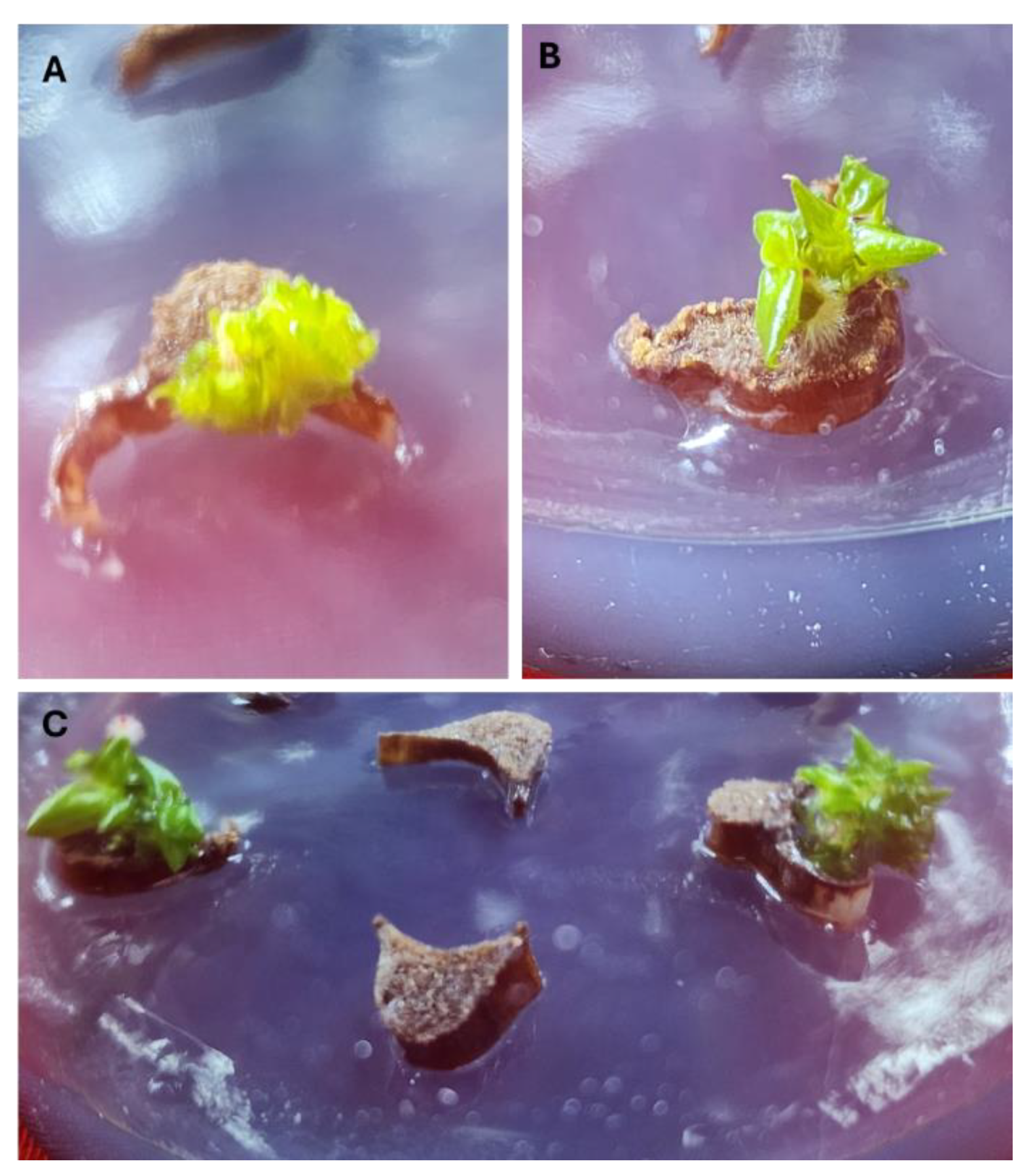
Figure 2.
Shoot multiplication, rooting and acclimatization. Shoot tips of Philodendron ‘White Knight’ developed multiple shoots on MS-B medium containing 5.0 µM 2-IP and 2.5 µM NAA after 6 weeks (A), and 13 weeks (B) of cultivation; C) Multiple shoot cluster; D) Regenerated shoots of Philodendron ‘White Knight’ developed roots on ½ MS medium with 10.0 µM IBA; D) Acclimatized Philodendron ‘White Knight’ plantlets. .
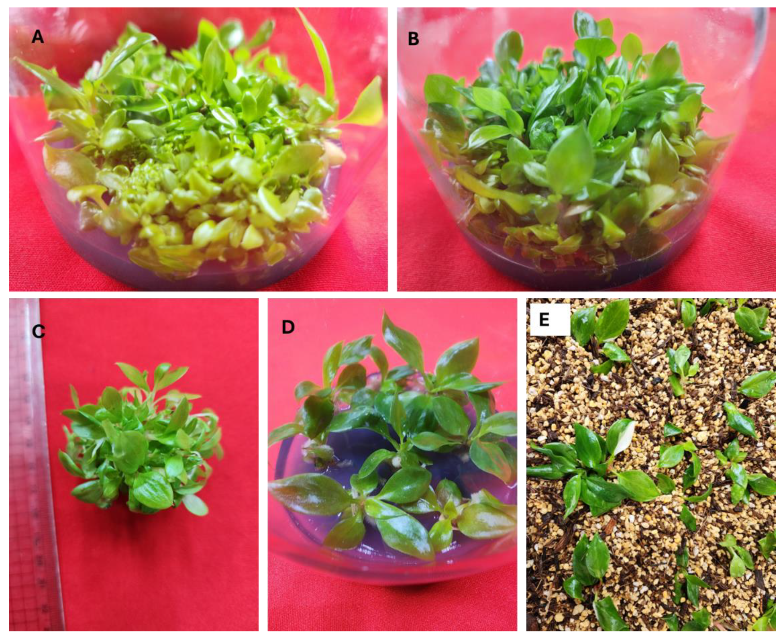
Figure 3.
In vitro regenerated normal and variegated plantlets of Philodendron ‘White Knight’.
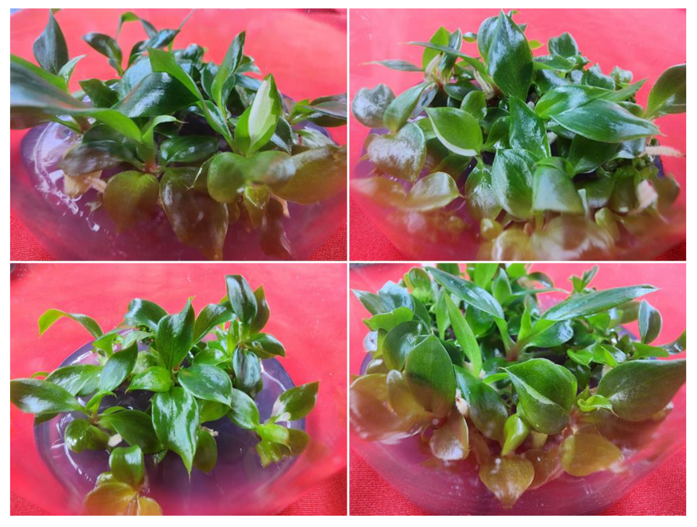

Table 1.
Effects of Ag NPs concentration and soaking period on the surface disinfection of Philodendron ‘White Knight’ Petiole Explants.
Table 1.
Effects of Ag NPs concentration and soaking period on the surface disinfection of Philodendron ‘White Knight’ Petiole Explants.
| Ag NPs (mg L-1) | Soaking Period (min) | Aseptic Explant (%) | Survival (%) |
|---|---|---|---|
| Control | 0 | 25.9 ± 1.6 h | 100 ± 0.0 a |
| 10 | 30 | 29.3 ± 1.2 h | 100 ± 0.0 a |
| 20 | 30 | 34.8 ± 1.2 g | 100 ± 0.0 a |
| 40 | 30 | 47.8 ± 1.3 e | 100 ± 0.0 a |
| 80 | 30 | 54.7 ± 1.7 d | 100 ± 0.0 a |
| 10 | 60 | 40.1 ± 1.4 f | 100 ± 0.0 a |
| 20 | 60 | 72.1 ± 2.1 c | 100 ± 0.0 a |
| 40 | 60 | 100 ± 0.0 a | 100 ± 0.0 a |
| 80 | 60 | 100 ± 0.0 a | 77.0 ± 3.0 d |
| 10 | 90 | 51.4 ± 1.5 d | 100 ± 0.0 a |
| 20 | 90 | 81.2 ± 1.9 b | 92.7 ± 1.5 b |
| 40 | 90 | 100 ± 0.0 a | 84.9 ± 2.4 c |
| 80 | 90 | 100 ± 0.0 a | 68.2 ± 2.3 e |
Means ± standard errors in a column that share the same letter(s) are not significantly different, as determined by DMRT at p ≤ 0.05.
Table 2.
Effect of different concentrations of 2-IP combined with NAA and IBA on adventitious shoot regeneration from petiole explants of Philodendron ‘White Knight’.
Table 2.
Effect of different concentrations of 2-IP combined with NAA and IBA on adventitious shoot regeneration from petiole explants of Philodendron ‘White Knight’.
| PGRs (µM) | Shoot Induction (%) | Number of Shoots/Explant | ||
|---|---|---|---|---|
| 2-IP | NAA | IBA | ||
| 0 | 0.0 | 0.0 | 0.0 ± 0.0 i | 0.0 ± 0.0 h |
| 5 | 2.5 | 0.0 | 0.0 ± 0.0 i | 0.0 ± 0.0 h |
| 10 | 2.5 | 0.0 | 15.0 ± 1.9 h | 2.8 ± 0.4 fg |
| 20 | 2.5 | 0.0 | 38.9 ± 1.7 bc | 7.0 ± 0.7 bc |
| 30 | 2.5 | 0.0 | 33.0 ± 1.4 de | 4.7 ± 0.3 de |
| 5 | 5.0 | 0.0 | 0.0 ± 0.0 i | 0.0 ± 0.0 h |
| 10 | 5.0 | 0.0 | 30.0 ± 2.2 ef | 4.2 ± 0.7 def |
| 20 | 5.0 | 0.0 | 52.6 ± 2.0 a | 13.9 ± 1.0 a |
| 30 | 5.0 | 0.0 | 42.2 ± 2.5 b | 8.4 ± 1.0 b |
| 5 | 0.0 | 2.5 | 0.0 ± 0.0 i | 0.0 ± 0.0 h |
| 10 | 0.0 | 2.5 | 14.5 ± 1.3 h | 1.9 ± 0.4 g |
| 20 | 0.0 | 2.5 | 21.2 ± 1.9 g | 5.4 ± 0.7 cd |
| 30 | 0.0 | 2.5 | 17.8 ± 1.8 gh | 3.5 ± 0.6 efg |
| 5 | 0.0 | 5.0 | 0.0 ± 0.0 i | 0.0 ± 0.0 h |
| 10 | 0.0 | 5.0 | 20.6 ± 1.2 g | 2.2 ± 0.4 g |
| 20 | 0.0 | 5.0 | 34.9 ± 2.0 cd | 6.3 ± 0.6 c |
| 30 | 0.0 | 5.0 | 28.2 ± 2.1 f | 3.4 ± 0.5 efg |
Means ± standard errors in a column that share the same letter(s) are not significantly different, as determined by DMRT at p ≤ 0.05.
Table 3.
Effect of different concentrations of BA combined with NAA and IBA on adventitious shoot regeneration from petiole explants of Philodendron ‘White Knight’.
Table 3.
Effect of different concentrations of BA combined with NAA and IBA on adventitious shoot regeneration from petiole explants of Philodendron ‘White Knight’.
| PGRs (µM) | Shoot Induction (%) | Number of Shoots/Explant | ||
|---|---|---|---|---|
| BA | NAA | IBA | ||
| 0 | 0.0 | 0.0 | 0.0 ± 0.0 i | 0.0 ± 0.0 g |
| 5 | 2.5 | 0.0 | 0.0 ± 0.0 i | 0.0 ± 0.0 g |
| 10 | 2.5 | 0.0 | 22.8 ± 1.9 d | 2.2 ± 0.3 de |
| 20 | 2.5 | 0.0 | 38.8 ± 2.0 b | 4.8 ± 0.4 b |
| 30 | 2.5 | 0.0 | 30.9 ± 1.2 c | 2.9 ± 0.4 cd |
| 5 | 5.0 | 0.0 | 0.0 ± 0.0 i | 0.0 ± 0.0 g |
| 10 | 5.0 | 0.0 | 10.8 ± 1.3 gh | 1.6 ± 0.2 ef |
| 20 | 5.0 | 0.0 | 16.7 ± 1.7 ef | 3.0 ± 0.4 cd |
| 30 | 5.0 | 0.0 | 8.3 ± 1.2 h | 1.2 ± 0.2 f |
| 5 | 0.0 | 2.5 | 0.0 ± 0.0 i | 0.0 ± 0.0 g |
| 10 | 0.0 | 2.5 | 18.9 ± 1.4 e | 1.8 ± 0.2 ef |
| 20 | 0.0 | 2.5 | 24.8 ± 1.6 d | 2.7 ± 0.2 cd |
| 30 | 0.0 | 2.5 | 13.9 ± 1.4 fg | 1.4 ± 0.2 ef |
| 5 | 0.0 | 5.0 | 0.0 ± 0.0 i | 0.0 ± 0.0 g |
| 10 | 0.0 | 5.0 | 32.2 ± 2.2 c | 3.3 ± 0.4 c |
| 20 | 0.0 | 5.0 | 46.1 ± 1.8 a | 7.1 ± 0.6 a |
| 30 | 0.0 | 5.0 | 39.3 ± 1.5 b | 4.6 ± 0.4 b |
Means ± standard errors in a column that share the same letter(s) are not significantly different, as determined by DMRT at p ≤ 0.05.
Table 4.
Effect of concentrations of 2-IP along with NAA on shoot multiplication.
| 2-IP (µM) | NAA (µM) | Number of Shoots/Explant | Shoot Length (cm) |
|---|---|---|---|
| 0.0 | 0.0 | 4.9 ± 0.7 d | 6.4 ± 0.7 a |
| 2.5 | 2.5 | 11.2 ± 1.0 c | 5.1 ± 0.7 ab |
| 5.0 | 2.5 | 34.0 ± 2.4 a | 3.7 ± 0.6 bc |
| 10.0 | 2.5 | 21.4 ± 1.5 b | 2.9 ± 0.5 c |
Means ± standard errors in a column that share the same letter(s) are not significantly different, as determined by DMRT at p ≤ 0.05.
Table 5.
Effect of Auxins on Rooting of Philodendron ‘White Knight’ Shoots.
| Auxin (µM) | Rooting (%) | Number of Roots/Explant | Plantlet Height (cm) | |
|---|---|---|---|---|
| IBA | NAA | |||
| 0.0 | 0.0 | 18.9 ± 2.0 f | 2.2 ± 0.1 d | 3.9 ± 0.3 ef |
| 2.5 | 0.0 | 52.2 ± 2.1 e | 3.6 ± 0.4 cd | 5.8 ± 0.2 cd |
| 5.0 | 0.0 | 75.1 ± 3.1 c | 4.7 ± 0.4 bc | 7.3 ± 0.4 b |
| 10.0 | 0.0 | 100 ± 0.0 a | 8.2 ± 0.7 a | 9.1 ± 0.3 a |
| 20.0 | 0.0 | 87.2 ± 2.6 b | 3.1 ± 0.3 d | 6.4 ± 0.3 c |
| 0.0 | 2.5 | 55.8 ± 3.2 e | 2.7 ± 0.4 d | 5.3 ± 0.5 d |
| 0.0 | 5.0 | 91.7 ± 1.7 b | 5.2 ± 0.3 b | 4.3 ± 0.2 e |
| 0.0 | 10.0 | 74.4 ± 1.8 c | 3.2 ± 0.5 d | 3.6 ± 0.2 ef |
| 0.0 | 20.0 | 65.9 ± 2.1 d | 2.9 ± 0.4 d | 3.1 ± 0.2 f |
Means ± standard errors (SE) in a column that share the same letter(s) are not significantly different, as determined by DMRT at p ≤ 0.05.
Disclaimer/Publisher’s Note: The statements, opinions and data contained in all publications are solely those of the individual author(s) and contributor(s) and not of MDPI and/or the editor(s). MDPI and/or the editor(s) disclaim responsibility for any injury to people or property resulting from any ideas, methods, instructions or products referred to in the content. |
© 2025 by the authors. Licensee MDPI, Basel, Switzerland. This article is an open access article distributed under the terms and conditions of the Creative Commons Attribution (CC BY) license (http://creativecommons.org/licenses/by/4.0/).
Copyright: This open access article is published under a Creative Commons CC BY 4.0 license, which permit the free download, distribution, and reuse, provided that the author and preprint are cited in any reuse.


