Preprint
Article

This version is not peer-reviewed.

3d Virtual Reconstruction: Usefulness In Preoperative Planning For Oncologic Renal Surgery In Children

Submitted:

28 February 2025

Posted:

03 March 2025

You are already at the latest version

Abstract

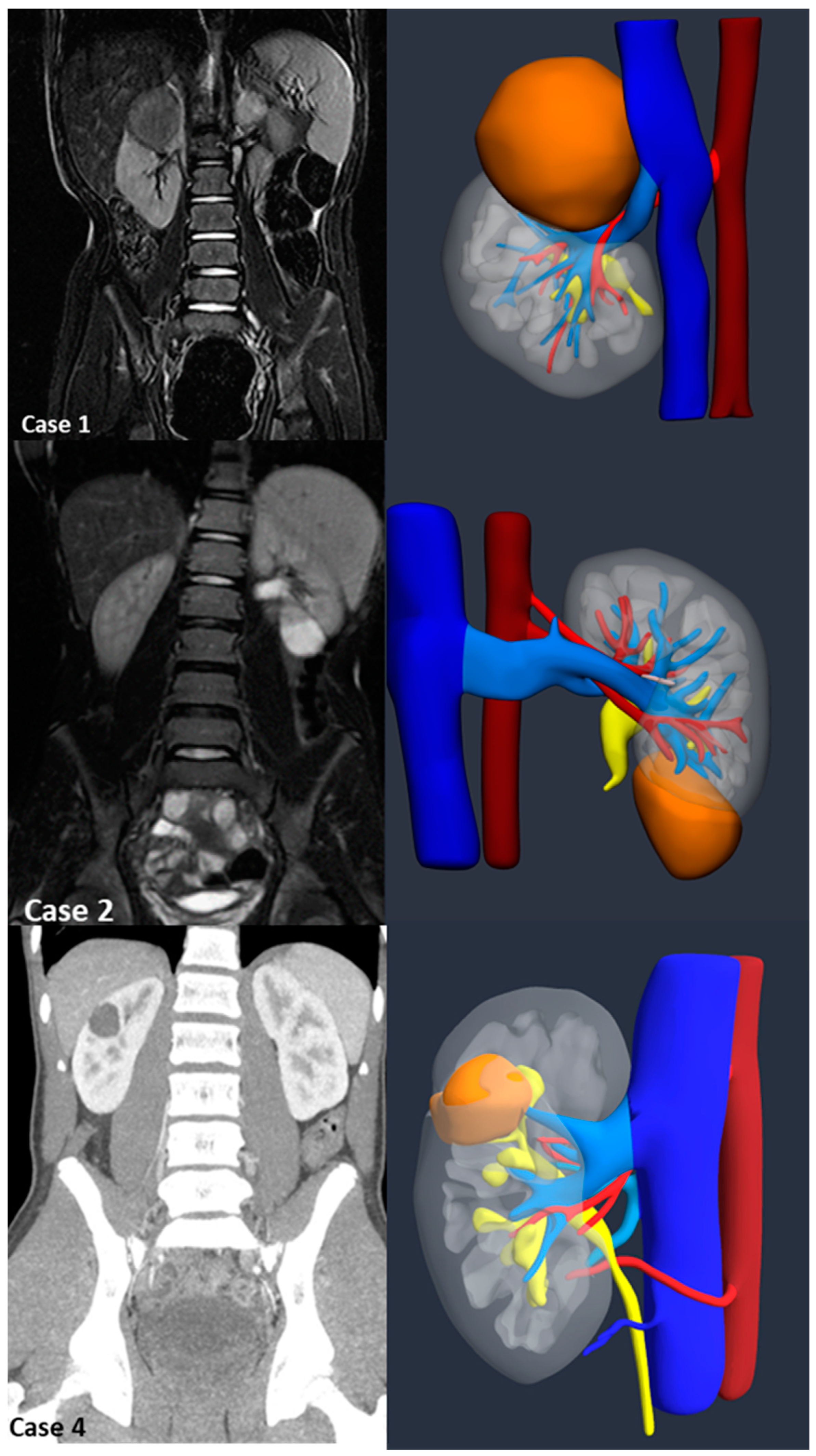
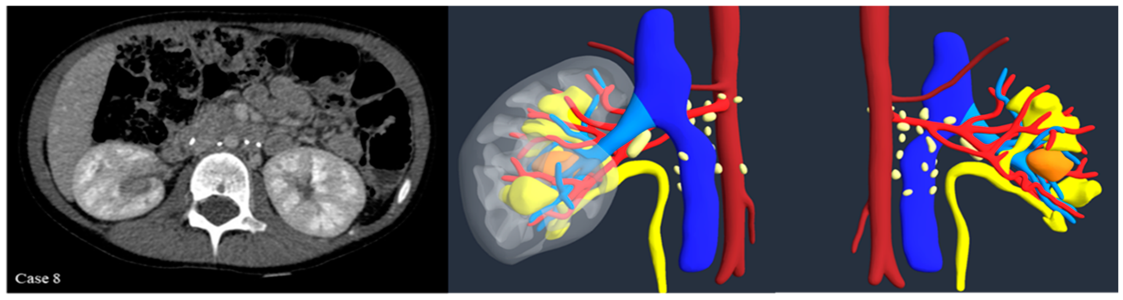
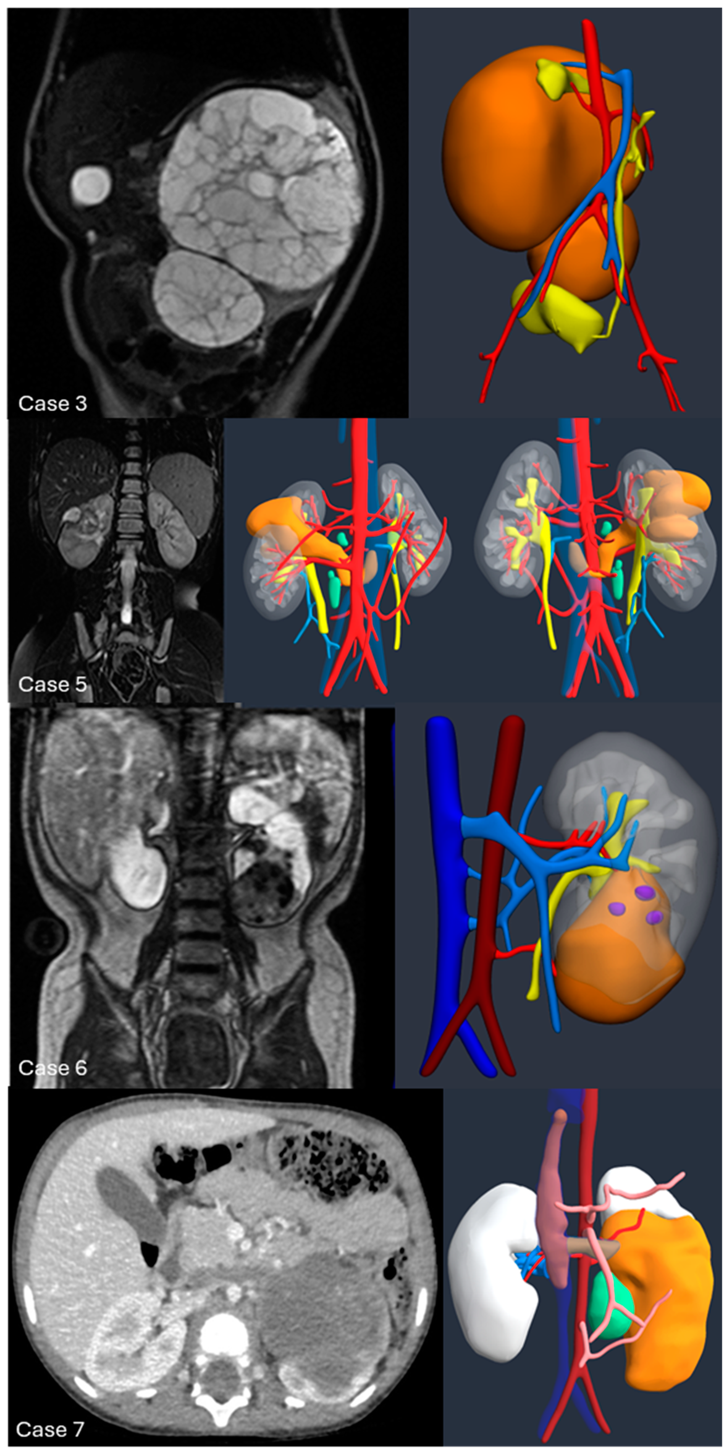
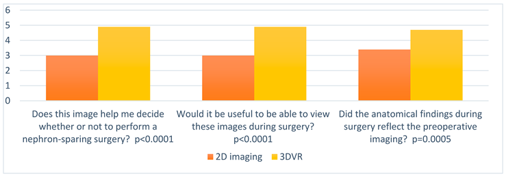
Introduction:The pre-operative planning for complex renal masses in children can be challenging, especially when nephron-sparing surgery (NSS) is recommended. We re-port our experience with the use of 3D-virtual reconstruction (3DVR) and its impact on surgical decision making. Materials and methods: Patients with complex renal masses underwent preoperative 3DVR. DICOM data were obtained from abdominal MRI and/or CT scans. 2D images segmentation was then performed. Three oncology surgeons were asked to individually evaluate each patient's preoperative MRI, CT and 3DVR. A questionnaire regarding the quality of conventional imaging compared to 3DVR was completed following surgery. Results: 8 patients (4♂,4♀) were included: Cases 1,2 and 4 were circumscribed tumours in the right upper pole, left lower pole (Bosniak cyst IV) and right mid-upper pole (Bosniak cyst IIF) respectively; Case 3 was a large hilar mass involving the whole kidney, unresponsive to chemotherapy; Case 5 and 7 were stage IV Wilms' tumour with venous thrombosis; case 6 was a left mid lower renal mass in a patient with WAGR syndrome and acute lymphoblastic leukae-mia (ALL) and case 8 was a recurrent central right WT after previous NSS in a child with Beckwith-Wiedemann syndrome(BWS). Four radical nephrectomies and three NSS were performed. In comparison to conventional imaging, the 3DVR models were judged to be superior by the expert reviewers for all anatomical structures except the urinary tract(p< 0.05). Conclusions: Our study suggests that 3DVR can be considered a useful tool in the pre-operative evaluation of children with complex renal masses and can facilitate NSS in selected patients.

Keywords: 
;  ;  ;  ;  ;  

1. Introduction

Renal tumours account for about 6% of all childhood malignancies. The most common is Wilms' tumour (WT) or nephroblastoma, which affects 1 in 10,000 children, with a median age at diagnosis of 3.5 years [1]. Other childhood kidney tumours include mesoblastic nephroma (MN) and malignant rhabdoid tumour of the kidney (MRTK), which are more common in infants, cystic nephroma (CN), angiomyolipoma and renal cell carcinoma (RCC), which is most common after the age of 14 years [2]. Radical nephroureterectomy with staging lymphnode sampling is the recommended surgical procedure for unilateral WT, which represents the vast majority of WT. However, according to the SIOP RTSG Umbrella Protocol 2016 [3,4], nephron-sparing surgery (NSS) must be pursued in specific cases such as bilateral WT, WT occurring in a single kidney, or WT occurring in the context of a syndromic or genetic predisposition (such as Beckwith-Wiedemann, Denys-Drash syndrome or WAGR syndrome) where an increased risk of bilateral tumors and eventual chronic kidney disease (CKD) can be predicted. Conventional imaging techniques, most commonly computed tomography (CT) and magnetic resonance imaging (MRI), have traditionally been used to define the preoperative surgical strategy [5]. However, conventional imaging techniques have many limitations in the paediatric setting: they provide only 2D images, often of limited quality; they have inaccuracies in anatomical definition of small structures that can be distorted by tumour growth; and they often require sedation in young children, which in turn affects image quality. In addition, protocols to reduce radiation exposure can affect image quality in CT. MRI that avoids radiation has a longer acquisition time and therefore more movement artefacts [6]. Overall, both MRI and CT may lack the ability to clearly define the extent of surgical resection, tumour volume and residual parenchyma when organ-sparing surgery is desired. In recent years, 3D reconstruction of organs and structures has emerged as a promising preoperative tool in many clinical settings and multiple disciplines, including urology, neurosurgery, general surgery, thoracic surgery, cardiac surgery, plastic surgery and maxillofacial surgery. It has been endorsed by both the American College of Radiology and the Radiological Society of North America [7]. In adult renal oncology, three-dimensional virtual reconstruction (3DVR) using dedicated software appears to provide a better understanding of the spatial relationships between tumour and adjacent structures (vessels and other organs) and allows interactive preoperative exploration of surgical anatomy. Few publications have reported the use of 3DVR in paediatric oncological surgery and even fewer in renal oncological surgery. [8]. The aim of our study was to analyse the feasibility of 3DVR in paediatric patients with complex renal masses and to evaluate the advantages and limitations of preoperative 3DVR in comparison to conventional 2D imaging. It is hypothesised that 3D models can be an important tool for preoperative planning, reducing complications and selecting patients for NSS.

2. Materials and Methods

From October 2021 to January 2025, preoperative 3DVR was performed in all patients referred to the Department of Oncology and the Division of Paediatric Urology with a renal mass deemed complex at the multidisciplinary meeting. Patients were selected for 3DVR reconstruction if they had a large tumour volume, infiltration of surrounding tissues, venous thrombosis equal to or greater than stage 2 according to the Daum classification [8], bilateral tumours, or if the patient was a potential candidate for NSS, as there is currently no defined tumour surgical complexity score for paediatric renal masses. Demographics (age, sex, weight), clinical characteristics, imaging (MRI, CT), tumour characteristics, surgical approach, complications and follow-up were retrospectively reviewed. Standard treatment according to the SIOP-RTSG UMBRELLA treatment protocol 2016 was provided to all patients considered as affected by WT. Data for 3DVR were obtained from pre-operative imaging studies in DICOM format. Children were either awake or sedated, depending on their ability to cooperate, for MRI and CT. All patients underwent a pre-operative MRI according to the SIOP-RTSG UMBRELLA protocol 2016. A 1.5 Tesla system (SIGNA™, GE HealthCare) was used in all patients. The imaging protocol consisted of a coronal fat-suppressed T2-weighted (-W) sequence, a sagittal T2-W sequence, an axial fat-suppressed T1-W sequence, a 3D fat-suppressed T1-W sequence and a diffusion-weighted sequence. After intravenous paramagnetic contrast injection (Gadovist 1.0 mmol/ml, Bayer Pharma, Berlin, Germany) at a dose of 0.1 ml/kg body weight, coronal, axial and 3d T1-W sequences were acquired and MRI angiography was performed to delineate the abdominal and pelvic vessels. All patients had unenhanced corticomedullary, nephrographic and urographic phases. For diagnosis and optimal tumour assessment, MRI was considered the preferred imaging modality. A high-resolution multiplanar scanner (Lightspeed 16-slice scanner, GE HealthCare) was used to acquire CT images. The images were acquired at 2.5 or 5 mm slice thickness, depending on weight, after intravenous iodinated contrast injection (Bracco Iopamiro 370 mg/ml) at a dose of 2 ml/kg body weight; images were also reconstructed at 1.25 mm slice thickness. The DICOM images were processed by Medics srl (www.medics3d.com, Turin, Italy) through dedicated software Mimics Medical 21.0 (Materialise, Leuven, Belgium) to perform Hyper Accuracy 3D reconstruction (HA3D®™). Both automatic and manual segmentation techniques were used to generate the 3DVR. When both CT scan and MRI were present, HA3D® was performed from the CT scan using MRI as a comparison, otherwise the T2 phase of the MRI was used. The renal pedicle and the tumour nourishing arteries were reconstructed using the dynamic region growing technique and subsequently manually refined, and the course of the extra- and intrarenal arteries up to the segmental arteries were reconstructed. The renal parenchyma was segmented by means of selective thresholding, in which different voxels were separated and grouped according to their grey value. The collecting system was segmented with the thresholding technique in the excretory phase and subsequently realigned in the arterial phase to fit the other reconstructed anatomical parts. Veins and tumour masses were instead reconstructed manually. Ultimately, the HA3D® reconstruction is achieved at the conclusion of the segmentation process and after proper post-processing using 3-matic Medical 13.0 (Materialise, Leuven, Belgium). To assess the accuracy of the models in comparison with the images obtained, the virtual models were jointly reviewed by bioengineers and urologists. The mathematical HA3D® model and the corresponding interactive 3D PDF file was generated to allow navigation during preoperative and intraoperative surgical planning. A panel of three surgeons with experience in oncological surgery was asked to individually evaluate the MRI and CT images and the 3D reconstructions for each patient prior to surgery and shortly after surgery. In the pre-operative phase, the surgeons assessed the reliability of the conventional diagnostic images compared to the 3DVR model in terms of specific anatomical details (tumour extent and density, extra- and intraparenchymal arterial and venous vascularisation, urinary tract). A specially designed seven-item questionnaire (Table 1) was employed. Each question had 5 possible options according to the Likert scale (1, disagree; 2, disagree; 3, neutral; 4, agree; and 5, strongly agree). On the day of surgery, a laptop with all relevant radiological findings was also available in the operating theatre. Intra-operative complications were documented. A post-operative questionnaire was completed at the end of surgery regarding the quality and usefulness of conventional imaging compared to 3DVR. The study was conducted according to good clinical practice guidelines. No formal institutional review board or ethics committee approval was required according to Italian law (Agenzia Italiana del Farmaco Guidelines for Observational Studies, 20 March 2008). Statistics were performed as appropriate: dichotomous variables were expressed as rates and percentages, while continuous variables were expressed as medians. Comparative analyses were performed using Mann-Whitney tests for continuous variables. P values < 0.05 were considered significant. Statistical analyses were performed with GraphPad Prism software (version 9, San Diego, CA).

3. Results

Eight patients were included in our study (4 ♂, 4 ♀) with a mean age of 74±66.03 (range 14-180) months and mean weight of 25.40±22.20 (range 9.4-70) kg. Patient demographics, tumor characteristics, and relevant technical outcomes for each patient are described in Table 2. Three patients (Case #3, #5 and #7) received neoadjuvant chemotherapy according to SIOP Umbrella 2016 protocol, while Case #6, a patient with WAGR syndrome, was treated with chemotherapy for acute lymphoblastic leukaemia (ALL) (Berlin Frankfurt-Munich BFM protocol). All 8 patients were evaluated with preoperative abdominal MRI, which required sedation in 6 (75%). CT scan was also performed in 7 (88%), 5 of which were potential candidates to NSS while 2 had complex vasculature anomalies and venous thrombosis. Of those patients candidated to NSS, three (case #1, #2 and #4) had a well-circumscribed and partially exophytic tumour respectively at the upper pole of the right kidney, the lower pole of the left kidney (Bosniak cyst IV) and the mid-upper pole of the right kidney (Bosniak cyst IIF), while the case # 6 had WAGR syndrome and case #8 had BWS with recurrent hilar-lower pole WT after NSS surgery and 7 months post-operative rest. Eligibility for NSS was confirmed in the first three patients after 3D vascular segmentation and assessment of the potential residual renal volume after surgery (Figure 1).
Excluded were case #6, who underwent total nephroureterectomy, and case #8, in whom, after initial evaluation of CT and MRI images, a 6-week course of chemotherapy (Vincristine VCR, Actinomycin D) was started, with subsequent re-evaluation of 2D and 3DVR images, which confirmed the impossibility of successfully performing NSS. It was therefore decided to continue chemotherapy and radiological surveillance in this last case (Figure 2).
Radical nephroureterectomy was the surgical choice in four patients (Figure 3):
-Case #3: One-year-old male child with a huge left renal mass with bilobed, inhomogeneous, cystic, multi-chamber MRI appearance with distortion of the vessels and urinary tract. The mass was unresponsive to neoadjuvat chemotherapy (VCR and Actinomycin D).
-Case #5: Four-year-old girl who came to Emergency for acute abdominal pain and macrohaematuria. CT and MRI showed bilateral lung metastases, a large heterogeneous renal mass of the right kidney and thrombosis of its venous system, together with congenital agenesis of the inferior VCI, right renal vein draining in azygos continuation, and thrombosis extending into the azygos vein. The patient underwent 6 cycles of chemotherapy according to the SIOP Umbrella 2016 protocol for stage IV (VCR, Actinomycin D, Doxorubicin). Restaging showed a good response to chemotherapy with an 87% reduction in initial mass volume before the procedure and unchanged thrombosis.
-Case # 6: Four-year-old girl with WAGR syndrome and ALL. Incidental finding at abdominal CT of a localised renal mass of the left kidney in middle-lower pole compatible with WT. The girls was treated for LLA with BFM protocol. During therapy an increased growth of the renal mass was observed. 3DVR highlightened the invasion of the pelvis, and the inability to preserve clinically significant residual parenchyma, therefore excluding the chance of NSS. Radical nephrectomy was performed, and infiltration of the pelvis confirmed at histology.
-Case #7: Two-year-old patient with occasional finding of a huge mass starting from the lower pole of the left kidney, on CT/MRI abdomen inhomogeneous structure with high cellularity and poor contrast enhancement and some necrotic colliquative areas with associated thrombosis of the left renal vein and inferior vena cava and secondary nodular localisations in the liver. Chemotherapy according to the SIOP 2016 protocol for metastatic WT was administered. The patient failed to respond and the disease progressed with secondary pulmonary lesions. Therefore, the decision was made to continue chemotherapy according to the Umbrella 4-Drug-Regimen (HR-2) protocol. Pre-operative re-evaluation using 2D imaging and 3DVR showed a significant reduction in pulmonary and hepatic secondary disease and an 84.5% reduction in renal mass volume.
The mean tumour volume at surgery was 26.67±5.50 ml in NSS versus 304.3±248.89 ml in radical nephroureterectomy (p=0.1181), with a 92% agreement between the calculated tumor volume on the 3D reconstruction and the histological tumor volume, compared with 66% with conventional imaging. Mean operative time was 186.7±32.14 (range 150-210) minutes for NSS and 330±87.55 (range 240-450) minutes for radical nephrourectomy (p=0.0454). No significant intraoperative bleeding was reported. A single intraoperative complication Clavien-Dindo grade 1 was reported, an inadverted lesion of the upper renal calyces which was soon sutured with no further complication. Postoperatively a single Clavien-Dindo grade 3b complication was recorded, a bowel obstruction on 5th postoperative day due to ileo-colic intussusception, which eventually required laparotomy and reduction. In cases #3, #5, #6 and #7 which underwent radical nephrectomy, transient antihypertensive therapy was started before surgery and was discontinued at discharge. Histological examination of the tumor revealed four WT (3 neproureterectomy and 1 NSS), two CN (1 nephroureterectomy and 1 NSS) and one case of tubulocystic renal cell carcinoma (TRCC) (NSS). Resection margins were unaffected in all cases. After NSS, ipsilateral renal function was preserved in all cases at DMSA renal scan. The results of the questionnaire submitted to the surgeons are reported in Table 3. The 3DVR showed a high grade of definition of the structure of the tumor (based on its varying density) and of its relationships with the surrounding organs, which was enhanced by the ability to make anatomical structures transparent or to eliminate them. 3DVR was regarded as superior to conventional images in order to understand the patient-specific anatomy and in matching the anatomical findings at surgery (p<0.0001). The possibility to move, rotate, zoom and make some structures transparent was indeed considered by the surgeons a major advantage of the 3D virtual model, which turned out to be very useful even during surgery. Furthermore, the 3D vessel models allowed to visualise the intraparenchymal arteries up to the second or third segmental arterial branch (p<0.0001), which was particularly important in NSS when accurate vessel segmentation allowed selective arterial clamping and obviated the need of warm ischemia of the kidney. The level of detail of the urinary collecting system was unfortunately significantly lower than the vessel model in both 3D and MRI/CT imaging modalities (p=0.0961). Overall surgeons considered the 3D models to be supportive and complementary to conventional imaging with the aim of improving and optimising the surgical treatment (p=0.0027, p<0.0001, p=0.0006 as shown in Table 4).

4. Discussion

Survival rates for paediatric renal tumours have improved considerably over the past decades. Currently, long-term survival for localised WT (stage I to III) with favourable hystology is expected to exceed 90% [9]. Despite the differences in treatment strategy between Europe and North America, surgery remains a fundamental step in the multimodal management of these tumours in accordance with the International Society for Paediatric Oncology (SIOP) and the National Wilms' Tumour Study Group/Children Oncology Group (NWTSG-COG). In unilateral WT, complete tumor nephroureterectomy through a transabdominal approach is the standard of surgical care for both SIOP and NWTS/COG treated patients, together with hilar and interaortocaval lymphnode sampling for staging purposes [10]. This surgery is not devoid of complications: a review showed an overall incidence of surgical complications of 19.8% and 12.7% in NWTS-3 and NWTS-4, respectively. The most common complication was bowel obstruction, which occurred in 5-6.7% of patients, followed by major intraoperative bleeding (1.9-6%), and intraoperative injury to other visceral organs (including the bowel, liver and spleen) and vascular structures (2%). An increased risk of surgical complications was associated with a tumour diameter of 10 cm or more. Patients who received pre-operative treatment, as according to SIOP protocols, had a lower incidence of surgical complications, estimated at around 8% [11]. Performance of NSS is a necessity in children presenting with bilateral WT, and is recommended in syndromic patients with an increased risk of metachronous development of ipsilateral or contralateral WT, in patients with a solitary kidney as well as in patients who are at risk of kidney failure. In non-syndromic unilateral WT, NSS is not a standard procedure. Potential benefits of NSS are the prevention of functional renal impairment and ESRD [12], and reduction of cardiovascular morbidity and mortality into adulthood, due to hypertension and the presence of a single kidney [13]. Despite some long-term follow-up data suggest good renal function after unilateral radical nephrectomy, long term kidney injury in patients with WT could be justified by reduced nephron mass by nephrectomy, nephrotoxic chemotherapy, and abdominal radiation. On the other hand, potential risks of NSS include risks of positive surgical margins and increased recurrence [14]. Recent protocols indicate nephron-sparing surgery (NSS) as an alternative to radical nephrectomy in selected cases [15]. NSS is a technically demanding procedure requiring a detailed preoperative knowledge of patient-specific renal anatomy and intraparenchymal vasculature [16]. In order to ensure radical surgery with low morbidity and good oncological outcome, careful patient selection and preoperative planning are essential. CT and MRI are the main imaging modalities for the evaluation of renal oncologic patients. In complex cases, where anatomical relationships are difficult to identify, multimodality imaging is used to provide a reliable diagnosis [17]. The gold standard for imaging of solid tumours is 2D planar imaging, which requires the surgeon to 'mentally create' a 3D model for surgical planning. In order to improve the accuracy of surgical resection and prevent potential surgical complications, modern post-processing techniques in combination with contrast agents can allow detailed assessment of the vasculature, definition of the tumour and precise definition of anatomical relationships [18]. 3D technology covers a wide range of possible applications, from realising a virtual 3D model, to printed 3D reconstruction in different materials, to the possibility of creating augmented reality (AR) where a 3D model is superimposed on a real-time surgical field, providing an exciting new opportunity to improve surgical practice [19]. Patient-specific 3D anatomical models are widely used for preoperative planning of NSS in adults. Several authors have evaluated the role of 3D virtual models before and during robotic partial nephrectomy and demonstrated how this technology is helpful in improving preoperative assessment of tumor complexity [20], avoiding global ischemia of the healthy renal remnant [21], and reducing the loss of renal function assessed by renal scan [22]. Benefits of 3D printed anatomical models have been further elucidated in adults, including reduced blood loss, intraoperative complications [23] and improved patient education [24]. Overall, adult NSS experience demonstrates the clinical role of novel 3DVR with perioperative and functional benefits. Further studies are ongoing in the adult literature that will not only provide high quality evidence to define the potential of 3DVR and printing for surgical planning, but will also create a new tool for intraoperative surgical navigation. On the basis of preoperative CT, high-resolution 3D models of the kidney and tumour are generated and then, thanks to a specially developed platform, the virtual models overlap with the in vivo anatomy in a static and dynamic way, allowing for an augmented reality robot-assisted partial nephrectomy [25]. This can offer an immersive surgical planning and training experience, and potentially revolution how surgeons view medical imaging, prepare surgical procedure and train young surgeons. This is a fascinating prospect. In children, there is a very limited amount of data available on the value of 3D reconstruction in solid tumours. A recent review of the use of 3D in paediatric oncology surgery [26] reported that the main benefits of 3D printing in complex neuroblastoma included estimating the total resectable tumour volume [27] and understanding the anatomical relationships between the tumour and vital structures [28]. The first case series on the use of 3D reconstruction for renal tumours in children was described by Schenk et al [29] in 7 children aged 1 month to 11 years: 3D visualisation with 3D animation of the situs helped to transfer important information from the paediatric radiologist to the paediatric surgeon and to optimise the surgical preparation. Giron-Vallejo et al. used MRI to develop a 3D printed model of a bilateral Wilms' tumor in a 10-month-old infant with suspected Danys-Drash syndrome: the model allowed a more precise assessment of the residual parechyma and led to a bilateral NSS, although it did not allow to avoid postoperative complications such as urinary fistula and renal atrophy due to vascular damage [30]. The same authors subsequently published a small series of 4 patients, 2 Wilms' tumours and 2 neuroblastomas, in which the 3D printed model helped the surgeons to accurately assess the expected risks during surgery, allowed them to practice before the planned surgery in order to reduce surgical complications and proved to be a useful tool for information and education of the family of the patients [31]. In these last two studies, MRI was used for 3DVR of Wilms' tumour, while Fuchs et al [32] preferred CT for the development of a model for preoperative 3DVR and virtual resections in 11 children with solid tumours, including 4 liver tumours and 4 unilateral WT. Girón-Vallejo [30] reported that one of the main limitations of their model was poor vascular information, not mentioned by other authors [32], but it is possible that poor quality of the original 2D image or segmentation process contributed to this limitation. Both the 3DVR and the printed models provide anatomical information about the tumour and the kidneys that may not be visible in the 2D image alone. The reconstructed models allowed a better visualisation of the preserved amount of parenchyma that was not correctly detected on MRI or CT in the studies by Girón-Vallejo [30] and Sánchez-Sánchez et al [31]. And Fuchs et al. reported that in one case, heminephrectomy seemed possible after ultrasound and CT, but 3DVR showed infiltration in the renal pelvis and central organ, indicating the need for tumour nephrectomy [32]. The same occurred in our series in case #6 with WAGR syndrome, where NSS was excluded only after 3DVR showed infiltration of the renal pelvis not seen on conventional imaging. Van der Zee et al [33] carried out retrospective 3DVR in 6 patients with WT who had already undergone NSS. CT was required to visualise vascular structures and the ureteral collecting system in 2 patients in addition to preoperative MRI. The virtual resection was performed after the actual resection was undertaken, and the surgeons found the virtual resection to be straightforward. There was a large variation between surgeons in whether real surgical tumour resection was considered difficult or not, and no clear opinion was derived for the usefulness of virtual resection, but it is possible that the retrospective model of the study influenced the surgeons' clinical relevance. Overall, according to the authors, 3D models and virtual resection allowed for a more accurate prediction of residual renal volume and postoperative renal function, thus influencing the choice of NSS, and could be used to predict possible surgical complications such as urinary leakage or positive resection margins. More recently, in 10 children with WT, Wellens et al. [34] used either MRI or CT to develop a 3D model. A 3D printed model was then created for each patient and compared to a mixed reality headset AR hologram and standard MRI/CT. In comparison to conventional imaging, the 3D print and AR hologram models were judged by surgeons to be superior for all anatomical structures. The largest series of 3DVR in pediatric oncology is the one described by Vinit N. et al [35], which created a 3D models from preoperative MRI in more than 150 patients, and among them 26 patients with pelvic tumors, 6 sacrococcygeal, 9 rhabdomyosarcomas (four bladder prostate, two vagina, one urachal and two pelvic), nine neurogenic tumors (seven neuroblastomas, two ganglioneuromas), two neurofibromas and ten Wilms tumors. The Authors built a dataset of 3 T MRI images and developed automatic segmentation tools based on deep learning approaches, which were particularly focused to obtain a more efficient representation of the peripheral nerve network (lumbosacral plexus). During surgery, the 3D model was projected onto screens in the operating room or integrated into the console of the robotic system. Surgeons and residents where then asked to evaluate their perception of the anatomy regarding organs, arteries, veins and tumors, the relationships between the tumor and the surrounding structures: especially residents agreed that 3D models improved their understanding of the anatomy and of the surgical procedure. The present series is a further confirmation of the usefulness of 3D in preoperative planning for WT in children. Due to non negligeable costs of 3D imaging, we decided to obtain a 3DVR only in selected patients with renal tumours, namely those with complex anatomy or who were potential candidates for NSS. Similarly to other Authors, we performed both MRI and CT in the majority of our patients in order to obtain high quality 2D images. An insufficient quality of the MRI or CT, in particular the lack of an exscretory phase, or difficulties in the segmentation process could have led to an insufficient 3DVR model. The selection criteria for NSS were strictly adhered to those defined in the Umbrella protocol (SIOP RTSG 2016 Umbrella) and were evaluated on the 3D reconstructions, namely tumour confined to one pole of the kidney or peripheral in the middle of the kidney, volume < 300 mL, no preoperative rupture, no intraluminal tumour on preoperative imaging in the renal pelvis, no invasion of surrounding organs, no thrombus in the renal vein or vena cava, excision feasible with oncologically safe margins, the renal remnant expected to have significant residual function. In agreement with previous Authors [36] who found 3DVR to be more accurate and reproducible than conventional 2D methods, in particular we found a 92% agreement between 3D and histological tumour volume compared to 66% using conventional imaging. Furthermore in two cases potentially candidate to NSS, this surgical option was deemed as not feasible after evaluation of 3DVR. The perception of surgeons was that 3DVR was a useful tool in defining the relationship of the tumour with the other organs, its vascular supply and its relationship to the urinary tract. In patients where NSS was performed, either in open or robotic assisted surgery, selective arterial clamping was always possible, therefore obviating the need of ischemia of the renal residual parenchyma. Furthermore, the assessment of surgical and patological resection margins showed no case of tumour breach. Throughout the whole series, including total nephrectomy and NSS, intraoperative complications were negligible, although the specific role of 3DVR cannot be assessed. We chose not to print the 3D model because of the need to limit costs and because virtual reconstruction has the ability to make some structures transparent and disappear, which was considered particularly valuable even intraoperatively to visualise deep structures. This study has some limitations which must be acknowledged. Firstly, the small number of patients did not allow a precise statistical analysis. However, this limited number was due to the strict selection of patients who were candidates for 3DVR and is not far from similar series, as the majority of papers dealing with 3D in renal tumours in children have a numerosity of less than 10 cases. Secondly, the comparison of 2D, 3DR and surgical findings was performed by the operating surgeons and not by an independent observer. The assessment of 3DVR in the questionnaire may have been biased as a result. However, similar questionnaires have been proposed, with similar results by Chaussy et al and Vinit et al [35,37]. The use of 3DVR technology is still not standardised in clinical practice and there are still challenges to its wider adoption due to high cost and expertise requirements. There is a urgent need for multicentre studies to realise the full potential of these technologies, with prospective evaluations of larger cohorts of patients.

5. Conclusions

Three-dimensional virtual models, which have gained popularity in adult oncological urology, are far from being the standard of care for children with WT. These models are indeed easy to understand, rich in anatomical details and may help surgeons to better anticipate the risks of surgery and to select patients candidates for NSS. They are a significat step towards precise patient-specific medicine. Our limited experience is consistent with the few data available in the literature and highlights the advantages of this technology, which can be applied in a wide range of settings, from preoperative surgical planning to training of young surgeons and communication with the patient and family. At present, the main barriers to the widespread use of 3D models are the need for standardisation of 2D imaging, automation of the segmentation process, high cost and the need for a high level of expertise. Being guided by 3DVR during surgery and performing procedures in augmented reality is an exciting prospect for the future. Standardised and widespread use of 3DVR in children with renal oncology, to familiarise with the new technology, can be the first step towards this future.

Author Contributions

Conceptualization, E.C., E.R and S.G.N; methodology, E.C. and E.T.; software,E.C. and E.T.; formal analysis, E.C. and L.C.; investigation, M.P., P.Q. and M.C; resources F.F and P.G.; data curation, E.R and M.S.; writing—original draft preparation, E.C. and S.G.N.; writing—review and editing, E.C. and S.G.N.; supervision, F.F. and P.G. All authors have read and agreed to the published version of the manuscript.

Funding

This research received no external funding.

Institutional Review Board Statement

Not applicable.

Informed Consent Statement

Not applicable.

Acknowledgements

For their professional technical support and continuous collaboration in improving our surgical work, we would like to thank Andrea Bellin and Simona Barbuto of Medics srl.

Conflicts of Interest

The authors declare no conflicts of interest

References

  1. Han Q, Li K, Dong K, Xiao X, Yao W, Liu G. Clinical features, treatment, and outcomes of bilateral Wilms' tumor: A systematic review and meta-analysis. J Pediatr Surg. 2018 Dec;53(12):2465-2469. Epub 2018 Sep 1. [CrossRef] [PubMed]
  2. Roy P, van Peer SE, de Witte MM, Tytgat GAM, Karim-Kos HE, van Grotel M, van de Ven CP, Mavinkurve-Groothuis AMC, Merks JHM, Kuiper RP, Hol JA, Janssens GOR, de Krijger RR, Jongmans MCJ, Drost J, van der Steeg AFW, Littooij AS, Wijnen MHWA, van Tinteren H, van den Heuvel-Eibrink MM. Characteristics and outcome of children with renal tumors in the Netherlands: The first five-year's experience of national centralization. PLoS One. 2022 Jan 13;17(1):e0261729. [CrossRef] [PubMed] [PubMed Central]
  3. De Kraker J, Graf N, van Tinteren H, Pein F, Sandstedt B, Godzinski J, Tournade MF; SIOP. Reduction of postoperative chemotherapy in children with stage I intermediate-risk and anaplastic Wilms' tumour (SIOP 93-01 trial): a randomised controlled trial. Lancet. 2004 Oct 2-8;364(9441):1229-35. [CrossRef] [PubMed]
  4. Theilen TM, Braun Y, Bochennek K, Rolle U, Fiegel HC, Friedmacher F. Multidisciplinary Treatment Strategies for Wilms Tumor: Recent Advances, Technical Innovations and Future Directions. Front Pediatr. 2022 Jul 14;10:852185. [CrossRef] [PubMed] [PubMed Central]
  5. Pereira HR, Barzegar M, Hamadelseed O, Esteve AV, Munuera J. 3D surgical planning of pediatric tumors: a review. Int J Comput Assist Radiol Surg. 2022 Apr;17(4):805-816. Epub 2022 Jan 18. [CrossRef] [PubMed]
  6. Günther P, Schenk JP, Wunsch R, Tröger J, Waag KL. Abdominal tumours in children: 3-D visualisation and surgical planning. Eur J Pediatr Surg. 2004 Oct;14(5):316-21. [CrossRef] [PubMed]
  7. Chepelev L, Wake N, Ryan J, Althobaity W, Gupta A, Arribas E, Santiago L, Ballard DH, Wang KC, Weadock W, Ionita CN, Mitsouras D, Morris J, Matsumoto J, Christensen A, Liacouras P, Rybicki FJ, Sheikh A; RSNA Special Interest Group for 3D Printing. Radiological Society of North America (RSNA) 3D printing Special Interest Group (SIG): guidelines for medical 3D printing and appropriateness for clinical scenarios. 3D Print Med. 2018 Nov 21;4(1):11. [CrossRef] [PubMed] [PubMed Central]
  8. Daum R, Roth H, Zachariou Z. Tumor infiltration of the vena cava in nephroblastoma. Eur J Pediatr Surg. 1994 Feb;4(1):16-20. [CrossRef] [PubMed]
  9. Owens CM, Brisse HJ, Olsen ØE, Begent J, Smets AM. Bilateral disease and new trends in Wilms tumour. Pediatr Radiol. 2008 Jan;38(1):30-9. Epub 2007 Nov 17. [CrossRef] [PubMed]
  10. Van den Heuvel-Eibrink MM, Hol JA, Pritchard-Jones K, van Tinteren H, Furtwängler R, Verschuur AC, Vujanic GM, Leuschner I, Brok J, Rübe C, Smets AM, Janssens GO, Godzinski J, Ramírez-Villar GL, de Camargo B, Segers H, Collini P, Gessler M, Bergeron C, Spreafico F, Graf N; International Society of Paediatric Oncology — Renal Tumour Study Group (SIOP–RTSG). Position paper: Rationale for the treatment of Wilms tumour in the UMBRELLA SIOP-RTSG 2016 protocol. Nat Rev Urol. 2017 Dec;14(12):743-752. Epub 2017 Oct 31. [CrossRef] [PubMed]
  11. Ritchey ML, Shamberger RC, Haase G, Horwitz J, Bergemann T, Breslow NE. Surgical complications after primary nephrectomy for Wilms' tumor: report from the National Wilms' Tumor Study Group. J Am Coll Surg. 2001 Jan;192(1):63-8; quiz 146. [CrossRef] [PubMed]
  12. Green, DM. Evaluation of renal function after successful treatment for unilateral, non-syndromic Wilms tumor. Pediatr Blood Cancer. 2013 Dec;60(12):1929-35. Epub 2013 Aug 19. [CrossRef] [PubMed]
  13. Cozzi DA, Ceccanti S, Cozzi F. Renal function up to the 5th decade of life after nephrectomy in childhood: A literature review. Nephrology (Carlton). 2018 May;23(5):397-404. [CrossRef] [PubMed]
  14. Long CJ, Mittal S, Kolon TF. Expanding the Use of Nephron-Sparing Surgery for Wilms Tumor. J Natl Compr Canc Netw. 2022 Feb 17;20(5):540-546. [CrossRef] [PubMed]
  15. Wilde JC, Aronson DC, Sznajder B, Van Tinteren H, Powis M, Okoye B, Cecchetto G, Audry G, Fuchs J, Schweinitz DV, Heij H, Graf N, Bergeron C, Pritchard-Jones K, Van Den Heuvel-Eibrink M, Carli M, Oldenburger F, Sandstedt B, De Kraker J, Godzinski J. Nephron sparing surgery (NSS) for unilateral wilms tumor (UWT): the SIOP 2001 experience. Pediatr Blood Cancer. 2014 Dec;61(12):2175-9. Epub 2014 Aug 23. [CrossRef] [PubMed]
  16. Klatte T, Ficarra V, Gratzke C, Kaouk J, Kutikov A, Macchi V, Mottrie A, Porpiglia F, Porter J, Rogers CG, Russo P, Thompson RH, Uzzo RG, Wood CG, Gill IS. A Literature Review of Renal Surgical Anatomy and Surgical Strategies for Partial Nephrectomy. Eur Urol. 2015 Dec;68(6):980-92. Epub 2015 Apr 22. [CrossRef] [PubMed] [PubMed Central]
  17. Schima W, Böhm G, Rösch CS, Klaus A, Függer R, Kopf H. Mass-forming pancreatitis versus pancreatic ductal adenocarcinoma: CT and MR imaging for differentiation. Cancer Imaging. 2020 Jul 23;20(1):52. [CrossRef] [PubMed] [PubMed Central]
  18. Pandey P, Lewis H, Pandey A, Schmidt C, Dillhoff M, Kamel IR, Pawlik TM. Updates in hepatic oncology imaging. Surg Oncol. 2017 Jun;26(2):195-206. Epub 2017 Apr 6. [CrossRef] [PubMed]
  19. Privitera L, Paraboschi I, Cross K, Giuliani S. Above and Beyond Robotic Surgery and 3D Modelling in Paediatric Cancer Surgery. Front Pediatr. 2021 Dec 20;9:777840. [CrossRef] [PubMed] [PubMed Central]
  20. Bianchi L, Schiavina R, Bortolani B, Cercenelli L, Gaudiano C, Carpani G, Rustici A, Droghetti M, Mottaran A, Boschi S, Salvador M, Chessa F, Cochetti G, Golfieri R, Bertaccini A, Marcelli E. Interpreting nephrometry scores with three-dimensional virtual modelling for better planning of robotic partial nephrectomy and predicting complications. Urol Oncol. 2021 Dec;39(12):836.e1-836.e9. Epub 2021 Sep 14. [CrossRef] [PubMed]
  21. Porpiglia F, Fiori C, Checcucci E, Amparore D, Bertolo R. Hyperaccuracy Three-dimensional Reconstruction Is Able to Maximize the Efficacy of Selective Clamping During Robot-assisted Partial Nephrectomy for Complex Renal Masses. Eur Urol. 2018 Nov;74(5):651-660. Epub 2018 Jan 6. [CrossRef] [PubMed]
  22. Amparore D, Pecoraro A, Checcucci E, Piramide F, Verri P, De Cillis S, Granato S, Angusti T, Solitro F, Veltri A, Fiori C, Porpiglia F. Three-dimensional Virtual Models' Assistance During Minimally Invasive Partial Nephrectomy Minimizes the Impairment of Kidney Function. Eur Urol Oncol. 2022 Feb;5(1):104-108. Epub 2021 Apr 24. [CrossRef] [PubMed]
  23. Wang Z, Qi L, Yuan P, Zu X, Chen W, Cao Z, Li Y, Wang L. Application of Three-Dimensional Visualization Technology in Laparoscopic Partial Nephrectomy of Renal Tumor: A Comparative Study. J Laparoendosc Adv Surg Tech A. 2017 May;27(5):516-523. Epub 2017 Feb 10. [CrossRef] [PubMed]
  24. Lupulescu C, Sun Z. A Systematic Review of the Clinical Value and Applications of Three-Dimensional Printing in Renal Surgery. J Clin Med. 2019 Jul 8;8(7):990. [CrossRef] [PubMed] [PubMed Central]
  25. Porpiglia F, Checcucci E, Amparore D, Piramide F, Volpi G, Granato S, Verri P, Manfredi M, Bellin A, Piazzolla P, Autorino R, Morra I, Fiori C, Mottrie A. Three-dimensional Augmented Reality Robot-assisted Partial Nephrectomy in Case of Complex Tumours (PADUA ≥10): A New Intraoperative Tool Overcoming the Ultrasound Guidance. Eur Urol. 2020 Aug;78(2):229-238. Epub 2019 Dec 30. [CrossRef] [PubMed]
  26. Pereira HR, Barzegar M, Hamadelseed O, Esteve AV, Munuera J. 3D surgical planning of pediatric tumors: a review. Int J Comput Assist Radiol Surg. 2022 Apr;17(4):805-816. Epub 2022 Jan 18. [CrossRef] [PubMed]
  27. Krauel L, Fenollosa F, Riaza L, Pérez M, Tarrado X, Morales A, Gomà J, Mora J. Use of 3D Prototypes for Complex Surgical Oncologic Cases. World J Surg. 2016 Apr;40(4):889-94. [CrossRef] [PubMed]
  28. Souzaki R, Kinoshita Y, Ieiri S, Kawakubo N, Obata S, Jimbo T, Koga Y, Hashizume M, Taguchi T. Preoperative surgical simulation of laparoscopic adrenalectomy for neuroblastoma using a three-dimensional printed model based on preoperative CT images. J Pediatr Surg. 2015 Dec;50(12):2112-5. Epub 2015 Sep 2. [CrossRef] [PubMed]
  29. Schenk JP, Waag KL, Graf N, Wunsch R, Jourdan C, Behnisch W, Tröger J, Günther P. 3-D-Visualisierung in der MRT zur Operationsplanung von Wilms-Tumoren [3D-visualization by MRI for surgical planning of Wilms tumors]. Rofo. 2004 Oct;176(10):1447-52. German. [CrossRef] [PubMed]
  30. Girón-Vallejo Ó, García-Calderón D, Ruiz-Pruneda R, Cabello-Laureano R, Doménech-Abellán E, Fuster-Soler JL, Ruiz-Jiménez JI. Three-dimensional printed model of bilateral Wilms tumor: A useful tool for planning nephron sparing surgery. Pediatr Blood Cancer. 2018 Apr;65(4). Epub 2017 Dec 12. [CrossRef] [PubMed]
  31. Sánchez-Sánchez Á, Girón-Vallejo Ó, Ruiz-Pruneda R, Fernandez-Ibieta M, García-Calderon D, Villamil V, Giménez-Aleixandre MC, Montoya-Rangel CA, Hernández Bermejo JP. Three-Dimensional Printed Model and Virtual Reconstruction: An Extra Tool for Pediatric Solid Tumors Surgery. European J Pediatr Surg Rep. 2018 Jan;6(1):e70-e76. Epub 2018 Oct 18. [CrossRef] [PubMed] [PubMed Central]
  32. Fuchs J, Warmann SW, Szavay P, Kirschner HJ, Schäfer JF, Hennemuth A, Scheel-Walter HG, Bourquain H, Peitgen HO. Three-dimensional visualization and virtual simulation of resections in pediatric solid tumors. J Pediatr Surg. 2005 Feb;40(2):364-70. [CrossRef] [PubMed]
  33. Van der Zee JM, Fitski M, Simonis FFJ, van de Ven CP, Klijn AJ, Wijnen MHWA, van der Steeg AFW. Virtual Resection: A New Tool for Preparing for Nephron-Sparing Surgery in Wilms Tumor Patients. Curr Oncol. 2022 Feb 1;29(2):777-784. [CrossRef] [PubMed] [PubMed Central]
  34. Davidoff AM, Fernandez-Pineda I. Complications in the surgical management of children with malignant solid tumors. Semin Pediatr Surg. 2016 Dec;25(6):395-403. Epub 2016 Oct 31. [CrossRef] [PubMed]
  35. Vinit N, Blanc T, Bloch I, Pio L, Kassir R, La Barbera G, Bonnot E, Gori P, Goulin J, Pire A, Boddaert N, Lozach C, Sarnacki S. Robotics and 3D modeling for precision surgery in pediatric oncology. EJC Paediatric Oncology 2024 Vol. 4. p.100181. [CrossRef]
  36. Durso TA, Carnell J, Turk TT, Gupta GN. Three-dimensional reconstruction volume: a novel method for volume measurement in kidney cancer. J Endourol. 2014 Jun;28(6):745-50. Epub 2014 Feb 27. [CrossRef] [PubMed]
  37. Chaussy Y, Vieille L, Lacroix E, Lenoir M, Marie F, Corbat L, Henriet J, Auber F. 3D reconstruction of Wilms' tumor and kidneys in children: Variability, usefulness and constraints. J Pediatr Urol. 2020 Dec;16(6):830.e1-830.e8. Epub 2020 Aug 28. [CrossRef] [PubMed]
Figure 1. Patients undergoing NSS, comparison of 2D and 3DR imaging.
Preprints 150912 g001
Figure 2. 3DVR was useful and decisive in this complex case #8 to rule out the possibility of performing an NSS given the location near the hilum, which would not have ruled out the need for a complete nephrectomy in a patient with BWS.
Preprints 150912 g002
Figure 3. Patients undergoing radical nephroureterectomy, comparison of 2D and 3DRV imaging.
Preprints 150912 g003
Table 1. Comparison questionnaire on the usefulness of 2D imaging versus 3DVR.
IMAGING EVALUATION QUESTIONNAIRE
(Likert scale 1-2-3-4-5)
Is it possible to clearly see the extent of the tumour and its characteristics?
Is it possible to clearly identify the arterial vasculature and its intraparenchymal branches?
Can the venous vasculature, intraparenchymal branches and thrombosis presence be clearly identified?
Is it possible to assess how the ureteral collecting system relates to the tumour and its possible invasion?
Does this image help me decide whether or not to perform a nephron-sparing surgery?
Would it be useful to be able to view these images during surgery?
Did the anatomical findings during surgery reflect the preoperative imaging?
Table 2. Demographic, diagnostic and surgical data of patients affected by compless renal mass.
Case 1 2 3 4 5 6 7 8
Sex (M/F) F M M M F F M F
Age (months) 48 180 14 178 50 42 28 52
Weight (kg) 15 50 9.4 70 18 10.8 13 17
Side Right Left Left Right Right Left Left Right
Position UP LP UP+MP+LP MP+UP UP MP+LP LP LP
Volume (ml) at surgery 33 24 597 23 421 63 136 -
Syndrome - - - - - WAGR - BWS
MRI X X X X X X X X
CT scan X X - X X X X X
Vein Thrombosis - - - - X - X -
UCSI - - - - - X - -
Surgery RAL NSS RAL
NSS
Open nephrectomy RAL NSS Open Nephrectomy Open Nephrectomy Open Nephrectomy -
Operative time (minutes) 210 200 240 150 450 310 320 -
Histology WT TRCC CN CN WT WT WT -
Tumor Volume calculated by elissoid volume formula ((width × length × height × 0.52); UP, upper pole, MP, mid pole, LP, lower pole; (X) yes, (-) no; UCSI, urinary collecting system invasion.
Table 3. Statistical analysis of the results of the questionnaire on the anatomical details obtained by comparing the conventional images and the 3DVR images.
Anatomical structure 2D imaging 3DVR P-value
Tumor 3.4 (3-4) 4.8 (4-5) < 0.0001
Arteries 3.6 (3-4) 4.9 (4-5) <0.0001
Veins 3.6 (3-4) 4.8 (4-5) .0004
Urinary collecting system 2.9 (2-4) 3.5 (3-5) .0961
Table 4. Evaluation of the accuracy and usefulness of imaging models to optimise the surgical approach.
Preprints 150912 i001
Disclaimer/Publisher’s Note: The statements, opinions and data contained in all publications are solely those of the individual author(s) and contributor(s) and not of MDPI and/or the editor(s). MDPI and/or the editor(s) disclaim responsibility for any injury to people or property resulting from any ideas, methods, instructions or products referred to in the content.
Copyright: This open access article is published under a Creative Commons CC BY 4.0 license, which permit the free download, distribution, and reuse, provided that the author and preprint are cited in any reuse.
Prerpints.org logo

Preprints.org is a free preprint server supported by MDPI in Basel, Switzerland.

Subscribe

Disclaimer

Terms of Use

Privacy Policy

Privacy Settings

© 2025 MDPI (Basel, Switzerland) unless otherwise stated