Preprint
Article

This version is not peer-reviewed.

Genomics Unravels Passion Fruit Viral Disease Complexity in Kenya

A peer-reviewed article of this preprint also exists.

Submitted:

13 December 2024

Posted:

13 December 2024

You are already at the latest version

Abstract

Passion fruit virus diseases (PWDs) pose a significant threat to Kenya's passion fruit industry. To unravel the complexity of these diseases, comprehensive virus surveys were conducted across major passion fruit-growing counties. PWD symptoms like fruit hardening, chlorotic mottling, and leaf distortion, were prevalent. The study unveiled the first 23 complete genomes of Ugandan Passiflora virus (UPV) and two East Asian Passiflora distortion virus (EAPDV) in Kenya. UPV showed 99% nucleotide (nt) match to a UPV genome from Uganda and 66% nt identity match to EAPDV. Phylogenetic analysis revealed distinct lineages (I-III), indicating potential multiple introductions into Kenya. Recombination analysis detected no significant breakpoints. However, the study proposed the renaming of EAPDV to passiflora distortion virus (PDV) and UPV to passiflora virus (PV) for neutral nomenclature. Additionally, the study highlighted the role of coinfections in symptom expression, suggesting a synergistic relationship between UPV, EAPDV (PDV), and other viruses. The results recommend for stringent management strategies and enhanced surveillance to mitigate the economic impact of these viruses to Kenyan passion fruit industry. Overall, the study highlights, need to strengthen phytosanitary measures and border surveillance to safeguard Kenya's agriculture from the threat of emerging plant viral diseases.

Keywords: 
;  ;  

1. Introduction

Passion fruit (Passiflora edulis) is a dicotyledonous perennial plant with shallow roots, and woody vines which climbs by use of tendrils. It belongs to the family Passifloraceae with a wide genetic base of approximately 525 species [1]. There are about 60 species of passion fruit producing edible fruits and the most important include the yellow passion fruit (Passiflora edulis var. flavicarpa) and the purple passion fruit (Passiflora edulis var. purplar) [2]. Passion fruit is an important fruit crop due to its aromatic (unique fragrance and flavor) edible fruits, medicinal properties (as sedatives, antiplasmodic, and antibacterial) and ornamental use. It is an important source of minerals and vitamins (such as ascorbic acid), phytoconstituents and phenolic compounds [3,4]. Passion fruit is thought to have originated within the latitudes of Tropical America probably in southern Brazil, Paraguay and Northern Argentina where it grows on the edges of rain forests [5,6]. The crop was introduced into England in 1810 and later spread to Australia, Hawaii, and South Africa in 1880 and Kenya in 1920. Today, passion fruit is widely cultivated in nearly all tropical and subtropical regions in the world [7].
The production potential of passion fruit in Kenya is estimated at 24 to 30 ton ha-1 [8,9]. However, the Kenya average yield is low (8 ton ha-1) mainly due to a significant number of pests and diseases and inadequate clean planting materials [9,10]. Important diseases of passion fruit include dieback (Fusarium spp and Phytopthora spp), brown spot (Alternaria passiflorae), collar and stem rot (Fusarium solani), anthracnose (Colletotrichum passiflorae), bacteriosis caused by Xanthomonas campestris pv. Passiflorae and woodiness viral disease complex [11]. Yield losses of 80-100% have been attributed mainly to multiple occurrences of those diseases [10,12,13] which contribute to lack of certified disease-free planting materials [14]. These constraints have also led to the reduction of the lifespan of the plants in the field from 7 years to an average of 1 to 2 years [9,15]. Of these constraints, viral diseases are the most devastating to the production of passion fruits.
Viruses are a major cause of diseases in passion fruit production and have posed a significant threat to fruit yields and quality [16]. Currently, more than 25 different viruses belonging to the genus Potyvirus, Cucumovirus, Begomovirus, Tymovirus, Cilevirus, Carlavirus, Tobamovirus and Nepovirus have been identified and characterized in passion fruit plants [7,17,18,19,20,21,22,23,24,25,26,27]. Of these, cowpea aphid borne mosaic virus (CABMV, genus Potyvirus), ugandan passiflora virus (UPV, genus Potyvirus) in East Africa and have been reported to be the causal agents of the most limiting disease (woodiness disease) in passion fruit production [20,21,28]. Other viral pathogens from other parts of the world have been designated as putative etiological agents of woodiness disease including passion fruit woodiness virus (PWV) in Australia and Brazil [29], East asian passiflora virus (EAPV) in Japan [29], South African Passiflora virus (SAPV) in South Africa [30], which was later found to be synonymous with CABMV [22,31]. Despite reports of passion fruit woodiness disease (PWD)-complex being the most important constraint in all passion fruit production areas in Kenya, affecting both the purple and the yellow forms [28,32,33], limited studies have been made to identify the viruses responsible for its epidemics. The most recently identified virus infecting passion fruit in Kenya is the potyvirus CABMV [21,28].
Given that a broad diversity of passion fruit-infecting viruses causing PWD has been found in other parts of the world [29,34,35], more etiological agents of PWD and passion fruit virome complexity remain to be undiscovered in East Africa. In addition, the lack of a holistic information on viruses infecting passion fruit in Kenya hinders the development of diagnostic tools, effective control measures, such as cross protection and creation of broad-spectrum virus-resistant passion fruit varieties through conventional breeding. High through-put sequencing (HTS) has immense potential in identifying unknown plant viruses, identify diverse strains or new variants [36,37,38,39]. This study explored: (i) the discovery and understanding of viruses causing passion fruit woodiness disease complex in major passion fruit growing regions of Kenya, (ii) provide first complete genomes of passion fruit related virome in Kenya and investigate whether any genetic relationship exist with other viruses in global database, (iii) whether any genetic recombination exist within the newly discovered virus and its role in driving virulence.

2. Materials and Methods

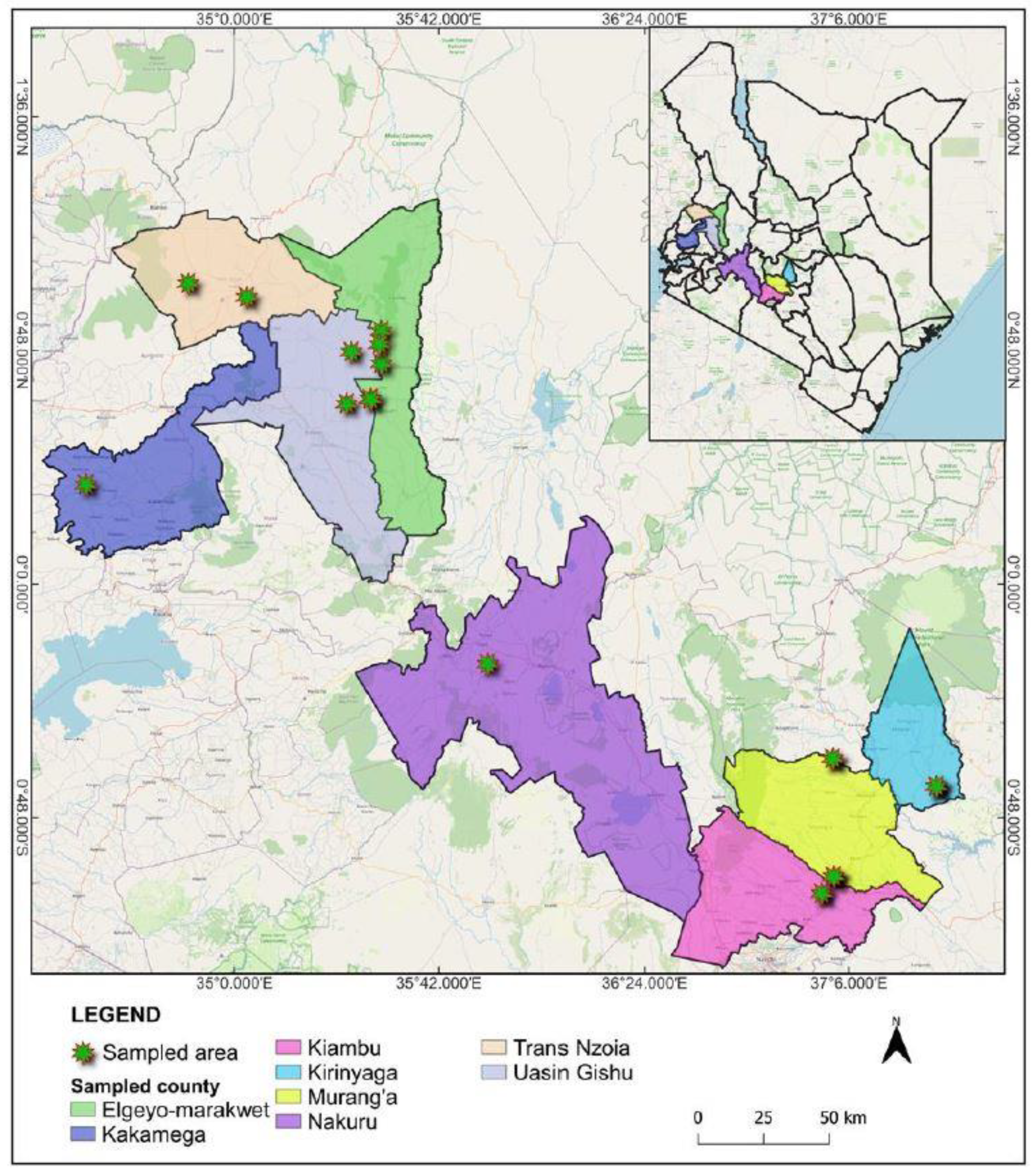
2.1. Collection of Passion Fruit Leaf Samples

Virus like and non-symptomatic passion fruit leaf samples were collected from small holder farms in major passion fruit growing areas in Kenya between August and November 2014 (Table 1; Figure 1). Five plants were sampled per field by collecting three leaf samples in each plant (from upper, middle, and lower leaves within a vine). The samples from each plant were bulked for virus testing. A total of 22 bulked samples were collected from Rift valley, (Nakuru (1), Trans Nzoia (3), Elgeyo Marakwet (5), Uasin Gishu (2)), Central (Kirinyaga (3), Muranga (2), Kiambu (5)) and Western (Kakamega (1) regions in Kenya placed and sealed in zip lock bags containing silica gel and preserved for at least one month prior to RNA extractions.

2.2. RNA Extraction

The RNA was extracted from the 22 samples preserved in silica gel using TRIzol reagent (Sigma-Aldrich, St. Louis, MO, USA), followed by RNase-free DNase (Invitrogen) treatment as described in [21]. The RNA quality control was performed using a Qubit (Invitrogen), and the integrity was further confirmed using RNA screentape (TapeStation 2200; Agilent Technologies). The extracted RNA was stored at -80 °C awaiting Library preparation.

2.3. High Throughput Sequencing

The extracted RNA with a concentration ranging from 0.1 - 1µg of total RNA was used for library preparation, using the TruSeq stranded Total RNA Sample Preparation kit with Ribozero Plant (Illumina, San Diego, CA). This involved subjecting the RNA to depleting Ribosomal RNA using rRNA binding beads. The RNA was then fragmented and primed with random hexamers. First strand cDNA synthesized using a mixture of Superscript II and Actinomycin D to allow RNA dependent synthesis and inhibit DNA dependent synthesis, respectively. The second strand cDNA synthesis was done using dUTP instead of dTTP to create stranded cDNA. The libraries were then adenylated at the 3’end using set A and B adaptors. The cDNA fragment was enriched by 15 cycles of PCR. The final size and concentration of each library was verified using Qubit and D1000 ScreenTape (TapeStation 2200, Agilent Technologies). Ten nanomolar library pools were prepared by mixing the libraries to achieve an equal molar concentration. The libraries were pooled and multiplexed in one lane, and a 1% PhiX v3 spike was included. Sequencing was conducted using MiSeq cycle 2 x 251 v3 kit (Illumina) to generate paired-end reads at Biosciences eastern and central Africa-International Livestock Research Institute (BecA-ILRI), Nairobi Kenya.

2.4. Sequence Analysis

Sequence reads were analyzed by VirusDetect v1.7 using default setting and plant virus database v232 and identified viral contigs extracted. In parallel the RNA-Seq raw reads were first trimmed using Trim Galore [40], with minimum sequence length set to 50bp and minimum required adapter overlap (stringency) set to 1bp. The de novo assembly was performed using the metaSPAdes version 3.13.0 genome assembler [41] with default settings. In addition, a second assembler, CLC Genomics Workbench 20 (CLCGW) (CLC bio, Qiagen, Redwood City, CA) was used with the quality scores limit set to 0.01, maximum number of ambiguities to two and removing any reads with <50 nt. De novo assembly was performed using CLCGW with settings of automatic word size, automatic bubble size, minimum contig length 800, mismatch cost two, insertion cost three, deletion cost three, length fraction 0.5 and similarity fraction 0.9 [42,43,44]. Contigs were sorted by length, examined individually and then subjected to a BLAST and PASC sequence comparisons [45,46]. In addition, trimmed reads from CLCGW were also imported into Geneious Prime (Biomatters Ltd, Auckland, New Zealand) [47], for reference mapping using minimum overlap 10%, minimum overlap identity 80%, allow gaps 10% and fine-tuning set to iterate up to 10 times. Virus coding regions were improved by aligning nt sequences to the aligned deduced amino acid sequences using MUSCLE available in Geneious. Open reading frames (ORFs) were predicted, and annotations made using Geneious. Consensus viral sequences assembled by both approaches were compared and all reads were re-mapped to the consensus in each sample to resolve conflicts and generate final consensus sequences by visual inspection using Geneious software. Reads were mapped back again at highest stringency to the final consensus sequences to determine read count and genome coverages.

2.5. Phylogenetic and Recombination Analysis

All available complete genome sequences of potyviruses reported to infect passionfruit were downloaded from NCBI and aligned with the potyvirus consensus sequences found in this study using the Muscle algorithm as implemented in MEGA-X [48]. Pairwise sequence distances and the optimal evolutionary model were then calculated for each alignment using MEGA-X. The optimal model for each alignment was then applied for phylogenetic analysis using the Maximum likelihood method in MEGA-X. Recombination analysis was performed using RDP version 4.97 [49] to detect putative recombination break points between 21 UPV and CABMV sequences. Any detected recombination signals flagged by RDP 4.97 as ‘potentially arising through evolutionary processes other than recombination’ were disregarded. Default parameters were used for the seven recombination detection methods implemented: RDP5.2 [50], GENECONV [51], Bootscan [52], MaxChi [53], Chimaera [54], 3Seq [55], and SiScan [56]. Only potential recombination events with an associated Bonferroni-corrected P value <0.05 of more than four recombination detection methods were considered credible evidence of recombination [57]. The BURT method of [49] was used to infer the locations and 95% confidence intervals of break point locations. Recombination breakpoint locations and origins of sequence regions potentially transferred during recombination were verified individually using the phylogenetic tools implemented in RDP 4.97.

3. Results

3.1. Virus Identification

A total of reads ranging from 1,465,364 and 3, 200,244 million reads were obtained from the HTS of different samples collected in Kenya passion fruit growing regions (Table 2). After de novo assembly, the contigs of interest had 4,424-232,782 reads mapping to the virus genome of interest, with an average depth of 61.9-3,159 and a normalized depth of 24.3-1,529. A total of 21 Uganda passiflora virus (UPV) and two east Asian Passiflora distortion virus (EAPDV) complete genomes were obtained. UPV and EAPDV coinfections were detected in sample 2 and 7. The UPV genomes collected from different Kenyan counties had 81 - 100% nucleotide identity (nt). Isolates 1KC, 3KE, 6KF, 7KH, 7KG and 8KI (Central region) and 11KJ, 12KK, 14KL, 18KN, 19KO, 12KD (Rift valley region) had 99 - 100% nt identity match between the isolates. Isolate KW from Uasin Gishu and isolate KM from Elgeyo Marakwet (Rift valley region) were identical but shared 86 % nt to any other closest isolate, and 81% nt identity match to KO and KN all from Rift valley region. In addition, isolate KW and KM shared 81% nt identity to 5 isolates from Central region. The highest UPV genome matched GenBank accession MK110656 from Uganda with 99% nucleotide identity (nt) to Rift valley and Central region isolates, but 81% nt identity to 9 isolates from Rift valley region. The two new EAPDV sequences (KA and KB) were both detected in Kirinyaga and Muranga county in Central region and had 65-66% nt identity match to the any of the new UPV genomes, but 77.2% nt identity to GenBank Japanese isolate PY-AK. Moreover, UPV variants and two partial passion fruit green spot virus (PFGSV) sequences and one partial (passiflora emaravirus) segment RNA1-5 (a new allexivirus and a new emaravirus, respectively (Supplementary Figure 1 and Figure 2) were detected. Passiflora virus A (PVA) was also detected co-infecting with UPV in samples 15 and 22 (Table 2). The complete genomes have been deposited in Genbank with accession MW355818 to MW355840.

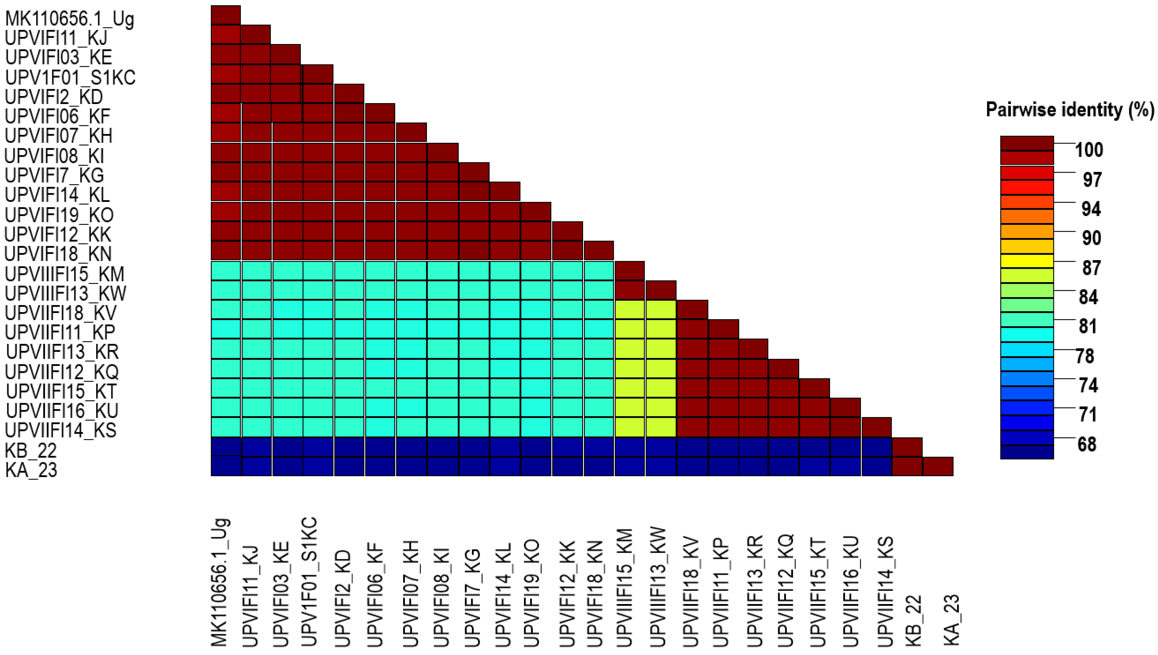
3.2. Phylogenetic and Recombination Analysis
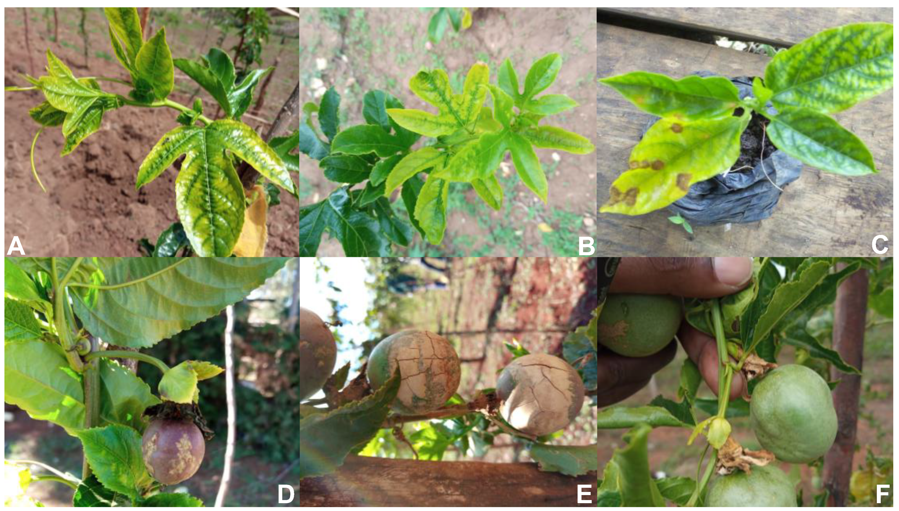
When all the new 21 UPV and two EAPDV (KA 23 and KB 22) genomes were compared with other closely related passiflora associated potyviruses from GenBank, the phylogenetic analysis several revealed distinct lineages (Figure 3). A distinct phylogroup consisted of UPV (I-III), EAPDV, passion fruit woodiness virus, passion fruit, severe mottle virus, bean common mosaic virus, soyabean mosaic virus, watermelon mosaic virus, and east Asian passiflora virus species. The UPV phylogroup I had sequences from Central, Rift valley and UPV-MK110656 from Uganda. The minor phylogroup II and III had all the UPV sequence from Rift valley. Notably, CABMV and bean common necrosis virus grouped separately forming a distinct phylogroup. The CABMV isolate 11KP from Kenya [21], grouped closely together with other eight CABMV sequences from different parts of the world (Figure 3). There was no significant putative recombination break points detected when all the new 21 UPV and the two EAPDV (KA 23 and KB 22) genomes obtained from this study.

3.3. Symptoms

The characteristic of virus-like symptoms of the passion fruit leaves samples included wrinkling, mosaic, chlorosis, vein clearing, ring spots and distorted leaves (Figure 2). However, these symptoms varied significantly within a region. For instance, the sample collected from Trans Nzoia revealed leaf yellow spots, flecks or mottling associated mosaic, rugosity and distortion which was associated with UPV-I. Kiambu samples had mosaic on the lower and upper surface of the leaves which was associated with confection of CABMV and UPV-I. In addition, passion fruit seedlings in Kiambu had mosaic on the leaves and found to be associated with UPV-I. The Muranga samples had thick hard and woody rind, cracked fruits which was observed and associated with UPV-I, KPV and CABMV. There were severe deformed fruits symptoms in the samples collected from Elgeyo Marakwet and found to be linked to UPV-I and UPV-II infections. In the neighboring Country of Uasin Gishu, severe mosaic on the upper leaves of passion fruit plant samples was observed and found to be associated with UPV-I, UPV-II and CABMV.

4. Discussion

To elucidate the passion fruit virus disease complexity in Kenya, virus surveys were conducted across major passion fruit growing Counties. Symptoms resembling PWD were prevalent, including conspicuous hardening and cracking of fruits, chlorotic mottle, and leaf distortion. Plant virus metagenomics has the capacity to detect viruses either as single or co-infections and can reveal the presence of novel or unsuspected plant viruses [39,58]. The study revealed the first full genomes of UPV and EAPDV in symptomatic passion fruit samples obtained from farms in Kenya. We detected mixed infections between UPV and EAPDV in two out of the 22 samples analysed by HTS. However, establishing associations between the specific viral infections detected and the symptoms expressed is complicated by mixed virus infections. Plant viruses co-infecting in the same host may interact either in a synergistic or antagonistic way [59] and this remains to be determined for viruses detected in passion fruit in Kenya. The discovery of UPV in major passion fruit growing regions of Kenya concurs with earlier studies reporting the presence of UPV in Uganda [20]. The detected UPV and EAPDV have close genetic link to a single UPV genome from Uganda. The UPV and EAPDV sequences grouped separately from the other passion fruit and potyviruses. A distinct lineage was formed when the 21 new UPV and EAPDV genomes were evaluated against other closely related potyvirus that are associated with the genus Passiflora. Recombination analysis found no significant putative recombination break points detected when all the new UPV and two EAPDV genomes were analysed. However, the CABMV had strong recombination hotspot towards the 5’UTR region of the virus.
Our results enabled the delineation of three UPV minor phylogroups named I – III. The minor phylogroup 1 had new sequences obtained from Central, Rift valley and Uganda, and the minor phylogroup II and III had all the UPV sequence from Rift valley only. The new EAPDV grouped separately to UPV but together with other four Japanese EAPDV sequences. All the other potential sequences of viruses associated passion fruit diseases retrieved from NCBI grouped separately from the Kenyan sequences. Since the UPV sequence nt identities of the samples from different passion fruit growing counties in Kenya ranged from 81 - 100%, support that the current Kenya UPV isolates belong to the same potyvirus species [20,60]. EAPDV sequences had 65 - 66% nt identity to the UPV genomes but 77.2% to the closest Japanese genome sequence isolate. Thus, neither the complete genome nor the CP comparisons found any evidence of close genomic relationship between the new UPV and EAPDV. Our analysis suggested that the observed nucleotide identities shared by the new UPV and EAPDV isolates from Kenya support the classification criteria of the new EAPDV from Kenyan as a distinct species within the genus potyvirus as previously found by [29].
The presence of UPV genomes sequences from Kenya within three minor phylogroup suggest multiple introductions might have been introduced to Kenya from the adjacent neighbouring countries, or elsewhere and the vice versa. The previous reports of UPV, genome sequences from Uganda and its high similarity to Kenyan isolate, could suggest its introduction was most likely from Uganda either directly or indirectly or vice versa. [61] reported occurrence of UPV from passionfruit virus infected samples from Rwanda with a high similarity to the UPV from Uganda based on the coat protein gene. This could be attributed to the complexity of countries in the East African Region geographic positions, such as land borders. There is therefore a need to strengthen quarantine measures due to the nature of the porous borders which could be playing a key role in the cross-border movement of the viruses with infected planting materials. For example, the Kenyan Western region borders with neighbouring countries such as Uganda with a porous land crossing border, where traders from both countries frequently interact. Unregulated trade practices, exemplified by activities such as cyclers or pedestrian trade transporters, may have inadvertently facilitated the dissemination of infected seeds or seedling planting materials along unmonitored routes, then dispersed to the rest of the country as seen in other parts of the world [61,62]. These reports of UPV infecting passion fruit in Kenya, Uganda and Rwanda suggests that UPV strains are spreading in East Africa and may be a problem more likely to emerge more widely in Africa. This indicates a threat to the passion fruit industry thus there is need to reinforce the control of PWD. Nonetheless, there is need for more sequencing to avail more complete genomes from East Africa region and other parts of the world. This will uncover UPV and EAPDV global distribution and enhance accurate determination of its origins, evolution, and molecular epidemiology towards improved passion fruit disease management. Further, availability of these genomes will facilitate development of sensitive diagnostic assays for use in virus indexing of planting materials in nurseries during certification programs as well as to support diagnostic laboratories that undertake plant health regulation mandate.
The study proposes that new currently known EAPDV “East Asian Passiflora distortion” be appropriately named as “Passiflora distortion virus (PDV)” to avoid geographical association. Likewise, UPV was previously detected in Uganda, and a neutral name “Passiflora virus” (PV) was proposed [20]. The new UPV sequences will equally follow the name (PV). This study has adopted latinised numerals in naming the phylogroups of these new sequences and other closely related species. Adopting neutral virus names deter misleading geographical names such as country names [37,63]. Neutral naming, hereafter such as “PV and PDV” allow a consideration of the only detected virus sequence that identifies the new virus, without a biased notion, associated with the first geographical detection locality, as the primary origin of the virus. This is because in several cases, very little surveillance and diagnostic is conducted prior to such naming being adopted. Consequently, it translates to lack of a broad spectrum understanding of the etiology of the new virus. For example, sweet potato virus East African strain was first identified in East Africa, and “East Africa (EA)” was adopted as strain name [64,65]. The “EA” is the phylogroup’s distribution originally considered restricted for this virus only, leading to the speculation that the EA is the subcenter of this virus diversity and evolution [66,67]. After 13 years, this virus was identified in East Timor grouping with many other sequences from southern Africa and Southeast Asia, and neutral naming (I) was adopted [68]. As such, in the current study we propose geographically neutral naming such as “PV and PDV”, since these viruses might be circulating in many other parts of the world and are yet to be detected. Notably, due to the increasing world trade in plants and plant products, it is only a matter of time before UPV and PDV are detected in additional countries. Therefore, such neutral names will minimize the unnecessary implications in virus research, i.e. evolution analysis, viral disease management, stigmatization of a particular region, international trade implications.
Thus, molecular, serological, and host range studies indicate the viral disease symptoms on passion fruit in Kenya are caused by a confection of multiple viruses with a limited molecular diversity among UPV isolates. Sequences corresponding to Normalized read depth between samples varied significantly indicating differences in virus titers in the plants. The PV occurring in Kenya was isolated from passion fruit leaves showing chlorotic mottle, distortion, thick, hard and woody cracked fruits, which are indicative of presence of PWD in Kenya. These symptoms have been previously found in passion fruit crops in Kenya [34] and neighboring countries including Uganda [7] and Rwanda [61]. Our findings suggest that these PWD distinct symptoms are contributed by synergistic coinfections of PV, CABMV and the EAPDV (PDV) within Kenya passion fruit production systems. Notably, CABMV has been associated PWD, which is linked to mosaic, blistering, and distortion on leaves, fruit woodiness, reduction in juice yield, and shorter lifespan [21,69]. Similarly, these viruses were also genetically distinct from other potyviruses infecting passion fruit as seen within the phylogenetic analysis. We therefore suggest that the new PV and PDV are distinct virus species associated with passion fruit woodiness disease, which has led to significant economic damages in Brazil, Japan, Taiwan, and East African countries [19,20,29,70]. The propagation nature of passion fruit in Kenya which mostly involves grafting also leads to accumulation of viruses [34]. As such stringent management measures including virus indexing of planting seedlings at nursery multiplication blocks is key to prevent further virus dissemination. This should be implemented to deter further impacts of these viruses and enhance passion fruit production. In addition, epidemiological studies are needed to understand the role or cowpea (Vigna unguiculata) crops in spreading CABMV to adjacent passion fruit growing hedges.
Recombination plays a pivotal role in viruses [71]. Most plant RNA viruses have high rates of recombination leading to an emergence of new virus species and their variants, a crucial factor in the emergence of resistance-breaking strains, virulence increase and change of host range as an adaptation strategy [37,72,73]. Despite, the critical role of recombination process in RNA viruses, the current new PV and PDV genomes isolated from Kenya passion fruit crops had no significant recombination break points. Although, recombination process varies extensively among viruses [71], genetic, biological, and epidemiological factors can affect the probability of virus recombination between different strains or species. This may include genetic homology, existing virus population size in the host, co-circulation in the same geographical area, and coinfection rate. The current sequences insufficient genetic diversity of 99-100% nt, could be attributed to the source of the nursery seedlings from a common seed stock multiplication and nursery centres then subsequently distributed to the rest of the country passion fruit growing regions. Furthermore, the large diversity of 34% nt genome difference between the new PDV and almost 20% nt between new PV isolates is likely to yield no viable offspring when recombining due to the large genetic distances. Importantly, viral recombination is replication-dependent, as such high viral titre increases the chances of exchanging genetic strands [74]. However, despite the remarkable symptoms observed in the field samples, it is unclear if the PWD viruses detected in this study existed with an overall high viral load. Future studies are required to decipher PWD viral load and its role in recombination.

5. Conclusions

These findings underscore the need to strengthen the current Kenya phytosanitary and border surveillance activities. This should include stringent quarantine measures over importation of plants and plant products to safeguard Kenya agriculture, and for horticultural industry to remain secure and free from new and severe viral diseases, that has potential to cripple the country’s economic backbone. Moreover, monitoring activities could prevent the spread of potential viral vectors through natural arrivals due to the unpredictable effects of climate change, as has already been the case with arrival of damaging insects like locusts.

Supplementary Materials

The following supporting information can be downloaded at the website of this paper posted on Preprints.org, Figure S1: Phylogenetic analysis of the new viruses and other potyviruses, (A and B) phylogeny of the new allexiviruses and cilevirus. Figure S1: Histograms of sequence coverage of consensus viral genomes (top) and variant assemblies with variant regions showing many folds higher sequence coverage. Bars at the bottom of the figure indicate position of variant regions.

Author Contributions

Conceptualization, FM. and T.H.; methodology, FM. S.M and T.H.; software, F.M and S.M.; validation, E.N., J.K. and Z.Z.; formal analysis, J.K and S.M.; investigation, F.M.; writing—original draft preparation, F.M., S.M;E.N writing—review and editing, All authors.; All authors have read and agreed to the published version of the manuscript.

Funding

We thank the Kenya Plant Health Inspectorate Service for supporting F. Munguti to undertake the Africa Biosciences Challenge Fund (ABCF) fellowship at BecA-ILRI hub. This project was supported by the Biosciences eastern and central Africa-International Livestock Research Institute, Nairobi (BecA-ILRI Hub), through the ABCF program. The ABCF program is funded by the Australian Department for Foreign Affairs and Trade through the BecA-CSIRO partnership, the Syngenta Foundation for Sustainable Agriculture, the Bill & Melinda Gates Foundation, the UK Department for International Development, and the Swedish International Development Cooperation Agency.

Data Availability Statement

The complete genomes have been deposited in NCBI with accession MW355818 to MW355840.

Conflicts of Interest

The authors declare no conflicts of interest.

References

  1. Cervi, A.C.I. , Daniela Cristina. A new species of Passiflora (Passifloraceae) from Mato Grosso do Sul, Brazil. Phytotaxa 2013, 103, 46–50. [Google Scholar] [CrossRef]
  2. Manicom, B.; Ruggiero, C.; Ploetz, R.C.; Goes, A.d. Diseases of passion fruit. Diseases of tropical fruit crops 2003, 413–441. [Google Scholar]
  3. Devi Ramaiya, S.; Bujang, J.S.; Zakaria, M.H.; King, W.S.; Shaffiq Sahrir, M.A. Sugars, ascorbic acid, total phenolic content and total antioxidant activity in passion fruit (Passiflora) cultivars. Journal of the Science of Food and Agriculture 2013, 93, 1198–1205. [Google Scholar] [CrossRef]
  4. Phamiwon Zas, P.Z.; John, S. Diabetes and medicinal benefits of Passiflora edulis. 2016.
  5. Castillo, N.R.; Ambachew, D.; Melgarejo, L.M.; Blair, M.W. Morphological and agronomic variability among cultivars, landraces, and genebank accessions of purple passion fruit, Passiflora edulis f. edulis. HortScience 2020, 55, 768–777. [Google Scholar] [CrossRef]
  6. Kumari, S.; Mishra, P. Passion fruit (Passiflora edulis Sims.)—An underexploited plant of nutraceutical value. Asian Journal of Medical and Health Research 2016, 1, 1–10. [Google Scholar]
  7. Ochwo-Ssemakula, M.; Sengooba, T.; Hakiza, J.; Adipala, E.; Edema, R.; Redinbaugh, M.; Aritua, V.; Winter, S. Characterization and distribution of a Potyvirus associated with passion fruit woodiness disease in Uganda. Plant Disease 2012, 96, 659–665. [Google Scholar] [CrossRef]
  8. AFA. AFA YEAR BOOK OF STATISTICS. 2022.
  9. Karani-Gichimu, C.; Mwangi, M.; Macharia, I. Assessment of passion fruit orchard management and farmers’ technical efficiency in Central-Eastern and North-Rift Highlands Kenya. Assessment 2013, 4, 183–190. [Google Scholar]
  10. Amata, R.; Otipa, M.; Waiganjo, M.; Wasilwa, L.; Kinoti, J.; Kyamanywa, S.; Erbaugh, M. Management strategies for fungal diseases in passion fruit production systems in Kenya. In Proceedings of the I All Africa Horticultural Congress 911; 2009; pp. 207–213. [Google Scholar]
  11. Gesimba, R.M. Screening Passiflora species for drought tolerance, compatibility with purple passion fruit, Fusarium wilt resistance and the relationship between irrigation, drenching and media composition in the control of Fusarium wilt. The Ohio State University, 2008.
  12. Amata, R.; Otipa, M.; Waiganjo, M.; Wabule, M.; Thuranira, E.; Erbaugh, M.; Miller, S. Incidence, prevalence and severity of passion fruit fungal diseases in major production regions of Kenya. 2009.
  13. Wangungu, C.; Mwangi, M.; Gathu, R.; Muasya, R.; Mbaka, J.; Kori, N. Reducing dieback disease incidence of passion fruit in Kenya through management practices. 2011.
  14. HCDA. NATIONAL HORTICULTURE VALIDATED REPORT. 2013.
  15. Morton, J. Jackfruit. Fruits of warm climates. Julia F. Morton, Miami, FL, 1987; 58–64. [Google Scholar]
  16. Baker, C.A.; Jeyaprakash, A.; Webster, C.G.; Adkins, S. Viruses infecting passiflora species in Florida; Florida Department of Agriculture and Consumer Services Tallahassee, FL, USA: 2014.
  17. Coutts, B.A.; Kehoe, M.A.; Webster, C.G.; Wylie, S.J.; Jones, R.A. Indigenous and introduced potyviruses of legumes and Passiflora spp. from Australia: Biological properties and comparison of coat protein nucleotide sequences. Archives of Virology 2011, 156, 1757–1774. [Google Scholar] [CrossRef]
  18. Fontenele, R.S.; Abreu, R.A.; Lamas, N.S.; Alves-Freitas, D.M.; Vidal, A.H.; Poppiel, R.R.; Melo, F.L.; Lacorte, C.; Martin, D.P.; Campos, M.A. Passion fruit chlorotic mottle virus: Molecular characterization of a new divergent geminivirus in Brazil. Viruses 2018, 10, 169. [Google Scholar] [CrossRef]
  19. Fukumoto, T.; Nakamura, M.; Rikitake, M.; Iwai, H. Molecular characterization and specific detection of two genetically distinguishable strains of East Asian Passiflora virus (EAPV) and their distribution in southern Japan. Virus Genes 2012, 44, 141–148. [Google Scholar] [CrossRef] [PubMed]
  20. Mbeyagala, E.; Maina, S.; Macharia, M.; Mukasa, S.; Holton, T. Illumina sequencing reveals the first near-complete genome sequence of Ugandan Passiflora virus. Microbiology Resource Announcements 2019, 8. [Google Scholar] [CrossRef] [PubMed]
  21. Munguti, F.; Maina, S.; Nyaboga, E.; Kilalo, D.; Kimani, E.; Macharia, M.; Holton, T. Transcriptome sequencing reveals a complete genome sequence of Cowpea aphid-borne mosaic virus from passion fruit in Kenya. Microbiology Resource Announcements 2019, 8. [Google Scholar] [CrossRef] [PubMed]
  22. Nascimento, A.V.S.d.; Santana, E.N.d.; Braz, A.S.K.; Alfenas, P.; Pio-Ribeiro, G.; Andrade, G.P.d.; De Carvalho, M.; Murilo Zerbini, F. Cowpea aphid-borne mosaic virus (CABMV) is widespread in passionfruit in Brazil and causes passionfruit woodiness disease. Archives of virology 2006, 151, 1797–1809. [Google Scholar] [CrossRef]
  23. Parrella, G.; Lanave, C. Identification of a new pathotype of Bean yellow mosaic virus (BYMV) infecting blue passion flower and some evolutionary characteristics of BYMV. Archives of virology 2009, 154, 1689–1694. [Google Scholar] [CrossRef]
  24. Polston, J.; Londoño, M.; Cohen, A.; Padilla-Rodriguez, M.; Rosario, K.; Breitbart, M. Genome sequence of Euphorbia mosaic virus from passionfruit and Euphorbia heterophylla in Florida. Genome announcements 2017, 5. [Google Scholar] [CrossRef]
  25. Song, Y.S.; Ryu, K.H. The complete genome sequence and genome structure of passion fruit mosaic virus. Archives of virology 2011, 156, 1093–1095. [Google Scholar] [CrossRef]
  26. Spiegel, S.; Zeidan, M.; Sobolev, I.; Beckelman, Y.; Holdengreber, V.; Tam, Y.; Bar Joseph, M.; Lipsker, Z.; Gera, A. The complete nucleotide sequence of Passiflora latent virus and its phylogenetic relationship to other carlaviruses. Archives of virology 2007, 152, 181–189. [Google Scholar] [CrossRef]
  27. Vaca-Vaca, J.C.; Carrasco-Lozano, E.C.; López-López, K. Molecular identification of a new begomovirus infecting yellow passion fruit (Passiflora edulis) in Colombia. Archives of Virology 2017, 162, 573–576. [Google Scholar] [CrossRef]
  28. Kilalo, D.; Olubayo, F.; Ateka, E.; Hutchinson, J.; Kimenju, J. Monitoring of aphid fauna in passionfruit orchards in Kenya. 2013.
  29. Iwai, H.; Yamashita, Y.; Nishi, N.; Nakamura, M. The potyvirus associated with the dappled fruit of Passiflora edulis in Kagoshima prefecture, Japan is the third strain of the proposed new species East Asian Passiflora virus (EAPV) phylogenetically distinguished from strains of Passion fruit woodiness virus. Archives of Virology 2006, 151, 811–818. [Google Scholar] [PubMed]
  30. Brand, R.; Burger, J.; Rybicki, E. Cloning, sequencing, and expression in Escherichia coli of the coat protein gene of a new potyvirus infecting South African Passiflora. Archives of Virology 1993, 128, 29–41. [Google Scholar] [CrossRef] [PubMed]
  31. McKern, N.; Strike, P.; Barnett, O.; Dijkstra, J.; Shukla, D.; Ward, C. Cowpea aphid borne mosaic virus-Morocco and South African Passiflora virus are strains of the same potyvirus. Archives of Virology 1994, 136, 207–217. [Google Scholar] [CrossRef] [PubMed]
  32. Kilalo, D. Molecular detection of viruses associated with passion fruit (Passiflora edulis sims) woodiness disease, monitoring and management of aphid vectors in Kenya. Doctoral dissertation, University of Nairobi. Homepage address: Erepository …, 2012.
  33. Njuguna, J.; Ndungu, B.; Mbaka, J.; Chege, B. Report on passion fruit diagnostic survey. A Collaborative Research of KARI/MOA/GTZ under promotion of private sector development in Agriculture (PSDA) programme, 2005. [Google Scholar]
  34. Asande, L.K.; Ombori, O.; Oduor, R.O.; Nchore, S.B.; Nyaboga, E.N. Occurrence of passion fruit woodiness disease in the coastal lowlands of Kenya and screening of passion fruit genotypes for resistance to passion fruit woodiness disease. BMC Plant Biology 2023, 23, 544. [Google Scholar] [CrossRef]
  35. Wu, W.; Ma, F.; Zhang, X.; Tan, Y.; Han, T.; Ding, J.; Wu, J.; Xing, W.; Wu, B.; Huang, D. Research Progress on Viruses of Passiflora edulis. Biology 2024, 13, 839. [Google Scholar] [CrossRef]
  36. Kreuze, J.F.; Perez, A.; Untiveros, M.; Quispe, D.; Fuentes, S.; Barker, I.; Simon, R. Complete viral genome sequence and discovery of novel viruses by deep sequencing of small RNAs: A generic method for diagnosis, discovery and sequencing of viruses. Virology 2009, 388, 1–7. [Google Scholar] [CrossRef]
  37. Maina, S.; Barbetti, M.J.; Edwards, O.R.; Minemba, D.; Areke, M.W.; Jones, R.A. Genetic connectivity between papaya ringspot virus genomes from Papua New Guinea and Northern Australia, and new recombination insights. Plant disease 2019, 103, 737–747. [Google Scholar] [CrossRef]
  38. Martin, R.R.; Constable, F.; Tzanetakis, I.E. Quarantine regulations and the impact of modern detection methods. Annual review of phytopathology 2016, 54, 189–205. [Google Scholar] [CrossRef]
  39. Villamor, D.; Ho, T.; Al Rwahnih, M.; Martin, R.; Tzanetakis, I. High throughput sequencing for plant virus detection and discovery. Phytopathology 2019, 109, 716–725. [Google Scholar] [CrossRef]
  40. Krueger, F. Trim galore. A wrapper tool around Cutadapt and FastQC to consistently apply quality and adapter trimming to FastQ files 2015, 516, 517. [Google Scholar]
  41. Nurk, S.; Meleshko, D.; Korobeynikov, A.; Pevzner, P.A. metaSPAdes: A new versatile metagenomic assembler. Genome research 2017, 27, 824–834. [Google Scholar] [CrossRef] [PubMed]
  42. Maina, S.; Coutts, B.A.; Edwards, O.R.; de Almeida, L.; Kehoe, M.A.; Ximenes, A.; Jones, R.A. Zucchini yellow mosaic virus populations from East Timorese and Northern Australian cucurbit crops: Molecular properties, genetic connectivity, and biosecurity implications. Plant Disease 2017, 101, 1236–1245. [Google Scholar] [CrossRef] [PubMed]
  43. Maina, S.; Coutts, B.A.; Edwards, O.R.; de Almeida, L.; Ximenes, A.; Jones, R.A. Papaya ringspot virus populations from East Timorese and Northern Australian cucurbit crops: Biological and molecular properties, and absence of genetic connectivity. Plant disease 2017, 101, 985–993. [Google Scholar] [CrossRef] [PubMed]
  44. Maina, S.; Edwards, O.R.; de Almeida, L.; Ximenes, A.; Jones, R.A. Metagenomic analysis of cucumber RNA from East Timor reveals an Aphid lethal paralysis virus genome. Genome Announcements 2017, 5. [Google Scholar] [CrossRef] [PubMed]
  45. Altschul, S.F.; Gish, W.; Miller, W.; Myers, E.W.; Lipman, D.J. Basic local alignment search tool. Journal of molecular biology 1990, 215, 403–410. [Google Scholar] [CrossRef]
  46. Bao, Y.; Chetvernin, V.; Tatusova, T. Improvements to pairwise sequence comparison (PASC): A genome-based web tool for virus classification. Archives of virology 2014, 159, 3293–3304. [Google Scholar] [CrossRef]
  47. Kearse, M.; Moir, R.; Wilson, A.; Stones-Havas, S.; Cheung, M.; Sturrock, S.; Buxton, S.; Cooper, A.; Markowitz, S.; Duran, C. Geneious Basic: An integrated and extendable desktop software platform for the organization and analysis of sequence data. Bioinformatics 2012, 28, 1647–1649. [Google Scholar] [CrossRef]
  48. Kumar, S.; Stecher, G.; Li, M.; Knyaz, C.; Tamura, K. MEGA X: Molecular evolutionary genetics analysis across computing platforms. Molecular biology and evolution 2018, 35, 1547–1549. [Google Scholar] [CrossRef]
  49. Martin, D.P.; Murrell, B.; Golden, M.; Khoosal, A.; Muhire, B. RDP4: Detection and analysis of recombination patterns in virus genomes. Virus evolution 2015, 1, vev003. [Google Scholar] [CrossRef]
  50. Martin, D.P.; Varsani, A.; Roumagnac, P.; Botha, G.; Maslamoney, S.; Schwab, T.; Kelz, Z.; Kumar, V.; Murrell, B. RDP5: A computer program for analyzing recombination in, and removing signals of recombination from, nucleotide sequence datasets. Virus Evolution 2021, 7, veaa087. [Google Scholar] [CrossRef]
  51. Padidam, M.; Sawyer, S.; Fauquet, C.M. Possible emergence of new geminiviruses by frequent recombination. Virology 1999, 265, 218–225. [Google Scholar] [CrossRef] [PubMed]
  52. Martin, D.P.; Posada, D.; Crandall, K.; Williamson, C. A modified bootscan algorithm for automated identification of recombinant sequences and recombination breakpoints. Microbiology 2005, 11. [Google Scholar] [CrossRef] [PubMed]
  53. Smith, J.M. Analyzing the mosaic structure of genes. Journal of molecular evolution 1992, 34, 126–129. [Google Scholar] [CrossRef] [PubMed]
  54. Posada, D.; Crandall, K.A. Evaluation of methods for detecting recombination from DNA sequences: Computer simulations. Proceedings of the National Academy of Sciences 2001, 98, 13757–13762. [Google Scholar] [CrossRef]
  55. Boni, M.F.; Posada, D.; Feldman, M.W. An exact nonparametric method for inferring mosaic structure in sequence triplets. Genetics 2007, 176, 1035–1047. [Google Scholar] [CrossRef]
  56. Gibbs, M.J.; Armstrong, J.S.; Gibbs, A.J. Sister-scanning: A Monte Carlo procedure for assessing signals in recombinant sequences. Bioinformatics 2000, 16, 573–582. [Google Scholar] [CrossRef]
  57. Ohshima, K.; Yamaguchi, Y.; Hirota, R.; Hamamoto, T.; Tomimura, K.; Tan, Z.; Sano, T.; Azuhata, F.; Walsh, J.A.; Fletcher, J. Molecular evolution of Turnip mosaic virus: Evidence of host adaptation, genetic recombination and geographical spread. Journal of General Virology 2002, 83, 1511–1521. [Google Scholar] [CrossRef]
  58. Maina, S.; Donovan, N.J.; Plett, K.; Bogema, D.; Rodoni, B.C. High-throughput sequencing for plant virology diagnostics and its potential in plant health certification. Frontiers in Horticulture 2024, 3, 1388028. [Google Scholar] [CrossRef]
  59. Syller, J. Facilitative and antagonistic interactions between plant viruses in mixed infections. Molecular plant pathology 2012, 13, 204–216. [Google Scholar] [CrossRef]
  60. Adams, M.; Antoniw, J.; Fauquet, C. Molecular criteria for genus and species discrimination within the family Potyviridae. Archives of virology 2005, 150, 459–479. [Google Scholar] [CrossRef]
  61. Bancy, W.W.; Dora, C.K.; Martina, K.; Mutuku, J.M. Molecular detection of Ugandan passiflora virus infecting passionfruit (Passiflora edulis sims) in Rwanda. Annual Research & Review in Biology 2019, 30, 1–10. [Google Scholar]
  62. Maina, S.; Jones, R.A. Enhancing biosecurity against virus disease threats to Australian grain crops: Current situation and future prospects. Frontiers in Horticulture 2023, 2, 1263604. [Google Scholar] [CrossRef]
  63. Jones, R.A.; Naidu, R.A. Global dimensions of plant virus diseases: Current status and future perspectives. Annual review of virology 2019, 6, 387–409. [Google Scholar] [CrossRef] [PubMed]
  64. Kreuze, J.; Savenkov, E.; Valkonen, J. Complete genome sequence and analyses of the subgenomic RNAs of Sweet potato chlorotic stunt virus reveal several new features for the genus Crinivirus. Journal of Virology 2002, 76, 9260–9270. [Google Scholar] [CrossRef]
  65. Wasswa, P. Sweet potato viruses in Uganda: Identification of a new virus, a mild strain of an old virus and reversion. University of Greenwich, 2012.
  66. Tairo, F.; Mukasa, S.B.; Jones, R.A.; Kullaya, A.; Rubaihayo, P.R.; Valkonen, J.P. Unravelling the genetic diversity of the three main viruses involved in sweet potato virus disease (SPVD), and its practical implications. Molecular Plant Pathology 2005, 6, 199–211. [Google Scholar] [CrossRef]
  67. Tugume, A.K.; Mukasa, S.B.; Kalkkinen, N.; Valkonen, J.P. Recombination and selection pressure in the ipomovirus Sweet potato mild mottle virus (Potyviridae) in wild species and cultivated sweetpotato in the centre of evolution in East Africa. Journal of general virology 2010, 91, 1092–1108. [Google Scholar] [CrossRef]
  68. Maina, S. Application of viral genomics to study the connectivity between crop viruses from northern Australia and neighbouring countries. 2018.
  69. Gonçalves, Z.S.; Lima, L.K.S.; Soares, T.L.; Abreu, E.F.M.; de Jesus Barbosa, C.; Cerqueira-Silva, C.B.M.; de Jesus, O.N.; de Oliveira, E.J. Identification of Passiflora spp. genotypes resistant to Cowpea aphid-borne mosaic virus and leaf anatomical response under controlled conditions. Scientia Horticulturae 2018, 231, 166–178. [Google Scholar] [CrossRef]
  70. Chong, Y.-H.; Cheng, Y.-H.; Cheng, H.-W.; Huang, Y.-C.; Yeh, S.-D. The virus causing passionfruit woodiness disease in Taiwan is reclassified as East Asian passiflora virus. Journal of General Plant Pathology 2018, 84, 208–220. [Google Scholar] [CrossRef]
  71. Pérez-Losada, M.; Arenas, M.; Galán, J.C.; Palero, F.; González-Candelas, F. Recombination in viruses: Mechanisms, methods of study, and evolutionary consequences. Infection, Genetics and Evolution 2015, 30, 296–307. [Google Scholar] [CrossRef]
  72. Gibbs, M.J.; Weiller, G.F. Evidence that a plant virus switched hosts to infect a vertebrate and then recombined with a vertebrate-infecting virus. Proceedings of the National Academy of Sciences 1999, 96, 8022–8027. [Google Scholar] [CrossRef]
  73. Lefeuvre, P.; Moriones, E. Recombination as a motor of host switches and virus emergence: Geminiviruses as case studies. Current Opinion in Virology 2015, 10, 14–19. [Google Scholar] [CrossRef] [PubMed]
  74. De Candia, C.; Espada, C.; Duette, G.; Ghiglione, Y.; Turk, G.; Salomón, H.; Carobene, M. Viral replication is enhanced by an HIV-1 intersubtype recombination-derived Vpu protein. Virology Journal 2010, 7, 1–9. [Google Scholar] [CrossRef] [PubMed]
Figure 1. Diversity in passion fruit virus symptoms observed in the field. (A) Chlorosis, mottling, (B) severe mosaic on the upper leaves, rugosity and distortion, (C) nursery seedling with leaf necrosis, mosaic on the lower surface of the leaves, (D) fruit deformation and discolored rind, (E) thick hard and woody rind, cracked fruits (F) deformed fruits.
Preprints 142786 g001
Figure 2. Map of Kenya showing the geographical field locations (counties) where all the passion fruit leaf samples with virus like symptoms were collected.
Preprints 142786 g002
Figure 3. Species demarcation tool (SDT) interface of the new passiflora virus genomes sequences obtained in this study. The colour-coded matrix of pairwise identity scores, show species nucleotide pairwise identity scores of the new 21 genomes and the Ugandan isolate currently available in GenBank.
Preprints 142786 g003
Figure 4. Phylogenetic tree analysis of 21 new UPV and two EAPDV (KA 23 and KB 22) isolates, along with other closely related potyviruses from GenBank. The tree was generated using MEGA using maximum likelihood method, and bootstrap values (≥ 50%) are shown at the nodes. Scale bar indicates evolutionary distance.
Preprints 142786 g004
Table 1. Cultivar type, symptoms and geographical information of passion fruit leaf samples that were used to uncover passion fruit viromes using high throughput sequencing (HTS) in Kenya. The samples were collected between August and November 2014.
Sample No. HTS No. Cultivar Fruit type Viral like disease symptoms County Region
K1 1_S1_L001 KPF 11 Yellow Symptomatic Kirinyaga Central
K2 2_S2_L001 KPF 11 Yellow Symptomatic Kirinyaga Central
K3 3_S3_L001 KPF4 Yellow Symptomatic Kirinyaga Central
K4 4_S4_L001 KPF 12 Yellow Symptomatic Kiambu Central
K5 5_S5_L001 KPF 4 Yellow Symptomatic Kiambu Central
K6 6_S6_L001 KPF4 Yellow Asymptomatic Muranga Central
K7 7_S7_L001 Unknown Hardy shell Symptomatic Muranga Central
K8 8_S8_L001 Unknown Purple Symptomatic Kiambu Central
K9 9_S9_L001 Unknown Sweet yellow Symptomatic Kiambu Central
K10 10_S10_L001 Unknown Sweet yellow Symptomatic Kiambu Central
K11 11_S11_L001 Unknown Purple Symptomatic Nakuru Rift Valley
K12 12_S12_L001 Unknown Purple Symptomatic Uasin Gishu Rift Valley
K13 13_S13_L001 Unknown Purple Symptomatic Uasin Gishu Rift Valley
K14 14_S14_L001 Unknown Yellow Symptomatic Elgeyo Marakwet Rift Valley
K15 15_S15_L001 Unknown Purple Symptomatic Elgeyo Marakwet Rift Valley
K16 16_S16_L001 Unknown Purple Symptomatic Elgeyo Marakwet Rift Valley
K17 17_S17_L001 Unknown Purple Symptomatic Elgeyo Marakwet Rift Valley
K18 18_S18_L001 Unknown Purple Symptomatic Elgeyo Marakwet Rift Valley
K19 19_S19_L001 Unknown Grafted Symptomatic Trans Nzoia Rift Valley
K20 20_S20_L001 Unknown Grafted Symptomatic Trans Nzoia Rift Valley
K21 21_S21_L001 Unknown Grafted Symptomatic Trans Nzoia Rift Valley
K22 22_S22_L001 Unknown Grafted Symptomatic Kakamega Western
Table 2. Viruses identified in different sample including total number of reads obtained, length of assembled genome sequences, number of sequences reads mapped, average and normalized sequencing depth.
Sample Raw reads Virus identified Contig length Sequence reads mapped Average depth normalized depth
1_KC 1,545,706 UPV-I 9,684 13,294 183 118.6
2_KA
2_KD
1,509,132 EAPDV 9,638 17,410 232.5 154.1
UPV-I 9,679 175,623 2,307. 1,529
3_KE 3,200,244 UPV-I 9,670 36,941 516.8 161.5
6_KF 2,547,696 UPV-I 9,650 4,424 61.9 24.3
7_KH
7_KB
1,149,924 UPV-I 9,779 66,140 939.9 817.4
EAPDV 9,638 11,276 162 141
8_KI 1,465,364 UPV-I 9,874 85,227 1,217 830.7
11_KJ
11_KP
2,578,474 UPV-I 9,669 9,217 117.9 45.8
UPV-II 9,654 14,188 181.7 70.5
CABMV (Munguti et al. 2020) 9,846 14,888 185.9 72.1
12_KK
12_KQ
2,706,154 UPV-I* 9,650 75,058 1,039.6 384.2
UPV-II* 9,669 32,348 431.3 159.4
13_KR
13_KW
1,779,280 UPV-II 9,877 81,444 1,151.8 647.4
UPV-III 9,683 43,291 620.2 348.6
14_KL
14_KS
2,974,246 UPV-I* 9,629 9,318 131.3 44.1
UPV-II 9,771 220,180 3,062.3 1,029.6
15_KT
15_KM
1,706,154 UPV-II 9,658 77,940 1,044.6 612.2
UPV-III 9,667 127,649 1,696.5 994.3
16_KU 1,627,772 UPV-II 9,677 41,050 557 342.3
17_KG 2,400,688 UPV-I 9,651 232,782 3,159 1,315.9
18_KN
18_KV
2,395,822 UPV-I 9,671 185,274 2,619 1,093.3
UPV-II 9,680 126,554 1,792.6 748
19_KO 1,847,440 UPV-I 9,636 55,234 735.9 398.4
* Additional variant of the virus identified, but could not be fully assembled. UPV - Uganda passiflora Virus (UPV). EAPDV - East Asian passiflora distortion virus. CABMV - Cowpea aphid borne mosaic virus. 2 -(Passiflora virus A) samples 4, 5, 9, 10, 20, 21 (incomplete UPV), 22 - (incomplete UPV and passion fruit green spot virus).
Disclaimer/Publisher’s Note: The statements, opinions and data contained in all publications are solely those of the individual author(s) and contributor(s) and not of MDPI and/or the editor(s). MDPI and/or the editor(s) disclaim responsibility for any injury to people or property resulting from any ideas, methods, instructions or products referred to in the content.
Copyright: This open access article is published under a Creative Commons CC BY 4.0 license, which permit the free download, distribution, and reuse, provided that the author and preprint are cited in any reuse.
Prerpints.org logo

Preprints.org is a free preprint server supported by MDPI in Basel, Switzerland.

Subscribe

Disclaimer

Terms of Use

Privacy Policy

Privacy Settings

© 2025 MDPI (Basel, Switzerland) unless otherwise stated