Preprint
Article

This version is not peer-reviewed.

Seasonal Variations in the Structure and Function of the Gut Flora in Adult Male Rhesus Macaques Reared in Outdoor Colonies

A peer-reviewed article of this preprint also exists.

Submitted:

03 December 2024

Posted:

04 December 2024

You are already at the latest version

Abstract

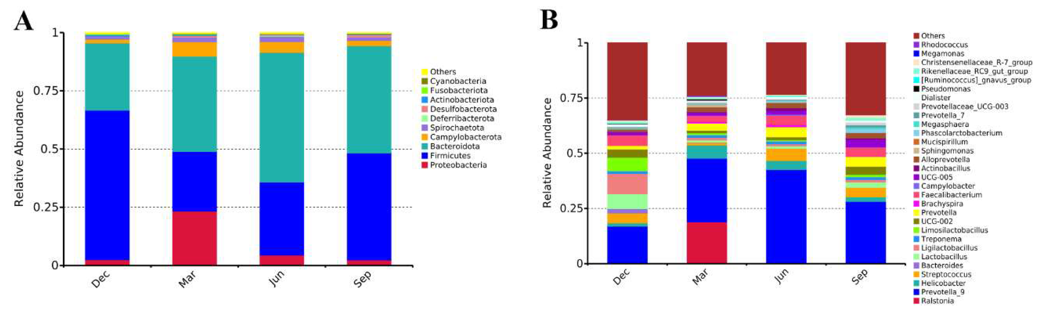
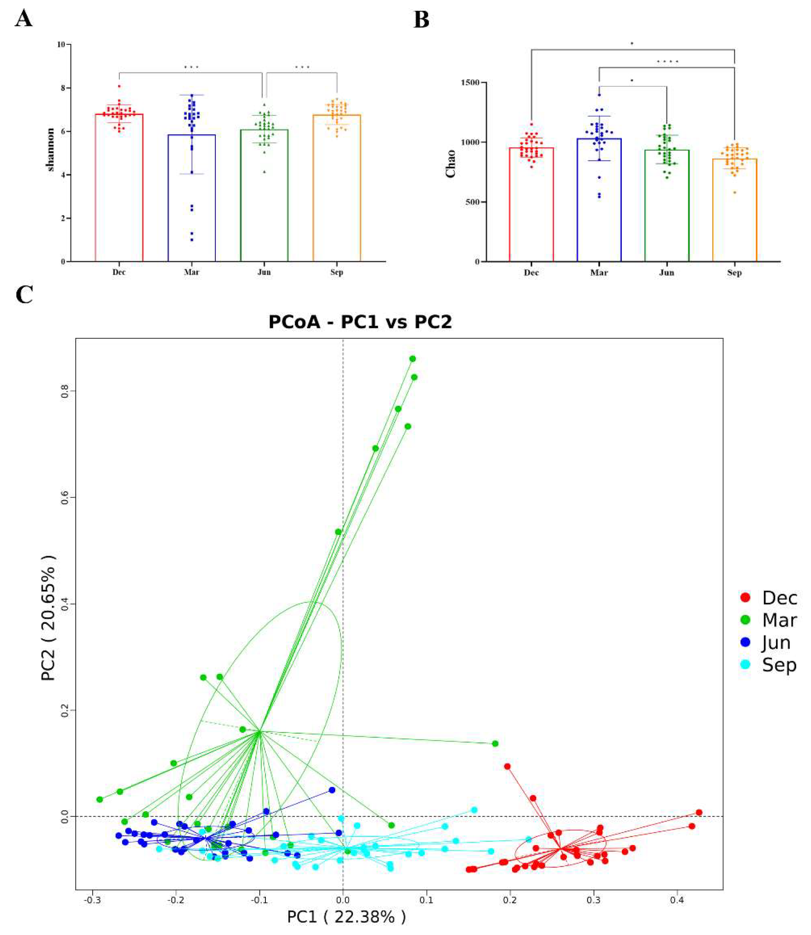
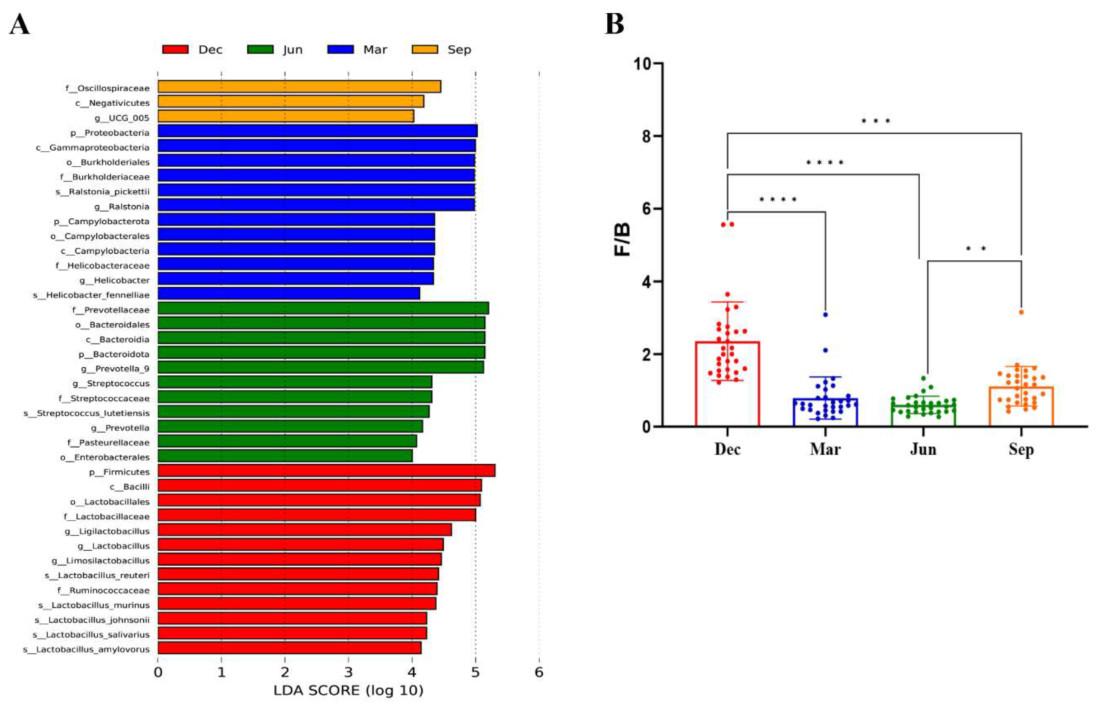
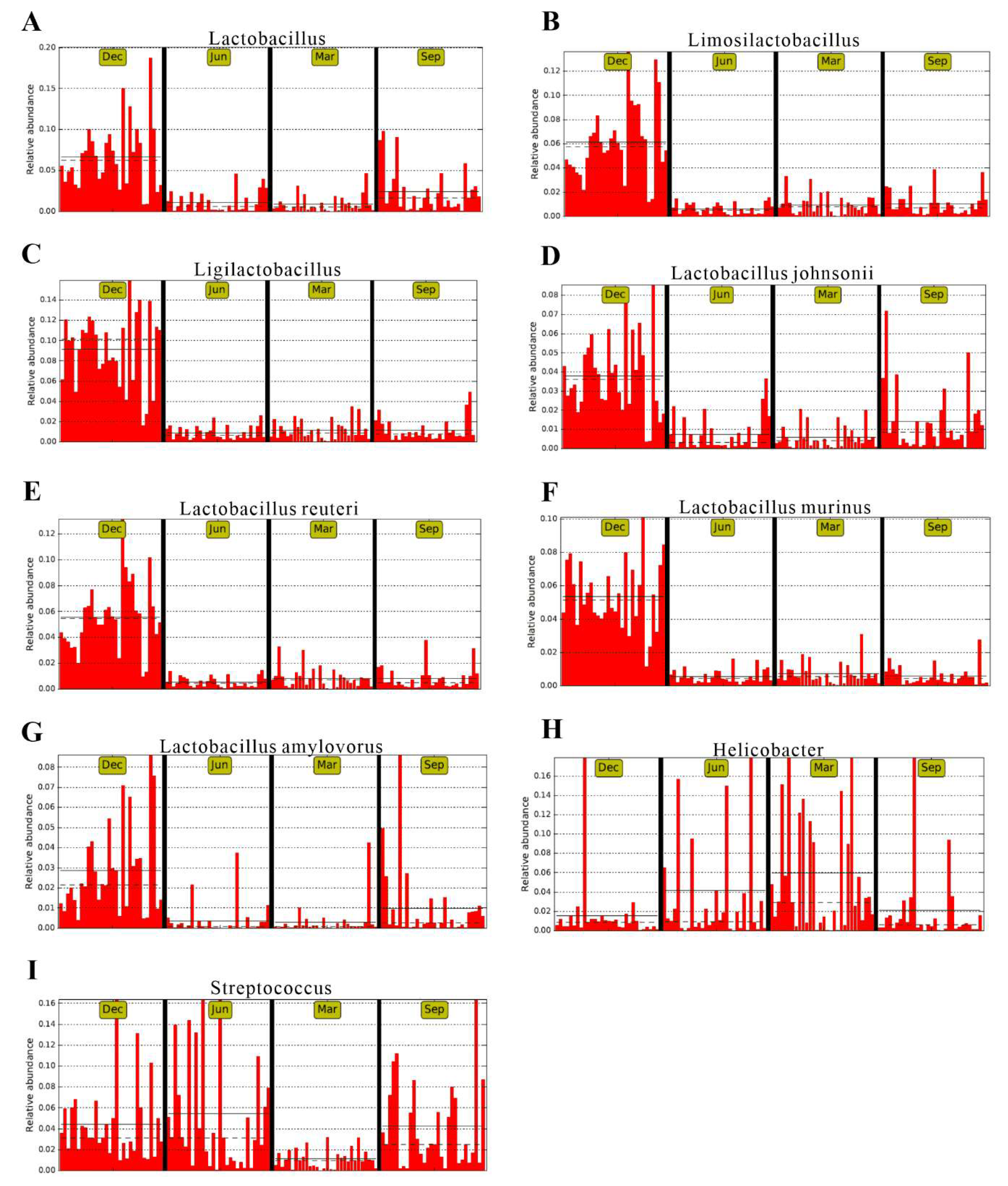
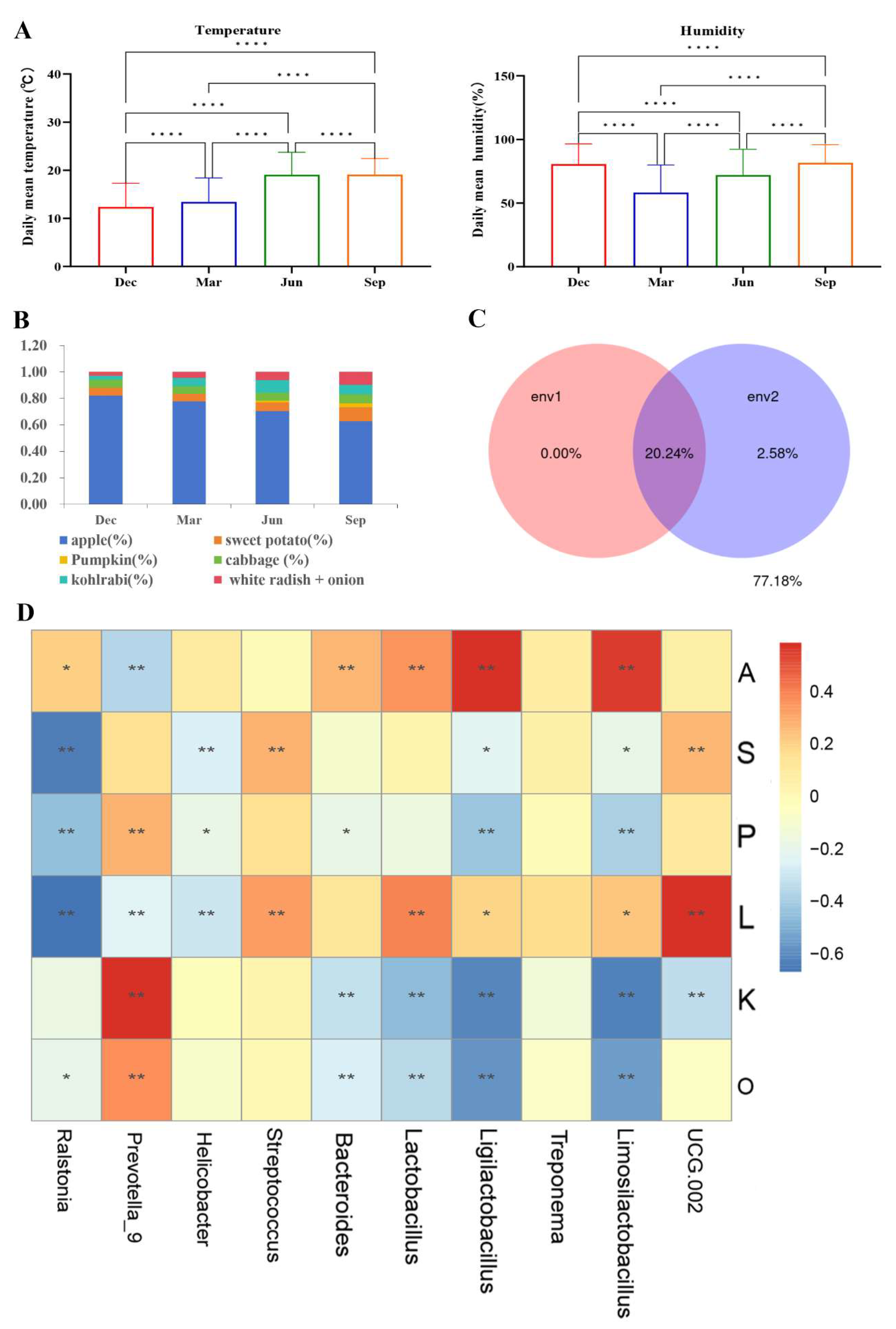
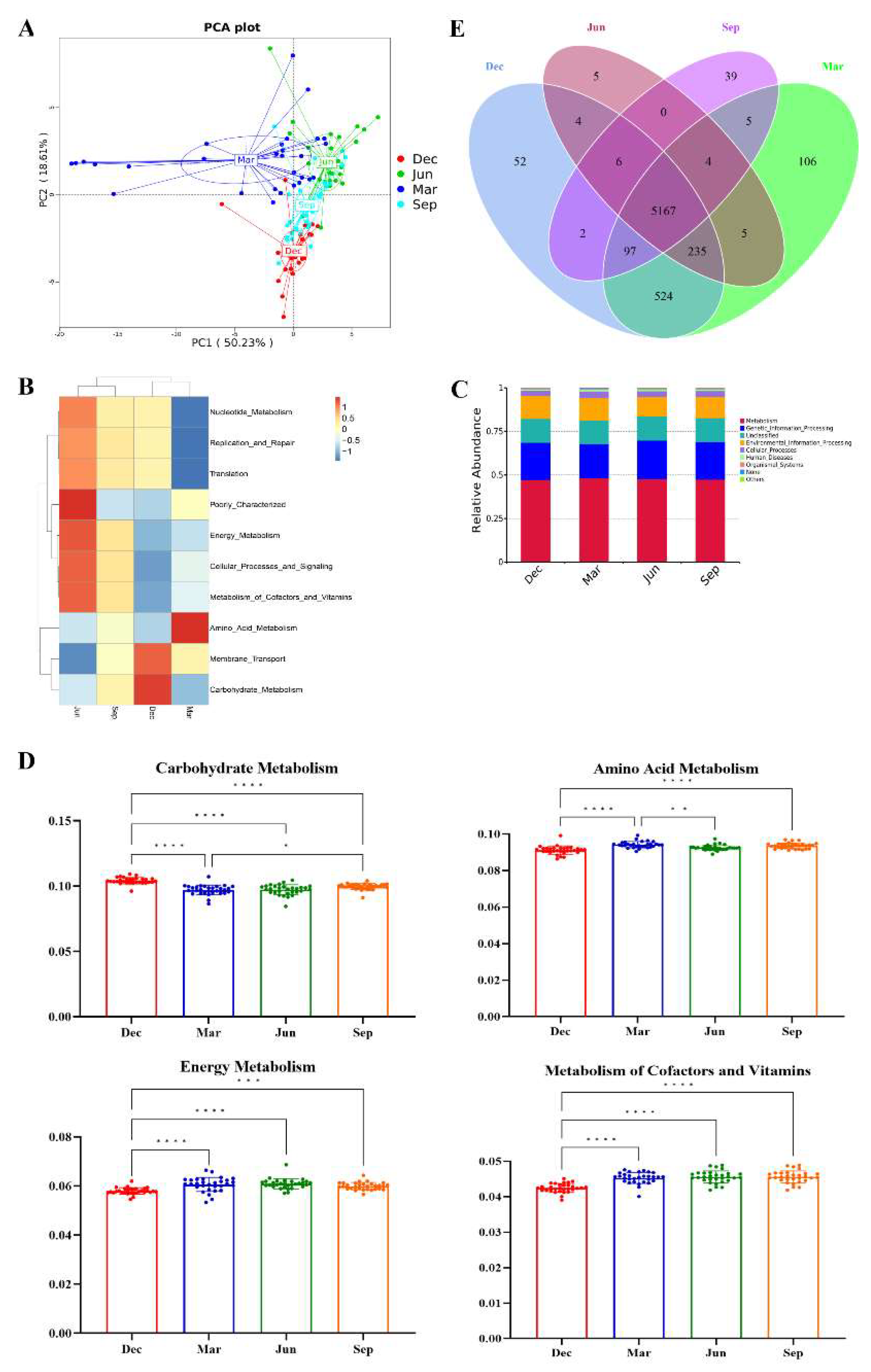
The seasonal variations that occur in the gut microbiota of healthy adult rhesus monkeys kept in outdoor groups under conventional rearing patterns, and how these variations are affected by environmental variables, are relatively poorly understood. In this study, we collected 120 fecal samples from 30 adult male rhesus monkeys kept in outdoor groups across four seasons and recorded the temperature and humidity of the housing facilities as well as the proportions of fruit and vegetables in their diet. 16S rRNA sequencing analysis showed that the alpha diversity of the gut microbiota of the rhesus monkeys was higher in winter and spring than in summer and autumn. Principal coordinate analysis (PCoA) also revealed significant seasonal differences in the structure and function of the gut microbiota in the rhesus monkeys. The phyla Firmicutes and Bacteroidetes and the genus Prevotella 9 were the significantly dominant groups in all 120 fecal samples from the rhesus monkeys. Linear Discriminant Analysis (LDA) Effect Size (LEfSe) analysis (LDA > 4) indicated that, at the phylum level, Firmicutes was significantly enriched in winter, Bacteroidetes was significantly enriched in summer, and Proteobacteria and Campylobacter were significantly enriched in spring. At the genus level, Helicobacter and Ralstonia were significantly enriched in spring; Prevotella 9, Streptococcus, and Prevotella were significantly enriched in summer, and UCG_005 was significantly enriched in autumn. The beneficial genera Lactobacillus, Limosilactobacillus, and Ligilactobacillus, and the beneficial species Lactobacillus johnsonii, Lactobacillus reuteri, Lactobacillus murinus, and Lactobacillus amylovorus, all showed the same seasonal trend, namely, their average relative abundance was significantly higher in winter than in other seasons. Compared with other seasons, carbohydrate metabolic function was significantly upregulated in winter (p < 0.01), amino acid metabolic function was relatively increased in spring, and energy metabolic function and the metabolic function of cofactors and vitamins were significantly downregulated in winter and relatively upregulated in summer. Variance partitioning analysis (VPA) and redundancy analysis (RDA) showed that the proportions of fruits and vegetables in the diet, but not climatic factors (temperature and humidity), significantly influenced the seasonal changes in the gut microbiota. These variations were related to changes in the proportions of fruits and vegetables. This study provides new evidence relating to how external environmental factors affect the intestinal environment of rhesus monkeys.

Keywords: 
;  ;  ;  

1. Introduction

The digestive tract of animals harbors a vast bacterial ecosystem that is in a delicate balance with the host environment. The host relies on the microbial community to degrade dietary fibers and other food components, thus playing a vital role in the host’s metabolism, immunity, and defenses against pathogens, among other physiological activities [1-3]. Conversely, the composition of the gut microbiota is influenced by factors such as host genetics, diet, seasons, age, social environment, antibiotic use, digestive tract structure, and growth stages [4-6]. Seasonal variations in gut microbial composition in animals are largely driven by seasonal shifts in diet [7,8]. Moreover, several longitudinal studies have suggested that gut microbial composition is not stable over time and is influenced by environmental fluctuations. In mammals such as black howler monkeys, white-faced capuchins, plateau pika, and musk, the gut microbiota exhibits seasonal dynamics [7,9-12], as does that of many fish [13], amphibians [14], and insects [15]The composition of the gut microbiota of non-human primates is significantly affected by lifestyle disruptions, such as long-term captive breeding, which represents a dietary alteration compared with wild populations [16]. While seasonal changes in the composition of the gut microbiome of animals are mainly driven by seasonal changes in diet, they can also be indirectly influenced by seasonal climatic fluctuations, such as changes in humidity and air temperature [17-19].
Rhesus macaques are closely related to humans[20]. Indeed, 93% of rhesus macaque genes are homologous to human genes, and the two species also share similar tissue structures and physiological and metabolic characteristics. Rhesus macaques have been widely used in research on brain function, cognition, neuropsychiatric diseases, stem cells, infectious disease mechanisms, and vaccine development[21]. With the advancement of biological and medical research, studies increasingly rely on the use of nonhuman primates, such as the rhesus macaque. The artificial breeding of rhesus macaques not only provides experimental animals with clear genetic backgrounds and stable physiological and biochemical indexes for medical biological research but also plays an important role in the protection of the ecological environment. Importantly, it is crucial to formulate suitable dietary plans and create appropriate environmental conditions in the artificial breeding of rhesus macaques. In artificial feeding, the diet usually consists of compound feed along with fruits and vegetables. Given that the compound feed is generally fixed, the choice and proportion of the fruit and vegetable feed assume special relevance. Monkeys are known to prefer food that is slightly sweet and soft and their intestines are well-suited to digesting foods rich in carbohydrates[22]. The main fruit in the diet is apples, while the vegetable component primarily comprises root vegetables such as sweet potatoes, pumpkins, cabbage, kohlrabi, white radishes, and onions, which, together, meet the nutritional needs and dietary habits of the macaques. However, how to scientifically and reasonably balance fruit and vegetable proportions is unclear, and requires further in-depth exploration. In artificial feeding and breeding, besides optimizing the dietary structure, it is equally important to maintain the appropriate environmental temperature and humidity. Both low and high temperatures can affect the reproductive function of the macaques[23]. Under high humidity, evaporation from the monkey’s body surface is inhibited, which frequently results in metabolic disorders, diminished resistance, and increased disease incidence. The incidence of intestinal diseases in macaques is relatively high during seasonal changes, with bacterial dysentery ranking first[24]. Some studies have suggested that insufficient absorption of folic acid and vitamin C from food can result in reduced resistance to dysentery, highlighting the need to provide rhesus monkeys with green fodder and fruits to enhance their resistance to this intestinal disease. The gut microbiome serves as a biological indicator of the health and welfare of rhesus monkeys. Nevertheless, how the seasonal changes in the intestinal flora of outdoor, group-housed rhesus monkeys maintained under conventional feeding patterns are affected by the proportion of fruits and vegetables provided or by temperature and humidity remains unclear. Therefore, discerning the changes in the diversity and composition of the gut microbiome of outdoor, group-housed macaques in the different seasons is crucial for assessing the suitability of the current feeding environment and diet, as well as for understanding whether these changes can affect the health of the animals.
In this study, we monitored 30 outdoor, group-housed adult male rhesus monkeys aged 10 to 15 years, The mean body weight was 11.35 ± 2.27 kg in winter, 10.57 ± 2.31 kg in spring, 10.47 ± 2.42 kg in summer, and 10.87 ± 2.62 kg in autumn. Feces were sampled from 30 individuals and the ratios of fruits and vegetables and the climatic conditions at the sampling site were surveyed over four seasons. Fecal microbiota profiling was conducted using 16S rRNA sequencing. Alpha diversity, dominant bacterial taxa, and microbial function were compared among the different seasons. The effect of environmental factors on gut microbial composition was evaluated. Finally, seasonal variations in gut microbial functions and the correlations between gut microbial indicators and environmental factors were assessed. The aim of this work was to identify differences in the composition and functional profiles of the gut microbiota in rhesus monkeys across the four seasons and determine the contribution of the proportions of fruits and vegetables provided in the diet to these differences. This study not only provides important information for understanding the seasonal changes and environmental adaptations in the gut microbiota in adult rhesus macaques raised in outdoor groups under artificial feeding conditions but also forms a basis for optimizing the selection and proportion control of fruits and vegetables in artificial diets. Our findings further address gaps in current domestic regulations and standards for laboratory primate feeds, which are limited to requirements for compound feeds, fruits and vegetables, and green fodder. As such, they lack objective evaluation criteria for the inclusion of fruits and vegetables in the diets of rhesus macaques, which may negatively affect the health and welfare of these animals.

2. Materials and Methods

2.1. Experimental Animals

Thirty healthy adult male rhesus macaques aged 10 to 15 years (Table 1) were weighed once at a fixed time each season. The mean body weight was 11.35 ± 2.27 kg in winter, 10.57 ± 2.31 kg in spring, 10.47 ± 2.42 kg in summer, and 10.87 ± 2.62 kg in autumn. There were no significant differences in body weight among the four seasons (p > 0.05) (Figure 1). The macaques were kept in the Laboratory Animal Center of Kunming Institute of Zoology, Chinese Academy of Sciences. The housing conditions and animal care were accredited by the Association for the Assessment and Accreditation of Laboratory Animal Care International (AAALAC) and were approved by the China National Accreditation Service for Conformity Assessment (CNAS). Briefly, the monkeys were provided standard sanitation, an adequate and regular diet, and a stable social structure. All macaques were housed in an outdoor monkey house and were not subjected to unnecessary manual intervention. Daily health checkups were performed by veterinarians. None of the 30 macaques exhibited diarrhea or other clinical symptoms and none received any medication or probiotics. They were also tested and found to be negative for simian retrovirus D (SRV), simian immunodeficiency virus (SIV), simian T-lymphotropic virus 1 (STLV-1), Cercopithecine Herpesvirus Type 1 (BV), and Mycobacterium tuberculosis (TB), as well as Salmonella and Shigella infection. The experiments were conducted at the Laboratory Animal Center of Kunming Institute of Zoological Research, Chinese Academy of Sciences [SYXK(Dian)K2022-0009], and this study was approved by the Ethics Committee for Animal Experiments of Kunming Institute of Zoological Research, Chinese Academy of Sciences (Ethics Approval No.PE-2021-07-002). The experiments were in accordance with the principles of the 3Rs.

2.2. Fecal Sample Collection

From December 2022 to September 2023, rectal swabs were collected from each of the 30 adult macaques, once in each of the four seasons. All the swabs were marked at 2.2 cm from the tip and then inserted into the rectum up to the marked line. The collected samples are placed in cryovials, rapidly frozen in liquid nitrogen, and stored at −80 °C for testing. Collections were made in December (winter), March (spring), June (summer), and September (autumn).

2.3. Environmental Factors

The Laboratory Animal Center of the Kunming Institute of Zoology, Chinese Academy of Sciences, is located in Huahongdong, in the western suburbs of Kunming, Yunnan, at an elevation of approximately 2,186 meters. The temperature and humidity in the breeding facilities are monitored by environmental sensors. The diet of the monkeys consisted of compound feed and fruit and vegetable feed at a ratio of 45% to 55%, respectively. All macaques were given the same compound feed with the following nutritional composition: crude protein 20.97%, crude fat 5.50%, crude fiber 3.30%, crude ash 6.20%, calcium 1.00%, and phosphorus 0.63%. The fruit and vegetable feed included seven types of food that were uniformly diced and mixed in different proportions according to the season—winter: 82% apple, 6% sweet potato, 0% pumpkin, 6% cabbage, 3% kohlrabi, and 3% white radish + onion; spring: 78% apple, 6% sweet potato, 0% pumpkin, 6% cabbage, 6% kohlrabi, and 4% white radish + onion; summer: 70% apple, 7% sweet potato, 1% pumpkin, 6% cabbage, 10% kohlrabi, and 6% white radish + onion; and autumn: 62% apple, 11% sweet potato, 3% pumpkin, 7% cabbage, 7% kohlrabi, and 10% white radish + onion.

2.4. DNA Extraction and 16S rRNA Gene Sequencing

Microbial community genomic DNA was extracted from all the fecal samples using the E.Z.N.A. soil DNA Kit (Omega Bio-Tek, Norcross, GA, USA) according to the manufacturer’s instructions. DNA concentration and purity were assessed on 1% agarose gels and the DNA was diluted to 1 ng/μL with sterile water. The V3-V4 hypervariable region of the bacterial 16S rRNA gene was amplified with the primer pair 341F (5′-CCTAYGGGRBGCASCAG-3′) and 806R (5′-GGACTACHVGGGTWTCTAAT-3′) using a Bio-Rad T100 gradient PCR thermocycler. PCR runs were carried out in a 30-μL reaction volume containing 15 μL of Phusion High-Fidelity PCR Master Mix (New England Biolabs), 0.2 μM each of forward and reverse primer, and 10 ng of template DNA. Thermal cycling consisted of an initial denaturation at 98 °C for 1 min, followed by 30 cycles of denaturation at 98 °C for 10 s, annealing at 50 °C for 30 s, and elongation at 72 °C for 30 s, with a final extension at 72 °C for 5 min. The same volumes of 1× loading buffer (containing SYB green) and PCR products were mixed and subjected to 2% agarose gel electrophoresis for band detection. The PCR products were mixed in equidensity ratios and then purified using a GeneJET Gel Extraction Kit (Thermo Fisher Scientific, MA, United States). Sequencing libraries were generated using a NEB Next Ultra DNA Library Prep Kit for Illumina (New England Biolabs, United States) following the manufacturer’s recommendations, and index codes were added. Library quantification and qualification were performed on a Qubit 2.0 Fluorometer (Thermo Fisher Scientific, MA, United States) and an Agilent Bioanalyzer 2100. Finally, the library was sequenced on the Illumina HiSeq platform, yielding 250-bp paired-end reads (Novogene Co., Ltd. Beijing)

2.5. Data Analysis

2.5.1. Data Quality Control

The paired-end reads were assigned to samples based on their unique barcodes, followed by the truncation of barcodes and primer sequences. The whole process was performed in Python (v.3.6.13) and the adapters and primer sequences were removed using cutadapt (v.3.3). The reads were merged using FLASH (v.1.2.11, http://ccb.jhu.edu/software/FLASH/) [25], generating raw reads. Quality filtering of the raw reads was performed using fastp (v.0.23.1) software to obtain high-quality clean tags[26]. These were then compared against reference databases (SILVA database [16S], https://www.arb-silva.de/; Unite database [ITS], https://unite.ut.ee/) to detect chimeric sequences. Effective tags were obtained by removing the chimeric sequences with the vsearch package (v.2.16.0, https://github.com/torognes/vsearch).

2.5.2. Bioinformatics Analysis

All data analysis was performed on the NovoMagic cloud-based platform (https://magic.novogene.com/customer/main#/homeNew). In brief, all effective tags were denoised using DADA2, yielding Amplicon Sequence Variants (ASVs). Species annotation was then performed based on the clustered ASV sequences using the SILVA database (release 138.1). Based on the ASV clustering results, abundance (expressed as mean ± standard deviation) and alpha and beta diversity indices were calculated using Qiime 1.9.1 software.
Principal coordinate analysis (PCoA) was used to analyze the beta diversity among the different groups using the “vegan” package in R software. The Bray–Curtis distance was used to calculate the similarities between samples at the operational taxonomic unit (OTU) level. Analysis of similarities (ANOSIM), a non-parametric statistical test, was employed to test for intergroup differences using R software (“anosim” function in the “vegan” package). The metabolic functions of the bacterial communities were predicted using Phylogenetic Investigation of Communities by Reconstruction of Unobserved States (PICRUSt) software based on the Kyoto Encyclopedia of Genes and Genomes (KEGG) databases using OTU species annotation and abundance information. Moreover, the Wilcoxon rank-sum test was used to analyze the seasonal differences in metabolism-related functions and the dominant bacteria in all the groups. The linear discriminant analysis effect size (LEfSe) was used to identify taxa with significant differences in abundance among the 4 seasons at various levels. To determine statistical significance, an LDA score greater than 4 and a p-value less than 0.05 were used. Using a distance-based linear model, redundancy analysis (RDA) and variance partitioning analysis (VPA) were employed to measure the effects of external factors on the gut microbiota, while Spearman’s correlation was applied to analyze the mutual relationships between species and environmental factors. The chi-square test and Fisher’s exact test were used to assess the differences in the unique microbiota.

3. Results

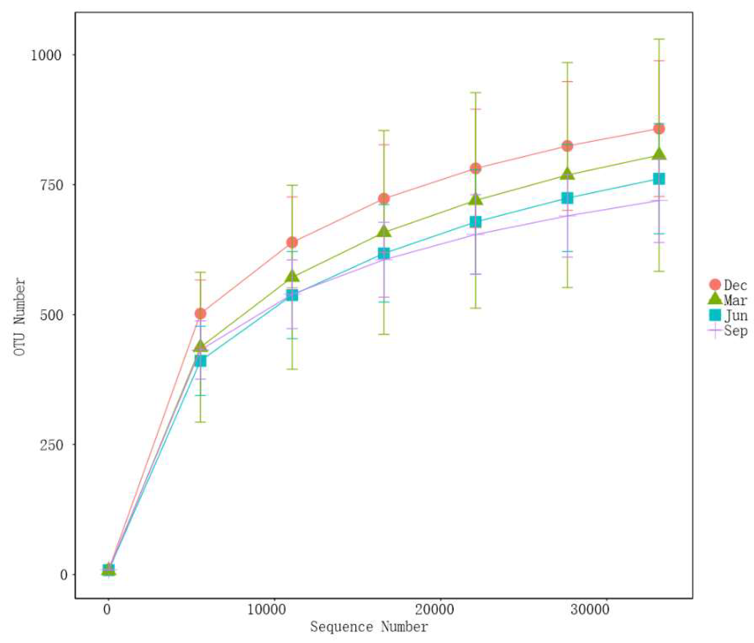
3.1. Assessment of Sequencing Data

A total of 9,647,634 high-quality clean reads (80,397 per sample) were obtained in all the samples. The rarefaction curves for Sobs and Shannon indexes at the OTU level gradually leveled off with increasing sequencing depth (Supplementary Figure 2). These results indicated that the sequencing depth was sufficient, as each fecal sample had enough OTUs to reflect the maximum level of bacterial diversity.
Following DADA2-based denoising, the sequences were clustered at 100% similarity, resulting in a total of 5,320 ASVs. These ASVs were annotated against the SILVA database (release 138), leading to the identification of 39 phyla, 78 classes, 182 orders, 309 families, and 666 genera.

3.2. Gut Microbiota Composition in Adult Rhesus Macaques Across Seasons

At the phylum level, Bacteroidetes and Firmicutes were dominant in different seasons, with average relative abundances of 42.85 ± 16.13% and 41.80 ± 18.09%, respectively, followed by Campylobacterota (3.66%) and Spirochaetota (1.80%) were also dominant (relative abundance >1%) (Figure 3A). The other six phyla were Proteobacteria, Actinobacteria, Desulfobacterota, Fusobacteriota, Acidobacteriota, and Verrucomicrobia. The relative abundance of the top10 phyla accounted for the majority (99%) of the detectable reads in all samples. At the genus level, Prevotella 9 (29.09%) and Ralstonia (4.74%) showed the greatest relative abundance. The other genera displaying average relative abundances greater than 1% (Figure 2C) were Faecalibacterium (3.90%), Streptococcus (3.82%), Helicobacter (3.43%), Prevotella (3.43%), Ligilactobacillus (3.08%), Lactobacillus (2.77%), UCG-002 (2.51%), Alloprevotella (2.04%), Limosilactobacillus (2.18%), UCG-005 (1.98%), and Treponema (1.22%) (Figure 3B). In summary, the dominant genera in rhesus monkeys across the four seasons belonged to the phyla Firmicutes and Bacteroidetes, with Prevotella 9 being a key microorganism closely related to a diet rich in dietary fiber.

3.3. Analysis of the Seasonal Differences in the Gut Microbiota

3.3.1. Seasonal Variation in Alpha Diversity

Shannon and Chao1 indices were employed to evaluate alpha diversity while the Wilcoxon rank-sum test was used to assess the significance of seasonal differences. We noted that the Shannon index of rhesus monkeys was significantly lower in summer than in autumn and winter (p < 0.001) (Figure 4A). Meanwhile, the Chao1 index was significantly higher in spring than in summer and autumn (p < 0.05) (Figure 4B). Overall, the alpha diversity of the gut microbiota in rhesus monkeys was higher in winter and spring than in autumn and summer.

3.3.2. Seasonal Variations in Beta Diversity

The Bray-Curtis distance algorithm was used to calculate the differences between rhesus monkey fecal samples in different seasons. ANOSIM was applied to test whether the differences between samples from different seasons were significantly greater than those within the same season. PCoA analysis showed that samples from different seasons formed distinct clusters, while samples from the same season clustered closely together, with a clear separation between winter and summer samples. This indicated that there were significant seasonal variations in the composition of the gut microbiota of rhesus monkeys (R = 0.4728, p = 0.001) (Figure 4C). ANOSIM results indicated that the differences between samples from different seasons were significantly greater than the differences between samples from the same season, with significant differences between summer and winter (R = 0.806, p = 0.001) and between spring and winter (R = 0.5899, p = 0.001) being noted (Table 2).

3.3.3. Seasonal Variations in Microbial Communities

LEfSe analysis (LDA threshold of 4) was used to identify taxa showing significant seasonal variations in abundance, with four groups being identified at the phylum level. Compared with that in other seasons, Firmicutes was significantly enriched in winter (LDA > 4, p < 0.05), Bacteroidetes was significantly enriched in summer, and Proteobacteria and Campylobacter were significantly enriched in spring. At the genus level, nine genera showed marked differences in relative abundance. Among these, Lactobacillus, Limosilactobacillus, and Ligilactobacillus were enriched in winter; Helicobacter and Ralstonia were enriched in spring; Prevotella 9, Streptococcus, and Prevotella were enriched in summer; and UCG_005 was enriched in autumn. At the species level, eight species showing significant differences were identified. These were Lactobacillus johnsonii, Lactobacillus reuteri, Lactobacillus murinus, Lactobacillus amylovorus, and Lactobacillus salivarius, which were enriched in winter; Helicobacter fennelliae and Ralstonia pickettii, which were enriched in spring; and Streptococcus lutetiensis, which was enriched in summer (Figure 5). Based on the Wilcoxon rank-sum test, the ratio of Firmicutes to Bacteroidetes (F/B ratio) was significantly higher in winter than in other seasons.

3.4. Analysis of Beneficial and Harmful Microorganisms

LEfSe analysis was used to compare the abundances of the differential taxa across different seasons. The beneficial bacterial genera (Lactobacillus, Limosilactobacillus, Ligilactobacillus) showed consistent seasonal changes (Figure 6A–C), with significantly higher average relative abundance in winter than in other seasons. The species Lactobacillus johnsonii, Lactobacillus reuteri, Lactobacillus murinus, and Lactobacillus amylovoru showed the same seasonal variation trend (Figure 6D–G). The main genera of conditionally pathogenic bacteria present in the intestinal tract of rhesus macaques are Streptococcus and Helicobacter. Here, we observed that the average relative abundance of Helicobacter in spring was higher than in other seasons (Figure 6H), while that of Streptococcus was higher in summer than in other seasons (Figure 6I).
In the feeding and management of rhesus macaques in this study, Salmonella, Shigella, and Clostridium difficile, common diarrhea-causing pathogenic bacteria in rhesus macaques, were not detected within the macaque population. The main pathogenic genera identified in the population were Pseudomonas, Campylobacter, Vibrio, and Escherichia–Shigella. Campylobacter was significantly more frequently detected in winter (100%, 30/30) than in autumn (100%, 30/30). Pseudomonas was significantly more prevalent in winter (100%, 30/30) and spring (80%, 24/30) than in summer (10%, 3/30) and autumn (6.67%, 2/30) (Table 3).

3.5. The effects of environmental factors on the seasonal variation in the gut microbiota of rhesus macaques

Temperature was significantly higher in summer and autumn than in spring and winter, while humidity was significantly higher in autumn and winter than in spring and summer (Figure 7A). The highest percentages of apples, cabbage, sweet potatoes, and white radishes with onions were provided in winter, summer, and autumn, respectively, and pumpkins were not given in winter and spring (Figure 7B).
A VPA was performed to quantify the relative contributions of the different environmental factors (climatic and dietary) to changes in the bacterial community structure using the “varpart” procedure. Climatic factors (temperature and humidity) alone explained 0% of the observed variation, while dietary factors (fruits and vegetables) alone explained 2.58% of the observed variation. A combination of both factors explained 20.24% of the observed variation (Figure 7C). This indicated that while these climatic factors alone could not explain the changes in the ecological community, they made a significant contribution to the observed changes when combined with dietary (fruit and vegetable-related) factors. The results of the RDA analysis also showed that temperature (r2 = 0.84, P = 0.58) and humidity (r2 = 0.99, P = 0.13) were not significantly correlated with the microbial community. Spearman’s correlation analysis was conducted to determine the relationship between the types of fruits and vegetables and the relative abundances of the gut microbiota (Figure 7D). The 10 most dominant genera were selected for this assessment. Apples displayed a positive correlation with the relative abundance of the genera Lactobacillus, Limosilactobacillus, and Ligilactobacillus and a negative correlation with the relative abundance of the genus Prevotella 9. The abundance of Prevotella 9 showed a positive correlation with kohlrabi, pumpkin, and white radish with onion.

3.6. Seasonal Differences in Gut Microbial Functions

Principal Component Analysis (PCA) was performed based on the statistical analysis of abundance from database-based functional annotations. The results showed that samples from different seasons formed separate clusters, while samples from the same season clustered together (Figure 8A). A clustering heatmap based on the relative abundance in level-2 KEGG pathways showed that monkeys that were not fed pumpkin but were fed a high proportion of apples in winter and spring were clustered into one group, while those that received fewer apples in summer and autumn were clustered into another group (Figure 8B). Functional prediction analysis in the KEGG database showed that the gut microbiota in outdoor-housed rhesus monkeys was mainly associated with membrane transport, translation, replication and repair, carbohydrate metabolism, amino acid metabolism, energy metabolism, and cofactors and vitamin metabolism (Figure 8C). The KEGG analysis showed that the magnitude of the four major metabolism-related functions of the gut microbiota of rhesus monkeys displayed seasonal differences. Overall, carbohydrate metabolism was significantly higher in winter than in the other seasons (p < 0.01), while amino acid metabolism was relatively higher in spring than in the other seasons. Energy metabolism and cofactors and vitamin metabolism were significantly lower in winter and relatively higher in summer than in spring or autumn (Figure 8D). The gut bacteria of the rhesus monkeys shared 4,574 KEGG Orthology (KO) terms over the seasons. In total, 87, 205, 13, and 40 KO terms were unique to December, March, June, and September, respectively (Figure 8E).

4. Discussion

In this study, Firmicutes and Bacteroidetes were the significantly dominant phyla (relative abundance >90%) among the 120 fecal samples obtained during the four seasons from rhesus macaques raised in large outdoor cages, which was consistent with previous reports [27,28]. Firmicutes and Bacteroidetes were reported to be the dominant phyla in non-human primates, with proportions of 70.50% to 98.30%[4], respectively. Firmicutes promotes fiber degradation in food and converts cellulose to volatile fatty acids, thereby enhancing food digestion and growth and development[29]. In this study, we observed that among the top 30 bacterial genera in the gut flora of rhesus macaques, 10 belonged to the Firmicutes, which is associated with carbohydrate metabolism and cellulose digestion and absorption. Additionally, 7 were in the phylum Bacteroidetes, the members of which are primarily associated with the digestion and absorption of proteins and carbohydrates in food, while also promoting the development of the gastrointestinal immune system. Studies have indicated that a diet rich in fiber can enhance the abundance of Prevotella in the gut[30]. Most of the seven above-mentioned genera were Prevotella, which may be related to the fact that the dietary structure of the rhesus macaque diets in this study was plant-based (fruits and vegetables). At the genus level, Prevotella 9, Ralstonia, Streptococcus, Helicobacter, Faecalibacterium, Prevotella, Ligilactobacillus, Lactobacillus, UCG-002, Alloprevotella, Limosilactobacillus, UCG-005, and Treponema were the most prevalent genera, all showing a relative abundance of >1%. Research has shown that Prevotella 9 breaks down sugars and carbohydrates, is a good source of vitamin B1, and can be used as a probiotic alternative to bacteria that break down sugars in food [30]. Faecalibacterium is a butyrate-producing bacterium that exerts a positive impact on human energy metabolism and possesses good anti-inflammatory characteristics[31]. UCG002 and UCG005 digest high-fiber foods and function as symbiotic bacteria in the gut[32]. The beneficial genera Ligilactobacillus, Lactobacillus, and Limosilactobacillus can inhibit the excessive growth of conditional pathogens in the gut by competing with them for adhesion sites as well as producing bacteriocins, organic acids, hydrogen peroxide, and other antimicrobial substances[33]. Their metabolic products are of great interest for their ability to regulate immune function, in addition to their anti-tumor activity, antibacterial properties, and preservative functions. Helicobacter is associated with diarrhea in rhesus monkeys and can cause disease under specific conditions, such as when the immune system is weakened or Helicobacter infection levels are high[34]. Additionally, the Ralstonia genus was detected in the rectal swabs of rhesus monkeys in our study, which aligns with the work of[35] which showed that this genus is present in the rectum of these macaques. Studies have indicated that this genus, which is widely found in nature, is an emerging pathogen, particularly in water sources, and can survive under poor nutritional conditions [11]. During the daily care and management of rhesus macaques, exposure to Ralstonia bacteria through the handling or ingestion of contaminated toys or food might allow the microbes to enter and colonize the macaque intestine. Accordingly, it is essential to strengthen the cleaning, disinfection, and hygiene management of fruits and vegetables in rhesus macaque rearing environments to reduce the health risks of potential pathogens such as Ralstonia.
PCoA analyses revealed that in rhesus monkeys, the gut microbial structure and composition vary significantly according to season, consistent with other reports [36,37]. Seasonal differences in the gut microbiome are closely related to food resources, dietary structure, nutrient utilization, and feeding pattern [38,39]. Here, we found that the alpha diversity of the gut microbiota exhibited marked seasonal variations in rhesus monkeys, as determined by the Wilcoxon rank-sum test. Overall, the alpha diversity of the gut microbiota was higher in winter and spring than in autumn and summer. It has been shown that an increase in alpha diversity leads to a more complex and stable intestinal microbiota composition, thereby enhancing resistance to external interference and adaptability, which is beneficial to the host’s health[40]. Changes in alpha diversity are associated with a variety of diseases [41]. Therefore, the observed increase in alpha diversity in the gut microbiota of the rhesus monkey in winter and spring may improve the resistance to, and reduce the influence of, adverse environmental factors, as well as promote the intake of fiber-rich food and nutrient absorption and utilization in the cold season. Increased bacterial diversity in the gut microbiota in winter and spring may be due to lower temperatures, higher humidity levels, and greater apple intake. Plateau pikas show an increase in alpha diversity in winter, influenced by a combination of low winter temperatures, drought, and dietary fiber content. It has also been suggested that the gut microbiota of animals can be influenced by physiological factors such as reproduction and metabolism, which also show seasonal variation [42]. For example, in the cave rat, the Chao1 index increases between estrus and non-estrus stages in response to changes in gut microbial structure and function[42].As macaques are characterized by seasonal reproduction [21], their reproductive cycles may affect their gut bacterial diversity.
LEfSe analysis showed that Firmicutes was significantly enriched in winter, while Bacteroidetes was significantly enriched in summer. In addition, the F/B ratio was markedly higher in winter than in other seasons. A high F/B ratio can reflect weight gain in birds [43]and mammals [44]. Here, we observed that the body weight of rhesus macaques was greater in winter than in the other seasons; however, the difference was not significant. One study showed that the abundance of Bacteroides in rats fed whole apples was lower than that in animals given the control diet[45]. In this study, although the proportion of apples provided in the diet was lower in summer than in winter, the abundance of Bacteroidetes was lower in winter than in summer. This suggested that the significant enrichment of Bacteroidetes observed in summer was related to apple intake. The relative abundance of Firmicutes in winter was higher than that in other seasons, which aided the adaptation of rhesus monkeys to the cold season by promoting the decomposition of cellulose and hemicellulose in feed, carbohydrate metabolism, and nutrient digestion and absorption. In addition, we found that Proteobacteria and Campylobacter were significantly enriched in spring relative to other seasons. The marked enrichment of Campylobacter in the intestinal tract of rhesus macaques was likely related to the increase in the abundance of the genus Helicobacter. The phylum Proteobacteria includes various pathogens, such as Escherichia coli and Salmonella. An increase in the relative abundance of Proteobacteria under conditions of intestinal dysbiosis often leads to diarrhea. Campylobacter spp. within the phylum Campylobacter is closely associated with diarrhea. However, the rhesus monkeys in this study were all healthy, indicating that healthy rhesus monkeys harbor varying levels of Proteobacteria and Campylobacter. The abundance of these bacteria is closely related to the immune status of animals. Diarrhea likely occurs only when the levels of bacteria within these phyla increase abnormally and immunity is compromised. In addition, during winter and spring, temperature changes and other environmental stresses pose thermoregulatory challenges for rhesus macaques. Under such conditions, bacteria that are not predominant in the intestinal tract, such as Proteobacteria and Campylobacter, proliferate, presumably due to trade-offs between adaptation to unfavorable environments and immune regulation. Additionally, beneficial genera such as Lactobacillus, Limosilactobacillus, and Ligilactobacillus, along with beneficial species such as Lactobacillus johnsonii, Lactobacillus reuteri, Lactobacillus murinus, and Lactobacillus amylovorus, showed seasonal variations and were significantly enriched in winter. The four beneficial species possess genes encoding mucus-binding proteins, which allow them to adhere to intestinal epithelial cells [22]. This suggests that the effective adhesion of probiotics to the gut is a fundamental mechanism underlying their actions, including immunomodulation of the host and inhibition of conditionally pathogenic bacteria. The intestines of macaques harbor beneficial probiotics, which help maintain a stable gut microbiome and alleviate intestinal inflammation. Bacteria of the Lactobacillus genus, which are prevalent in rhesus monkeys, are considered potential probiotics that can prevent diarrhea. Moreover, the highest contents of probiotic Lactobacillus and related species were observed in the winter, indicating that rhesus monkeys are in a state of constant self-regulating dynamic balance. When conditionally pathogenic bacteria proliferate, beneficial bacteria compete with them to maintain a healthy internal environment.
The function prediction analysis based on the KEGG database showed that the gut microorganisms in rhesus monkeys are mainly involved in carbohydrate metabolism, amino acid metabolism, energy metabolism, and cofactor and vitamin metabolism. These magnitudes of these metabolic functions also displayed seasonal differences and were significantly correlated with core bacteria phyla and genera. Meanwhile, the seasonal changes in the core bacteria were also closely associated with the seasonal function changes.
The VPA showed that the proportion of fruits and vegetables significantly affected the microbial community, while the impact of climatic factors on microbial community changes was mixed. Climatic factors alone could explain 0% of the variation in the gut microbiota of rhesus monkeys, while the combined effect of climatic and dietary compositional factors explained 20.24% of the observed variance. In addition, only a few of the floral constituents exhibited a substantial change in abundance between the coldest and hottest seasons. This implies that changes in the proportion of fruits and vegetables play an important role in driving rhesus macaque gut microbial communities. Climatic factors did not significantly affect gut microbial community structure, which may be related to both the fact that temperature and humidity in the four seasons remained within the range suitable for the healthy growth of rhesus monkeys and the good hygienic welfare of the rhesus macaques under captive conditions. In the present study, apples displayed a positive correlation with the relative abundance of the genera Lactobacillus, Limosilactobacillus, and Ligilactobacillus, and a negative correlation with the relative abundance of the genus Prevotella 9. The abundance of Prevotella 9 showed a positive correlation with kohlrabi, pumpkin, and white radish with onion. This is indicative of the potential of apples in promoting gut health. Apples are rich in nutrients such as sugars, organic acids, and vitamins, as well as dietary fiber. Studies have shown that the dietary fiber in apples promotes intestinal peristalsis and has laxative effects, while their abundant polyphenols effectively scavenge free radicals[46].These active ingredients help maintain a dynamic balance in the intestinal flora, thereby exerting health-promoting effects. [47]demonstrated that consuming two apples a day for 2 weeks increased the abundance of Bifidobacterium and Lactobacillus spp. in the feces of eight healthy adults. In addition, it has been shown that apple fiber isolate increases both fecal output (weight, frequency) in humans and rats [48] and gut transit time in humans, which may be helpful in constipation. In our study, the relative abundance of Lactobacillus, Limosilactobacillus, and Ligilactobacillus was higher in winter than in the other seasons, likely due to the increased intake of apples during this season. Our findings also suggested that under artificial feeding conditions with fixed compound feeds, different fruit and vegetable compositions and ratios have varying degrees of influence on the composition and function of the intestinal flora of rhesus macaques, which subsequently impacts their metabolism and health. This highlights the importance of a reasonable mix of fruits and vegetables. Their types and proportions can be flexibly adjusted according to seasonal changes to meet the nutritional needs and taste preferences of the macaques. Ensuring the hygiene and quality of these foods is also important for protecting the health of rhesus monkeys

Author Contributions

LL: conceived and designed the experiments, Funding acquisition, Resources, and Supervision, and Writing—review and editing. FZ: Conceptualization, Methodology, Investigation, Writing – original draft, Writing – review & editing. HZ and WX: Investigation, Methodology, and Validation. YH and WW: Conceptualization, Methodology, Investigation. ZZ and FZ: Investigation. QD: Conceptualization, Writing – review & editing, Supervision. XT: Conceptualization, Funding acquisition, Project administration, Resources, Methodology, Supervision, and Writing—review and editing. All authors have read and approved the final manuscript.

Funding

This research was funded by the National Key Research and Development Plan Program (2022YFF0710900 to L. L.), National Natural Science Foundation of China (82160923, 82374425, 82260929), Academician Expert Workstation of Yunnan Kunming (YSZJGZZ-2022063), Technology Talent and Platform Plan of Yunnan Province (202305AF150160).

Data Availability Statement

We encourage all authors of articles published in MDPI journals to share their research data. In this section, please provide details regarding where data supporting reported results can be found, including links to publicly archived datasets analyzed or generated during the study. Where no new data were created, or where data is unavailable due to privacy or ethical restrictions, a statement is still required. Suggested Data Availability Statements are available in section “MDPI Research Data Policies” at https://www.mdpi.com/ethics.

Conflicts of Interest

The authors declare no conflicts of interest.

References

  1. Shan, Y.; Lee, M.; Chang, E.B. The Gut Microbiome and Inflammatory Bowel Diseases. Annual Review of Medicine 2022, 73, 455–468. [Google Scholar] [CrossRef] [PubMed]
  2. Duan, J.; Yin, B.; Li, W.; Chai, T.; Liang, W.; Huang, Y.; Tan, X.; Zheng, P.; Wu, J.; Li, Y.; et al. Age-related changes in microbial composition and function in cynomolgus macaques. Aging (Albany NY) 2019, 11, 12080–12096. [Google Scholar] [CrossRef] [PubMed]
  3. Ni, Q.; He, X.; Zeng, B.; Meng, X.; Xu, H.; Li, Y.; Yang, M.; Li, D.; Yao, Y.; Zhang, M.; et al. Variation in Gut Microbiota of Captive Bengal Slow Lorises. Current Microbiology 2020, 77, 2623–2632. [Google Scholar] [CrossRef]
  4. Yildirim, S.; Yeoman, C.J.; Sipos, M.; Torralba, M.; Wilson, B.A.; Goldberg, T.L.; Stumpf, R.M.; Leigh, S.R.; White, B.A.; Nelson, K.E. Characterization of the Fecal Microbiome from Non-Human Wild Primates Reveals Species Specific Microbial Communities. Plos One 2010, 5. [Google Scholar] [CrossRef]
  5. Hao, W.-Z.; Li, X.-J.; Zhang, P.-W.; Chen, J.-X. A review of antibiotics, depression, and the gut microbiome. Psychiatry Research 2020, 284. [Google Scholar] [CrossRef]
  6. Ochman, H.; Worobey, M.; Kuo, C.H.; Ndjango, J.B.; Peeters, M.; Hahn, B.H.; Hugenholtz, P. Evolutionary relationships of wild hominids recapitulated by gut microbial communities. PLoS Biol 2010, 8, e1000546. [Google Scholar] [CrossRef] [PubMed]
  7. Amato, K.R.; Leigh, S.R.; Kent, A.; Mackie, R.I.; Yeoman, C.J.; Stumpf, R.M.; Wilson, B.A.; Nelson, K.E.; White, B.A.; Garber, P.A. The gut microbiota appears to compensate for seasonal diet variation in the wild black howler monkey (Alouatta pigra). Microb Ecol 2015, 69, 434–443. [Google Scholar] [CrossRef]
  8. Carey, H.V.; Walters, W.A.; Knight, R. Seasonal restructuring of the ground squirrel gut microbiota over the annual hibernation cycle. Am J Physiol Regul Integr Comp Physiol 2013, 304, R33–42. [Google Scholar] [CrossRef]
  9. Orkin, J.D.; Campos, F.A.; Myers, M.S.; Cheves Hernandez, S.E.; Guadamuz, A.; Melin, A.D. Seasonality of the gut microbiota of free-ranging white-faced capuchins in a tropical dry forest. Isme j 2019, 13, 183–196. [Google Scholar] [CrossRef]
  10. Fan, C.; Zhang, L.; Jia, S.; Tang, X.; Fu, H.; Li, W.; Liu, C.; Zhang, H.; Cheng, Q.; Zhang, Y. Seasonal variations in the composition and functional profiles of gut microbiota reflect dietary changes in plateau pikas. Integr Zool 2022, 17, 379–395. [Google Scholar] [CrossRef]
  11. Rajni, E.; Goel, P.; Sarna, M.K.; Jorwal, A.; Sharma, C.; Rijhwani, P. The genus Ralstonia: The new kid on the block. J R Coll Physicians Edinb 2023, 53, 44–52. [Google Scholar] [CrossRef] [PubMed]
  12. Jiang, F.; Gao, H.; Qin, W.; Song, P.; Wang, H.; Zhang, J.; Liu, D.; Wang, D.; Zhang, T. Marked Seasonal Variation in Structure and Function of Gut Microbiota in Forest and Alpine Musk Deer. Front Microbiol 2021, 12, 699797. [Google Scholar] [CrossRef] [PubMed]
  13. Karlsen, C.; Ottem, K.F.; Brevik Ø, J.; Davey, M.; Sørum, H.; Winther-Larsen, H.C. The environmental and host-associated bacterial microbiota of Arctic seawater-farmed Atlantic salmon with ulcerative disorders. J Fish Dis 2017, 40, 1645–1663. [Google Scholar] [CrossRef] [PubMed]
  14. Tong, Q.; Hu, Z.F.; Du, X.P.; Bie, J.; Wang, H.B. Effects of Seasonal Hibernation on the Similarities Between the Skin Microbiota and Gut Microbiota of an Amphibian (Rana dybowskii). Microb Ecol 2020, 79, 898–909. [Google Scholar] [CrossRef] [PubMed]
  15. Kesnerova, L.; Emery, O.; Troilo, M.; Liberti, J.; Erkosar, B.; Engel, P. Gut microbiota structure differs between honeybees in winter and summer. Isme Journal 2020, 14, 801–814. [Google Scholar] [CrossRef]
  16. Guo, W.; Mishra, S.; Wang, C.; Zhang, H.; Ning, R.; Kong, F.; Zeng, B.; Zhao, J.; Li, Y. Comparative Study of Gut Microbiota in Wild and Captive Giant Pandas (Ailuropoda melanoleuca). Genes (Basel) 2019, 10. [Google Scholar] [CrossRef]
  17. Bestion, E.; Jacob, S.; Zinger, L.; Di Gesu, L.; Richard, M.; White, J.; Cote, J. Climate warming reduces gut microbiota diversity in a vertebrate ectotherm. Nat Ecol Evol 2017, 1, 161. [Google Scholar] [CrossRef]
  18. Siles, J.A.; Margesin, R. Seasonal soil microbial responses are limited to changes in functionality at two Alpine forest sites differing in altitude and vegetation. Sci Rep 2017, 7, 2204. [Google Scholar] [CrossRef]
  19. Liu, H.; Li, Y.; Liang, J.; Nong, D.; Li, Y.; Huang, Z. Evaluation of Gut Microbiota Stability and Flexibility as a Response to Seasonal Variation in the Wild François' Langurs (Trachypithecus francoisi) in Limestone Forest. Microbiol Spectr 2023, 11, e0509122. [Google Scholar] [CrossRef]
  20. O'Sullivan, A.; He, X.; McNiven, E.M.; Hinde, K.; Haggarty, N.W.; Lönnerdal, B.; Slupsky, C.M. Metabolomic phenotyping validates the infant rhesus monkey as a model of human infant metabolism. J Pediatr Gastroenterol Nutr 2013, 56, 355–363. [Google Scholar] [CrossRef]
  21. Li, Y.; Wang, J.; Liu, Q.; Yang, F.; Chen, L.; Xu, H.; Jin, W.; Liu, Y.; Zhang, W.; Yang, F.; et al. Composition of the intestinal microbiota of infant rhesus macaques at different ages before and after weaning. Heliyon 2023, 9, e13915. [Google Scholar] [CrossRef] [PubMed]
  22. Bailey, M.T.; Coe, C.L. Intestinal microbial patterns of the common marmoset and rhesus macaque. Comp Biochem Physiol A Mol Integr Physiol 2002, 133, 379–388. [Google Scholar] [CrossRef] [PubMed]
  23. Baniel, A.; Amato, K.R.; Beehner, J.C.; Bergman, T.J.; Mercer, A.; Perlman, R.F.; Petrullo, L.; Reitsema, L.; Sams, S.; Lu, A.; et al. Seasonal shifts in the gut microbiome indicate plastic responses to diet in wild geladas. Microbiome 2021, 9, 26. [Google Scholar] [CrossRef] [PubMed]
  24. Breed, M.W.; Perez, H.L.; Otto, M.; Villaruz, A.E.; Weese, J.S.; Alvord, G.W.; Donohue, D.E.; Washington, F.; Kramer, J.A. Bacterial Genotype, Carrier Risk Factors, and an Antimicrobial Stewardship Approach Relevant To Methicillin-resistant Staphylococcus Aureus Prevalence in a Population of Macaques Housed in a Research Facility. Comp Med 2023, 73, 134–144. [Google Scholar] [CrossRef]
  25. Magoč, T.; Salzberg, S.L. FLASH: fast length adjustment of short reads to improve genome assemblies. Bioinformatics 2011, 27, 2957–2963. [Google Scholar] [CrossRef]
  26. Bokulich, N.A.; Subramanian, S.; Faith, J.J.; Gevers, D.; Gordon, J.I.; Knight, R.; Mills, D.A.; Caporaso, J.G. Quality-filtering vastly improves diversity estimates from Illumina amplicon sequencing. Nat Methods 2013, 10, 57–59. [Google Scholar] [CrossRef]
  27. Hu, X.; Liu, G.; Shafer, A.B.A.; Wei, Y.; Zhou, J.; Lin, S.; Wu, H.; Zhou, M.; Hu, D.; Liu, S. Comparative Analysis of the Gut Microbial Communities in Forest and Alpine Musk Deer Using High-Throughput Sequencing. Front Microbiol 2017, 8, 572. [Google Scholar] [CrossRef]
  28. Zhao, G.; Ma, T.; Tang, W.; Li, D.; Mishra, S.K.; Xu, Z.; Wang, Q.; Jie, H. Gut Microbiome of Chinese Forest Musk Deer Examined across Gender and Age. Biomed Res Int 2019, 2019, 9291216. [Google Scholar] [CrossRef]
  29. Wang, Y.; Cao, P.; Wang, L.; Zhao, Z.; Chen, Y.; Yang, Y. Bacterial community diversity associated with different levels of dietary nutrition in the rumen of sheep. Appl Microbiol Biotechnol 2017, 101, 3717–3728. [Google Scholar] [CrossRef]
  30. Xie, X.; Yang, C.; Guan, L.L.; Wang, J.; Xue, M.; Liu, J.X. Persistence of Cellulolytic Bacteria Fibrobacter and Treponema After Short-Term Corn Stover-Based Dietary Intervention Reveals the Potential to Improve Rumen Fibrolytic Function. Front Microbiol 2018, 9, 1363. [Google Scholar] [CrossRef]
  31. Martín, R.; Rios-Covian, D.; Huillet, E.; Auger, S.; Khazaal, S.; Bermúdez-Humarán, L.G.; Sokol, H.; Chatel, J.M.; Langella, P. Faecalibacterium: a bacterial genus with promising human health applications. FEMS Microbiol Rev 2023, 47. [Google Scholar] [CrossRef] [PubMed]
  32. Matulova, M.; Nouaille, R.; Capek, P.; Péan, M.; Delort, A.M.; Forano, E. NMR study of cellulose and wheat straw degradation by Ruminococcus albus 20. Febs j 2008, 275, 3503–3511. [Google Scholar] [CrossRef] [PubMed]
  33. O'Callaghan, J.; O'Toole, P.W. Lactobacillus: host-microbe relationships. Curr Top Microbiol Immunol 2013, 358, 119–154. [Google Scholar] [CrossRef]
  34. Compo, N.R.; Mieles-Rodriguez, L.; Gomez, D.E. Fecal Bacterial Microbiota of Healthy Free-Ranging, Healthy Corralled, and Chronic Diarrheic Corralled Rhesus Macaques (Macaca mulatta). Comp Med 2021, 71, 152–165. [Google Scholar] [CrossRef]
  35. Cui, Y.F.; Wang, F.J.; Yu, L.; Ye, H.H.; Yang, G.B. Metagenomic comparison of the rectal microbiota between rhesus macaques (Macaca mulatta) and cynomolgus macaques (Macaca fascicularis). Zool Res 2019, 40, 89–93. [Google Scholar] [CrossRef]
  36. Maurice, C.F.; Knowles, S.C.; Ladau, J.; Pollard, K.S.; Fenton, A.; Pedersen, A.B.; Turnbaugh, P.J. Marked seasonal variation in the wild mouse gut microbiota. Isme j 2015, 9, 2423–2434. [Google Scholar] [CrossRef] [PubMed]
  37. Trosvik, P.; de Muinck, E.J.; Rueness, E.K.; Fashing, P.J.; Beierschmitt, E.C.; Callingham, K.R.; Kraus, J.B.; Trew, T.H.; Moges, A.; Mekonnen, A.; et al. Multilevel social structure and diet shape the gut microbiota of the gelada monkey, the only grazing primate. Microbiome 2018, 6, 84. [Google Scholar] [CrossRef]
  38. Xue, Z.; Zhang, W.; Wang, L.; Hou, R.; Zhang, M.; Fei, L.; Zhang, X.; Huang, H.; Bridgewater, L.C.; Jiang, Y.; et al. The bamboo-eating giant panda harbors a carnivore-like gut microbiota, with excessive seasonal variations. mBio 2015, 6, e00022–00015. [Google Scholar] [CrossRef]
  39. Kartzinel, T.R.; Hsing, J.C.; Musili, P.M.; Brown, B.R.P.; Pringle, R.M. Covariation of diet and gut microbiome in African megafauna. Proc Natl Acad Sci U S A 2019, 116, 23588–23593. [Google Scholar] [CrossRef]
  40. Stoffel, M.A.; Acevedo-Whitehouse, K.; Morales-Durán, N.; Grosser, S.; Chakarov, N.; Krüger, O.; Nichols, H.J.; Elorriaga-Verplancken, F.R.; Hoffman, J.I. Early sexual dimorphism in the developing gut microbiome of northern elephant seals. Mol Ecol 2020, 29, 2109–2122. [Google Scholar] [CrossRef]
  41. Rogers, M.B.; Firek, B.; Shi, M.; Yeh, A.; Brower-Sinning, R.; Aveson, V.; Kohl, B.L.; Fabio, A.; Carcillo, J.A.; Morowitz, M.J. Disruption of the microbiota across multiple body sites in critically ill children. Microbiome 2016, 4, 66. [Google Scholar] [CrossRef] [PubMed]
  42. Wu, X.; Shang, Y.; Wei, Q.; Chen, J.; Zhang, H.; Chen, Y.; Gao, X.; Wang, Z.; Zhang, H. Gut Microbiota in Dholes During Estrus. Front Microbiol 2020, 11, 575731. [Google Scholar] [CrossRef]
  43. Zhang, L.; Li, J.; Yun, T.T.; Qi, W.T.; Liang, X.X.; Wang, Y.W.; Li, A.K. Effects of pre-encapsulated and pro-encapsulated Enterococcus faecalis on growth performance, blood characteristics, and cecal microflora in broiler chickens. Poult Sci 2015, 94, 2821–2830. [Google Scholar] [CrossRef] [PubMed]
  44. Ley, R.E.; Bäckhed, F.; Turnbaugh, P.; Lozupone, C.A.; Knight, R.D.; Gordon, J.I. Obesity alters gut microbial ecology. Proc Natl Acad Sci U S A 2005, 102, 11070–11075. [Google Scholar] [CrossRef] [PubMed]
  45. Licht, T.R.; Hansen, M.; Bergström, A.; Poulsen, M.; Krath, B.N.; Markowski, J.; Dragsted, L.O.; Wilcks, A. Effects of apples and specific apple components on the cecal environment of conventional rats: role of apple pectin. BMC Microbiol 2010, 10, 13. [Google Scholar] [CrossRef]
  46. Katsirma, Z.; Dimidi, E.; Rodriguez-Mateos, A.; Whelan, K. Fruits and their impact on the gut microbiota, gut motility and constipation. Food Funct 2021, 12, 8850–8866. [Google Scholar] [CrossRef]
  47. Shinohara, K.; Ohashi, Y.; Kawasumi, K.; Terada, A.; Fujisawa, T. Effect of apple intake on fecal microbiota and metabolites in humans. Anaerobe 2010, 16, 510–515. [Google Scholar] [CrossRef]
  48. Cummings, J.H.; Branch, W.; Jenkins, D.J.; Southgate, D.A.; Houston, H.; James, W.P. Colonic response to dietary fibre from carrot, cabbage, apple, bran. Lancet 1978, 1, 5–9. [Google Scholar] [CrossRef]
Figure 1. Changes in mean body weight of rhesus monkeys over four seasons.
Preprints 141686 g001
Figure 2. Rarefaction curves of the observed indexes.
Preprints 141686 g002
Figure 3. The relative abundance of microbial communities in rhesus monkey fecal samples at the phylum and genus levels. (A) The relative abundance of the dominant phyla (top 10). (B) The relative abundance of the dominant genera (top 30).
Preprints 141686 g003
Figure 4. Analysis of the alpha diversity and beta diversity of the gut microbiome of rhesus macaques in different seasons. (A) Seasonal differences in the Shannon index. (B) Seasonal differences in the Chao1 index. (C) Principal Coordinate Analysis (PCoA) of gut microbial composition in rhesus monkeys in different seasons.
Preprints 141686 g004
Figure 5. Variations in the bacterial composition of the gut microbiota of rhesus macaques across seasons. (A)The phyla, genera, and species present in each season were identified by Linear Discriminant Analysis (LDA) Effect Size (LEfSe) analysis (LDA > 4, p < 0.05). (B) Seasonal differences in the ratio of Firmicutes to Bacteroidetes. ∗p < 0.05, ∗∗p < 0.01, ∗∗∗p < 0.001, ∗∗∗∗p < 0.0001 (Wilcoxon rank-sum test).
Preprints 141686 g005
Figure 6. Linear discriminant analysis effect size (LEfSe) analysis of fecal samples in the different seasons. (A) Lactobacillus, (B) Limosilactobacillus, (C) Ligilactobacillus, (D) Lactobacillus johnsonii, (E) Lactobacillus reuteri, (F) Lactobacillus murinus, (G) Lactobacillus amylovorus, (H) Helicobacter, and (I) Streptococcus.
Preprints 141686 g006
Figure 7. The effects of environmental factors on the seasonal variation in the gut microbiota of rhesus monkeys. (A) Environmental factors in the different seasons (mean ± SEM). (B) The ratio of fruits and vegetables provided in the different seasons. (C) Variance partitioning analysis (VPA) of the two types of environmental factors (env2: dietary andenv1: climatic). (D) Spearman’s correlations of the types of fruits and vegetables with the dominant genera (*p < 0 .05, **p < 0.01).
Preprints 141686 g007
Figure 8. Seasonal differences in gut microbial functions. (A) Principal Component Analysis (PCA) of abundance statistics based on functional annotations against the KEGG database. (B) A heatmap of the cluster analysis of functions. (C) The relative abundance in level-2 KEGG pathways. (D) Seasonal differences in the main functions of the gut microbiota of the rhesus monkeys based on the KEGG database. ∗p < 0.05, ∗∗p < 0.01, ∗∗∗p < 0.001, ∗∗∗∗p < 0.0001 (Wilcoxon rank-sum test). (E) Venn diagram showing the distribution of the KEGG Orthology (KO) terms among the seasons.
Preprints 141686 g008
Table 1. Selected experimental monkeys.
Rhesus Monkey Number Age Salmonella Diarrhoea Five-pathogens negative Have not used any medication or probiotics
/Shigella
1 15 Negative No Yes Yes
2 15 Negative No Yes Yes
3 14 Negative No Yes Yes
4 13 Negative No Yes Yes
5 13 Negative No Yes Yes
6 10 Negative No Yes Yes
7 10 Negative No Yes Yes
8 10 Negative No Yes Yes
9 10 Negative No Yes Yes
10 10 Negative No Yes Yes
11 11 Negative No Yes Yes
12 11 Negative No Yes Yes
13 11 Negative No Yes Yes
14 10 Negative No Yes Yes
15 10 Negative No Yes Yes
16 10 Negative No Yes Yes
17 10 Negative No Yes Yes
18 10 Negative No Yes Yes
19 10 Negative No Yes Yes
20 12 Negative No Yes Yes
21 15 Negative No Yes Yes
22 15 Negative No Yes Yes
23 11 Negative No Yes Yes
24 11 Negative No Yes Yes
25 10 Negative No Yes Yes
26 10 Negative No Yes Yes
27 12 Negative No Yes Yes
28 12 Negative No Yes Yes
29 12 Negative No Yes Yes
30 12 Negative No Yes Yes
Five- pathogens negative monkeys: TB, SRV, STLV, SIV and BV.
Table 2. Analysis of similarities (ANOSIM) in the gut microbiota between every two seasons in rhesus monkeys.
R-value P-value
December versus June 0.806 0.001
December versus September 0.5652 0.001
March versus December 0.5899 0.001
March versus September 0.2947 0.001
March versus June 0.2035 0.001
June versus September 0.3278 0.001
Table 3. The proportions of detected harmful genera.
December (%) March (%) June (%) September (%)
Escherichia-Shigella 17/30 (56.67) 13/30 (43.33) 8/30 (26.67)# 7/30 (23.33)#
Pseudomonas 30/30 (100) 24/30 (80) 3/30 (10)+$ 2/30 (6.67)+$
Campylobacter 30/30 (100) 27/30 (90) 28/30 (93.33) 23/30 (76.67)&
Vibrio 2/30 (6.67) 9/30 (30) 0/30 (0) 1/30 (3.33)
Escherichia-Shigella: #represents a significant difference (p < 0.05) between the December and June, as well as between the December and September. Pseudomonas: ‡represents a significant difference (p < 0.05) between the December and March. +represents a significant difference (p < 0.0001) between the December and June, as well as between the December and September. $represents a significant difference (p < 0.0001) between the March and June, as well as between the March and September. Campylobacter: &represents a significant difference (p < 0.05) between the December and September.
Disclaimer/Publisher’s Note: The statements, opinions and data contained in all publications are solely those of the individual author(s) and contributor(s) and not of MDPI and/or the editor(s). MDPI and/or the editor(s) disclaim responsibility for any injury to people or property resulting from any ideas, methods, instructions or products referred to in the content.
Copyright: This open access article is published under a Creative Commons CC BY 4.0 license, which permit the free download, distribution, and reuse, provided that the author and preprint are cited in any reuse.
Prerpints.org logo

Preprints.org is a free preprint server supported by MDPI in Basel, Switzerland.

Subscribe

Disclaimer

Terms of Use

Privacy Policy

Privacy Settings

© 2026 MDPI (Basel, Switzerland) unless otherwise stated