Preprint
Article

This version is not peer-reviewed.

Detection of Kelch13 and Coronin Genes in Colpodella sp. Atcc 50594

A peer-reviewed article of this preprint also exists.

Submitted:

02 December 2024

Posted:

03 December 2024

You are already at the latest version

Abstract

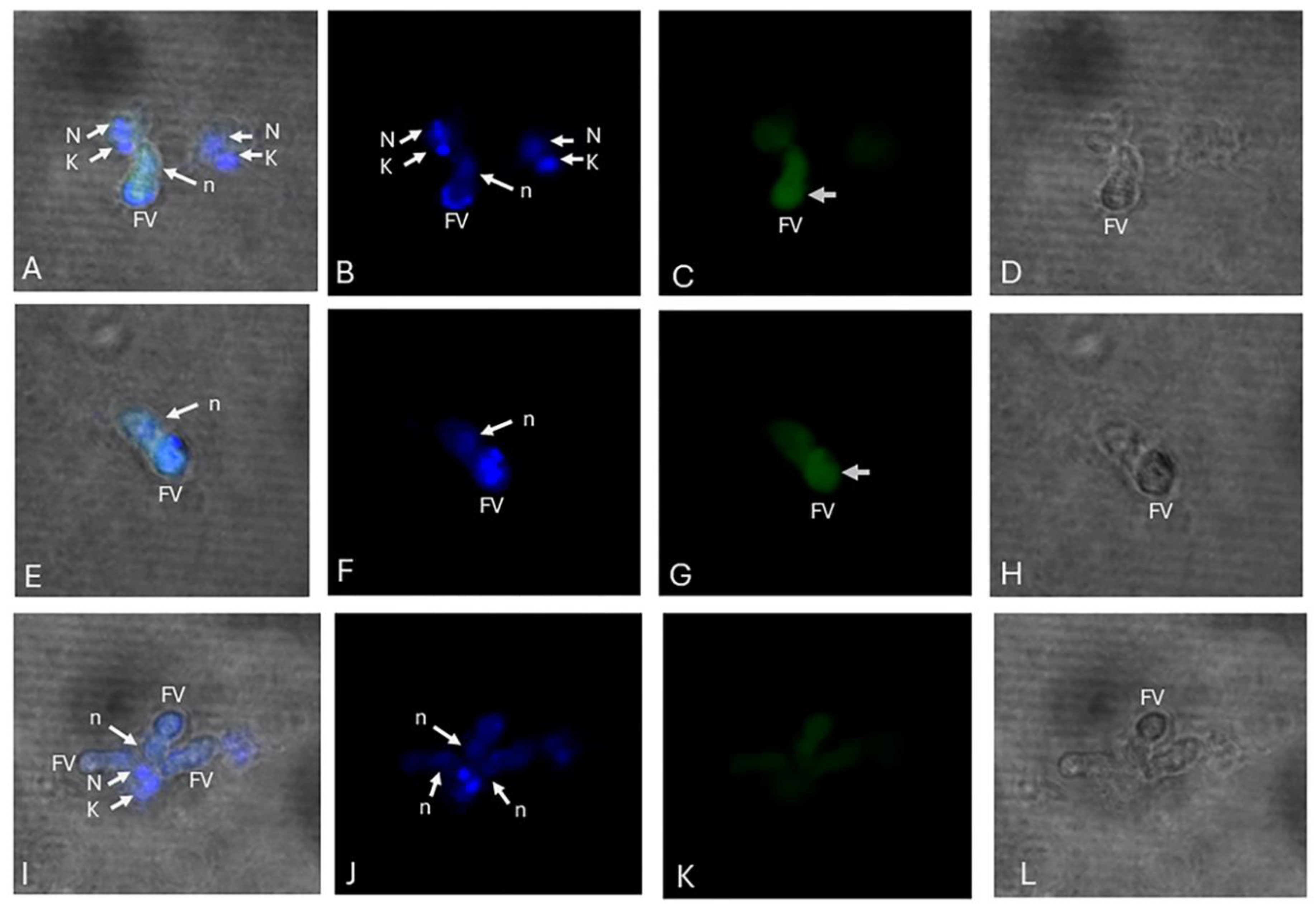
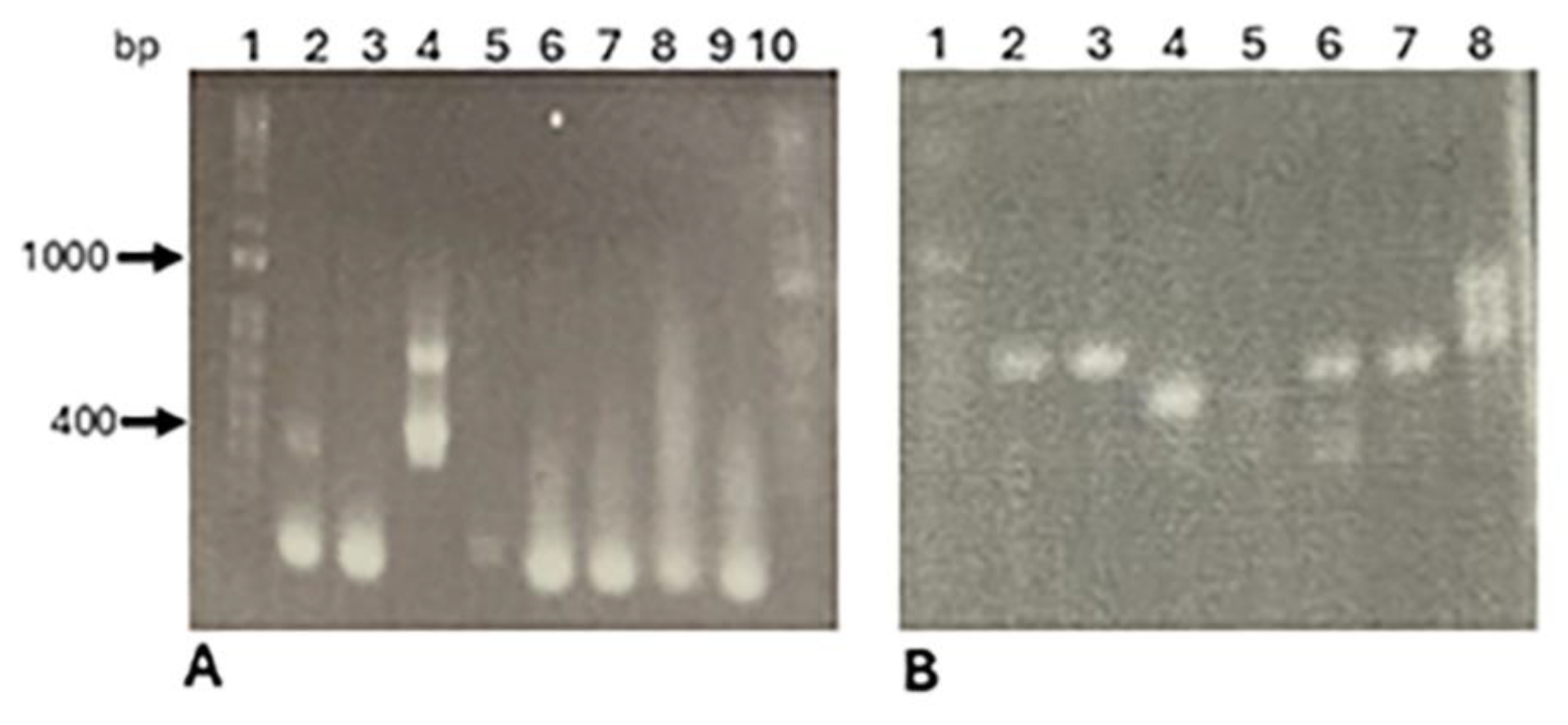
Colpodella species are predatory biflagellates phylogenetically related to pathogenic Apicomplexans like Plasmodium spp., Cryptosporidium spp., Babesia spp. and Theilaria spp. Colpodella species have been reported in human and animal infections. Trophozoites of Colpodella sp. ATCC 50594 obtain nutrients through myzocytosis and endocytosis. Following attachment of Colpodella sp. to its prey Parabodo caudatus, cytoplasmic contents of the prey are aspirated into a posterior food vacuole that initiates encystation. Unattached trophozoites also endocytose nutrients as demonstrated by the uptake of 40 and 100 nm nanoparticles. Cytochalasin D treatment was shown to distort the tubular tether formed during myzocytosis showing that actin plays a role in myzocytosis. Markers associated with myzocytosis, endocytosis and food vacuole formation are unknown. Furthermore, the relationship between the model Colpodella sp. ATCC 50594 and Colpodella sp. identified in arthropods, human and animal hosts are unknown. In this study we investigated the conservation of the coronin and Kelch 13 genes in Colpodella sp. ATCC 50594 using polymerase chain reaction (PCR). Kelch 13 distribution in Colpodella sp. ATCC 50594 life cycle stages was investigated using anti-Kelch 13 antibodies by immunofluorescence and confocal microscopy. Both genes were amplified from genomic DNA extracted from diprotist culture containing Colpodella sp. and P. caudatus but not from monoprotist culture containing P. caudatus alone. We amplified DNA encoding 18s rRNA with similarity to 18s rRNA amplified using piroplasm primers from the Italian Colpodella sp. identified in cattle and ticks. Detection of the coronin and Kelch genes in Colpodella sp. provides for the first time markers for actin binding and endocytosis in Colpodella species that can be investigated further to gain important insights into the mechanisms of myzocytosis, endocytosis and food vacuole formation in Colpodella sp.

Keywords: 
;  ;  ;  ;  ;  ;  ;  ;  

1. Introduction

Apicomplexans pathogenic in humans and animals are intensely investigated to understand the mechanisms of transmission and pathogenesis, and to discover avenues for treatment and prevention of infection. Apicomplexans such as Plasmodium spp., Babesia spp., Theilaria spp., Cryptosporidium spp. and Toxoplasma gondii infect host cells, reside intracellularly within host cells and obtain nutrients through a variety of structures including the cytostome and micropore [1,2]. Cryptosporidium spp. develop a feeder organelle at the interface of the parasite and the host epithelial cell [1]. Colpodella species are free-living relatives of the apicomplexa, that prey on bodonids, ciliates and algae [3,4] and have been described from marine, fresh water and soil environments [3,4,5]. Oligonucleotide primers targeting 18s rRNA from Cryptosporidium spp., Theilaria spp and Babesia spp. amplify 18s rRNA from Colpodella species identified in several species of ticks, biting flies associated with humans, goats, cattle, camels, horses, dogs, cats, ducks, foxes and pangolins [6], https://www.ncbi.nlm.nih.gov/nuccore/?term=Colpodella]. Colpodella spp. have also been detected in blood from human and horse infections as well as from fecal samples in ruminants and a tiger [7,8,9,10]. Reports of Colpodella spp. detection now span a wide geographic area globally, that includes countries in Africa, Asia, Europe, Australia, North and South America, Supplementary Table 1 (Table S1), [https://www.ncbi.nlm.nih.gov/nuccore/?term=Colpodella]. Colpodella gonderi and C. tetrahymenae prey on ciliates, remain on the ciliates for prolonged periods as ectoparasites, before encysting in the case of C. tetrahymenae [5,11]. Colpopdella species are increasingly reported in various species of ticks and biting flies that are associated with the “transmission” of Colpodella spp. into animals such as goats, cats, dogs [6] and into humans [7,12]. Colpodella spp. have also been identified in fecal samples. Oligonucleotide primers targeting the 18s rDNA of Cryptosporidium spp. and Babesia spp. identified Colpodella spp. in routine screenings for Cryptosporidium spp. and piroplasms in animals. Reported cases of Colpodella spp. infection in humans and animals suggest both tick-borne and direct transmission mechanisms. However, the specific mechanisms of transmission are currently unknown, and markers associated with transmission, nutrient uptake and survival of Colpodella spp. within the vertebrate and arthropod hosts are unknown.
Among the Myzozoans which comprise apicomplexans, dinoflagellates and the chrompodellids such as Colpodella spp., Voromonas pontica and Chromera spp., myzocytosis for nutrient uptake has been described [1]. Myzocytosis is a type of endocytosis, where the prey attaches to its prey, takes up the plasma membrane of the prey, degrades the membrane and aspirates the cytoplasmic contents of the prey as described for Colpodella sp. ATCC 50594 [13]. Among apicomplexans, nutrients are obtained within the host cells by endocytosis along with food vacuole formation such as the process described in Plasmodium spp. and in gregarines. The life cycle of Colpodella spp. consists of a trophozoite and a cyst stage [4,5,14]. A posterior food vacuole is formed in Colpodella sp. at the conclusion of myzocytosis followed by encystation in species that encyst [4,5,14]. The trophozoite stages of Colpodella sp. carry out endocytosis in addition to myzocytosis [15,16] and actin was shown to be involved in nutrient uptake [15]. However, it is unclear whether endocytosis alone can lead to encystation and development of juvenile trophozites to continue the life cycle. Additional methods of nutrient uptake have been described such as phagocytosis and apical phagotrophy [1,17,18] but the mechanisms are unclear and no markers have been identified. This is an area that needs further investigation. Markers of endocytosis have been identified in Plasmodium falciparum and T. gondii and include Kelch 13, AP-2µ, UBP1 and Eps-15 [19,20]. Kelch 13 has been identified in Myzozoans, including the apicomplexans, dinoflagellates and chrompodellids [18]. Conserved domains in the Kelch 13 gene are shared among ciliates and other eukaryotes [18]. A stable endocytotic complex identified in T. gondii and located in the IMC has molecular conservation to the cytostome described in Plasmodium spp. [18]. In P. falciparum, the protein VPS45 also plays a role in hemoglobin uptake [20]. Inactivation of VPS45 and Kelch 13 genes leads to decreased hemoglobin uptake and Kelch 13 depletion disrupts endocytosis and plasma membrane homeostasis in T. gondii [20]. Mutations in the Kelch 13 gene in P. falciparum results in resistance to artemisinin combination therapies (ACT) bringing focus to the importance of the Kelch 13 gene [19,21,22]. In addition to Kelch 13, mutations in the coronin gene are also associated with resistance to ACT in P. falciparum infections [23,24]. Both Kelch13 and coronin proteins have roles as actin binding proteins with the WD40 domain of P. falciparum coronin binding to F-actin and having additional functions in phagocytosis, locomotion and proliferation [25,26]. Due to the involvement of actin in myzocytosis in Colpodella sp. ATCC 50594, formation of the posterior food vacuole and the demonstration of nutrient uptake by endocytosis in Colpodella sp. ATCC 50594 trophozoites [15], we investigated the conservation of the Kelch 13 and coronin genes in Colpodella sp. ATCC 50594 and evaluated the expression and distribution of the Kelch 13 protein in the life cycle stages of Colpodella sp. ATCC 50594.

2. Materials and Methods

Diprotist culture conditions. The predator Colpodella sp. (ATCC 50594) and its prey Parabodo caudatus in a diprotist culture were obtained from the American type culture collection (ATCC) (Manassas, Virginia, USA). Both protists were cultured in Hay medium (Wards Scientific Rochester, New York, USA) bacterized with Enterobacter aerogenes as described previously [27]. The prey species Parabodo caudatus (ATCC 30905) was also maintained in Hay medium, bacterized with E. aerogenes. Both protists in tissue culture flasks were observed using an inverted microscope.
Genomic DNA isolation. Diprotist cultures containing Colpodella sp. and B. caudatus was centrifuged at 1475xg for 15 min, the supernatant was discarded and the pellet used for genomic DNA (gDNA) isolation. Plasmodium falciparum schizont pellets were obtained as described previously [27] following hypotonic lysis in 10 mM Tris buffer, pH 8.8. Diprotist and P. falciparum schizont pellets were homogenized in a solution of 10mM Tris-HCl pH 7.6, 50 mM EDTA pH 8.0, 0.1% SDS, and 1 mg/ml proteinase K by passing the pellet suspension 30x through a 25G needle. The homogenate was incubated overnight (O/N) in a 50ºC water bath and processed for DNA extraction as described previously [27].
Polymerase chain reaction and DNA sequencing. Conventional and nested Polymerase chain reaction (PCR) was performed using oligonucleotide primers targeting P. falciparum Kelch 13 and coronin genes. PCR was performed in a total volume of 25 µl containing 13 µl Dream Taq green master mix, 2 µl each of Forward and Reverse Primer, 3-4 µl of template DNA and nuclease free water to make up 25ul for the total reaction volume. For nested PCR, 13 µl Dream Taq green Master Mix, 2 µl each of Forward and reverse primers, and 8 µlof DNA from the first round of PCR. Amplified products were separated by electrophoresis on 1 %, 1.5 % or 2 % agarose gels containing ethidium bromide for visualization. Gene Ruler 1 kb Plus DNA ladder and 100 bp ladder (ThermoFischer) were used as standards. Plasmodium falciparum genomic DNA was used as a positive control and nuclease free water added to reaction tubes instead of template DNA served as a negative control. Amplified products showing single DNA bands on agarose gels were sequenced. Sequencing was performed by MCLAB (mclab@mclab.com, San Francisco, CA). Identity of nucleotide sequences was searched by BLAST in the databases at NCBI (www.ncbi.nlm.nih.gov)
Primers for18S rDNA for Colpodella spp. [28]; 18s colpo SY F=AATACCCAATCCTGACACAGGG; R=TTAAATACGAATGCCCCCAAC were used in PCR as described using the cycling parameters: Initial denaturation at 94 degrees for 2min then 30 cycles of denaturation at 94 degrees for 30 seconds, annealing at 60 degrees for 30 seconds, extension at 72 degrees for 1 minute. Then a final extension for 7 minutes at 72 degrees. Primers for Kelch 13 were used in a nested PCR. Kelch 13 nested PCR; Primary pair of primers designated: (5a) Kelch13 F for SY= GGGAATCTGGTGGTAACAGC; (5a) Kelch13 R for SY= CGGAGTGACCAAATCTGGGA were used in first round of PCR using the cycling conditions; denaturation at 95 °C for 1 minute, followed by 35 cycles at 95°C for 20 seconds, 58 °C for 20 seconds, and 60 °C for 1 minute, with a final extension at 60 °C for 3 minutes. For nested PCR, a nested pair of primers (5b) Kelch13 (S) F for SY= GCCTTGTTGAAAGAAGCAGA; (5b) Kelch13 (S) R for SY= GCCAAGCTGCCATTCATTTG were used for nested PCR cycling conditions; denaturation at 95 °C for 1 minute, followed by 35 cycles at 95 °C for 20 seconds, 56 °C for 20 seconds, and 60 °C for 1 minute, with a final extension at 60 °C for 3 minutes. Primers for Kelch 13 [29, Kelch 13 direct] were used for conventional PCR; Kelch13 Direct PCR; Forward— 5′-CTATACCCATACCAA AAGATTTAAGTG-3′, reverse—5′-GCTTGGCCCATCTTTATTAGTTCC C-3′), (from codon 412 to codon 723). The PCR cycling conditions were denaturation at 94 °C 3 min, followed by 10 cycles of 94 °C for 30 s, 57 °C for 30 s, 72 °C for 30 s then 30 cycles of 94 °C for 30 s, 55 °C for 30 s, 72 °C for 30 s with a final extension at 72 °C for 3 min.
Primers for Kelch 13 were used for nested PCR as described [30]. The primers ACT treated Kelch (F-A)CGGAGTGACCAAATCTGGGA; ACT treated Kelch (R-A) GGGAATCTGGTGGTAACAGC were used for the primary reactions and ACT treated Kelch (F-B) GCCAAGCTGCCATTCATTTG and ACT treated Kelch (R-B) GCCTTGTTGAAAGAAGCAGA were used for the nested reaction. PCR cycling conditions for both primary and nested reactions included 5 minutes of initial denaturation at 95°C followed by 40 cycles of 30s denaturation at 94°C, 90 s annealing at 60°C, 90s extension at 72°C and a 10-minute final extension at 72°C.
Primers for Kelch 13 nested Propeller were used for PCR as described [31]. Kelch 13 Propeller (F-A) CGGAGTGACCAAATCTGGGA; Kelch 13 Propeller (R-A) GGGAATCTGGTGGTAACAGC; Kelch 13 Propeller (F-B) TCAACAATGCTGGCGTATGTG; Kelch 13 Propeller (R-B) TGATTAAGGTAATTAAAAGCTGCTCC. For the first round, DNA was denatured at 95 °C for 5 min, followed by 40 cycles of denaturation at 94 ° C for 30 min, 60 °C for 1 min 30 s, and 72 °C for 1 min 30 s, and final elongation at 72 °C for 10 min. For the second round of PCR, the same amplification parameters were used with 5 μl of the first PCR product used as template.
Primers for coronin were used in conventional PCR as described [23], ACT Coronin] F:ATGGCAAGTTGAAGGGGGAG; R:TTGTCTTCACCACCAAATCCA, using the cycling parameters for amplification with denaturation at 95°C for 5 min, followed by 40 cycles of denaturation at 95°C for 30 s, annealing at 56°C for 45 s and extension at 68°C for 30 s, followed by final extension at 72°C for 7 min.
Coronin nested PCR; First round of PCR: 3D7 Coronin (F-A)-TGATTTGTTCATATTATA GGTAC-3′ and 3D7 Coronin (R-A)-TATTCTGAC AAGTTCCACTTAATA. First round of PCR: 30s at 90°C; followed by 30 cycles of 20 s at 90°C, 30 s at 45°C, 1.30 min at 68°C. And for the nested PCR: 3D7 Coronin (F-B)-CATATTATAGGTACCATG GCAAGTT and 3D7 Coronin (R-B)-AGGCTT CTTCTCATTTTCTATATC. For the nested PCR, primary PCR products were amplified under the same conditions using the following cycling program: 30 s at 90°C; followed by 45 cycles of 15s at 94°C, 30s at 50°C, 1 min at 68°C. All PCR products were analyzed by electrophoresis on agarose gels (Table S2).
Immunofluorescence and confocal microscopy. Immunofluorescence and confocal microscopy was performed on Colpodella sp. and B. caudatus diprotist culture as described previously [27] using 5 % formalin fixed cells. Formalin-fixed cells from diprotist cultures were permeabilized with 0.1 % Triton X-100. Briefly, smears were incubated with an anti-Kelch 13 mouse monoclonal antibody Mab K13, clone D9 (a gift from Dr. David Fidock, Columbia University, Irving Medical Center, New York, NY ) diluted 1:500 and 1:1000. Incubation with the primary antibody was carried out for 1 h at 37°C; slides were washed three times with 1× PBS followed by incubation for 1 h with secondary rabbit anti-mouse antibody conjugated to Alexa 488 diluted 1:500 (Molecular Probes, ThermoFischer Scientific). The smears were washed three times with 1× PBS followed by one wash in distilled water. Fluoroshield mounting medium containing 4′, 6-diamidino-2-phenylindole (DAPI; www.abcam) or ProLong Diamond Antifade Mountant with DAPI Invitrogen by ThermoFisher Scientific, Life Technologies Corporation, Eugene, Oregon, USA) was used to mount the slides. Normal (preimmune) mouse serum (NMS), was used as a negative control for IFA. Images were collected using a Leica TCS-SP5II upright laser scanning confocal microscope (Leica Microsystems, GmbH, Wetzlar, Germany). In addition, an SP8 True Scanning Confocal (TCS) on a DMI8 inverted microscope was used to generate differential interference contrast (DIC) images. Sam-Yellowe’s trichrome stained and confocal images were adjusted to 300 dpi using the CYMK color mode and RGB color mode on Adobe photo shop (CS6). Confocal microscopy was performed at the Cleveland Clinic, Lerner Research Institute Imaging Core, Cleveland, OH, USA.

3. Results

During the life cycle of Colpodella sp. ATCC 50594, trophozoites prey on trophozoites of Parabodo caudatus and aspirate cytoplasmic contents of the prey into a posterior food vacuole in a process of myzocytosis. Using Sam-Yellowe’s trichrome staining for light microscopy, the enlarged posterior food vacuole can be observed while the Colpodella trophozoite is still attached to its prey (Figure 1 A and B, yellow arrows). Following myzocytosis, the pre-cyst stage still containing the food vacuole with aspirated contents from the prey (Figure 1A, C and D, orange arrows) will develop into a cyst.
We investigated the conservation of Kelch 13 and coronin genes in Colpodella sp. ATCC 50594 using oligonucleotide primers targeting Kelch 13 and coronin from P. falciparum using conventional and nested PCR. We also investigated the relationship of Colpodella sp. ATCC 50594 to the recently identified Colpodella species from ticks reported from Italy and using Colpodella sp. ATCC 50594 DNA templates extracted from two different diprotist cultures and a monoprotist culture containing only P. caudatus. Figure 2 A shows amplified coronin DNA bands of approximately 368 bp from Colpodella sp. ATCC 50594 identified in lane 2 and from P. falciparum (strain HB3) in lane 3. The Kelch 13 gene was also amplified from P. falciparum (HB3) in lanes 2 and 3 as well as in Colpodella sp. using nested PCR in lane 4 (Figure 2B). Primers targeting a partial region of 18s rRNA genes for piroplasm parasites [28] amplified DNA bands of approximately 408 bp shown in lanes 6 and 7 (Figure 2B). BLAST (BLASTn) searches of DNA sequences were performed using the NCBI databases (https://blast.ncbi.nlm.nih.gov/Blast) to identify homologous DNA sequences. Nucleotide sequence identity of 100 %, 99.67 % and 99.01 % was obtained with Colpodella sp. ATCC 50594 amplified using the Piro primers in the current study. Sequence identity of 97.82% and 97.55 % was obtained with Colpodella isolates 103 and 115, respectively identified from horse blood and 89.2 % sequence identity to Cryptosporidium sp. isolate NJ3559, from BLAST searches.
DNA sequence for the amplified coronin DNA BLAST searched in the database matched the coronin gene of P. falciparum. The nucleotide sequence of the coronin gene showed 99.34 % sequence identity to P. falciparum coronin gene and 96.39%, 93.73% and 93.07% sequence identity, respectively to P. reichenowi, P. gaboni and Plasmodium sp. gorilla clade G2 coronin gene. A BLAST search of the nucleotide sequences obtained for the Colpodella sp. ATCC 50594 Kelch13 gene showed 100% sequence identity to P. falciparum Kelch 13.
Phylogenetic tree reconstruction was performed using the Colpodella sp. 18S rRNA, coronin and Kelch 13 DNA sequences obtained and DNA sequences retrieved from the NCBI database in order to determine the phylogenetic relationship between the Colpodella DNA sequences with DNA sequences from related Apicomplexan species. Phylogenetic tree analysis of Colpodella sp. ATCC 50594 18S rRNA was performed along with 14 sequences retrieved from the NCBI database shown in supplementary Figures 1 (S1) and 2. The DNA sequence obtained from the current study groups with most of the 18S rRNA gene sequences in a large clade identified from sheep and dog ticks, pangolin ticks and from ticks associated with relapsing fever in two human cases in a tree analysis inferred by Maximum likelihood using PhyML (aLRT) program (http://www.phylogeny.fr/index.cgi). The Colpodella angusta (isolate Zotu 1986) 18S rRNA gene sequences branched separately from the rest of the sequences. A larger phylogenetic tree reconstruction (Figure S2) performed within NCBI BLAST distance tree analysis shows a clade containing Cryptosporidium spp. sequences most closely related to Colpodella sp. ATCC 50594 than Theilaria spp. sequences. Colpodella sp. clone Kc2-17 branched separately from the DNA sequences from Colpodella sp. ATCC 50594. Phylogenetic tree analysis of Colpodella sp. ATCC 50594 coronin along with 5 sequences retrieved from the NCBI database along with P. falciparum HB3 coronin DNA sequenced in the current study and inferred by Maximum likelihood using PhyML showed both sequences clustered within a clade containing P. falciparum isolates and next to a second clade also containing P. falciparum isolates. DNA sequences from Dictyostelium discoideum were branched separately from the Plasmodium and Colpodella sequences (Figure S3). In a more expanded phylogenetic tree reconstruction performed using the NCBI distance tree construction, the Colpodella sp. ATCC 50594 coronin gene was closed clustered with Plasmodium falciparum Pf coronin gene alleles KG278d14 and KG278d0 and more distantly related to the coronin genes from P. reichenowi, P. gaboni and the Plasmodium sp. gorilla clade (Figure S4). Phylogenetic tree reconstruction was performed using the DNA sequences obtained for Colpodella sp. ATCC 50594 Kelch 13 gene along with 4 sequences retrieved from NCBI and P. falciparum HB3 sequences obtaibed in the current study. Following analysis by PhyML, all sequences were seen to be closely related (Figure S5). An expanded phylogenetic tree analysis showed Colpodella sp. ATCC 50594 Kelch 13 gene in the same cluster with 25 Plasmodium falciparum isolates. Plasmodium falciparum isolate KBG-05-15 was separate from the 25 isolates as seen in Figure S6. In order to determine the localization of the Kelch 13 protein in Colpodella sp. ATCC 50594 life cycle stages, we performed immunofluorescence and confocal microscopy using an anti-PfKelch 13 monoclonal antibody. Figure 6, panels A, C, E and G show the antibody reactivity with the posterior food vacuole of Colpodella trophozoites attached to prey (6A, C) and in the pre-cyst stage (6E, G). DAPI stained nuclear and kinetoplast aspirated from the prey was detected in the food vacuole of Colpodella sp. (Figure 6 A, B, E, F, I, J).
Figure 3. Confocal and differential interference contrast (DIC) microscopy of 5% formalin-fixed Colpodella sp. ATCC 50594 reacted with Mab K13, clone D9 in IFA. The food vacuole showing cross reactivity with Mab K13, clone D9 is shown in panels C and G. A-D, Colpodella trophozoite with a large food vacuole (FV) attached to P. caudatus in myzocytosis. The DAPI stained nucleus (N) and kinetoplast (K) of P. caudatus are shown. The nucleus (n) of Colpodella is also shown. E-H, A pre-cyst stage unattacched following feeding is shown. The FV in panels A, B, E and F show the DAPI-stained aspirated nucleus and kinetoplast aspirtated from the prey. I-L, Normal mouse serum negative control.
Preprints 141588 g003

Discussion

In this study we investigated the conservation of the Kelch 13 and coronin genes in Colpodella sp. ATCC 50594. The 18S rRNA gene from Colpodella sp. ATCC 50594 was also investigataed to determine the phylogenetic relationship to 18S rRNA genes identified from ticks associated with various animals, birds and two human cases. Colpodella sp. ATCC 50594 obtain nutrients by myzocytosis and endocytosis. Colpodella trophozoites attach to P. caudatus trophozoites in a process known as myzocytosis by engulfing the plasma membrane of the prey, degrading the membrane and aspirating the cytoplasmic contents of the prey into a posterior food vacuole [13]. Colpodella trophozoites also perform endocytosis for nutrient uptake and were shown to take up nanobeads of 40 and 100 nm [15]. Following myzocytosis encystation occurs in cyst forming Colpodella species [4,5,14]. The mechanisms of nutrient uptake are unknown among Colpodella species. However, the process of attachment and tether formation by Colpodella trophozoites is actin-mediated due to the distortion of the tether observed following cytochalasin D treatment [13]. Markers of endocytosis and myzocytosis have not been identified in Colpodella species. In P. falciparum several molecules have been identified as markers of endocytosis. These include Kelch 13, adaptor protein-2µ (AP-2µ), ubiquitin carboxyl-terminal hydrolase (UBP1), epidermal growth factor receptor substrate-15 (Eps15) [20], coronin [25] and VPS45 a protein directly involved in host cell cytosol uptake in P. falciparum [20,32]. Inactivation of VPS45 results in decreased uptake of cytoplasmic contents into the digestive vacuole (food vacuole) [20,32]. Of these proteins, Kelch 13 is reported to be a conserved protein associated with endocytosis among myzozoans [18]. However, the gene has not been reported in Colpodella species. Similarly, the coronin gene has not been reported in Colpodella species. Cellular sturctures used for endocytosis have been identified in P. falciparum such as the cytostome [19] and micropores in both P. falciparum and Toxoplasma gondii [2,33]. Pores present on the parasitophorous vacuole membrane (PVM) of T. gondii and P. falciparum could allow for the transport of nutrients and other metabolites across the PVM [34]. The endosomal and phagosomal pathways utilized for nutrient uptake and degradation among the apicomplexa remain areas that are poorly understood and merit further investigationse. In the current study, anti-P. falciparum Kelch 13 antibodies cross-reacted with Colpodella sp. proteins in the food vacuole. However, the role of Kelch 13 in the formation of the myzocytic aperture or uptake of the prey’s cytoplasmic contents is unknown. Antibodies specific for the coronin protein were unavailable for use in coronin localization in this study. Therefore, it is unclear whether Kelch 13 and coronin are present in the same compartments or whether both proteins function at the same time periods in the life cycle during myzocytosis and food vacuole formation. Colpodella species have been identified in several tick species and biting flies associated with cattle, small ruminants, dogs, cats, camels, pangolins, and two cases of relapsing fever in humans [6, Table S1]. Whether the ticks are true vectors capable of transmitting Colpodella infection is presently unknown. Colpodella spp. have also been identified from animal skin, blood, fecal samples and in different environmental sources [Table S1]. The potential for zoonotic infections caused by Colpodella species poses a public health concern. Therefore, being able to identify species and strains using specific markers is crucial. The morphology of the different Colpodella species identified by PCR is unknown and none have been cultured. In our previous studies, we have used Sam-Yellowe’s trichrome staining to identify and differentiate different life cycle stages of Colpodella sp. ATCC 50594. This has aided confocal and electron microscopic analysis of Colpodella sp. morphology [13,16]. The phyogenetic relationship obtained by analysis of the 18S rRNA of Colpodella species shows that different species and strains of Colpdella are represented in the DNA sequences obtained and some of these sequences demonstrate close relationships with the apicoamplexans Cryptosporidium spp. and Theilaria spp. Primers for Cryptosporidium spp. and piroplasms such as Theilaria spp. and Babesia spp. have amplified Colpodella species 18S rRNA from ticks and from fecal sampes [8,10,35,36,37]. Detection of antibody reactivity with the food vacuole of Colpodella sp. represents the first time a marker for endocytosis has been identified within the food vacuole in Colpodella species. Kelch 13 specific antibody reactivity was identified within the digestive vacuole, cytoplasm, plasma membrane, and endoplasmic reticulum (ER) of P. falciparum [38] demonstrating that structures involved with the uptake of nutrients into the food vacuole can be identified. Electron microscopy will be required to confirm the specific structures involved and their distribution within the trophozoite. The demonstration that the coronin and Kelch 13 genes are conserved in Colpodella sp. ATCC 50594 will pave the way for the identification of other important genes that will provide insights into life cycle stage transitions and the process of nutient uptake in culture, in the environment and within the arthropod and vertebrate hosts infected by Colpodella species.

Supplementary Materials

The following supporting information can be downloaded at the website of this paper posted on Preprints.org, Supplementary Figure 1. 18S rRNA sequences retrieved from the NCBI database were used to construct a phylogenetic tree to determine the relationship of the genes. Phylogenetic analysis was inferred by Maximum likelihood using PhyML (aLRT) program (https://www.phylogeny.fr/) following sequence alignment by MUSCLE. DNA sequence from 18S rRNA from 14 sequences retrieved from NCBI BLAST search were aligned with the DNA sequence obtained from the current study. Numbers at the nodes represent posterior probabilities. Branch length scale bar values are shown. Supplementary Figure 2. 18S rRNA sequences retrieved following a BLAST search of Colpodella sp. ATCC 50594 18S rRNA sequences was aligned for distance tree analysis to show the relationship between Colpodella sequences identified from ticks and different vertebrate hosts and other apicomplexa such as Theilaria spp. and Cryptosporidium spp. Supplementary Figure 3. Coronin gene sequences retrieved from the NCBI database were used to construct a phylogenetic tree to determine the relationship of the genes. Phylogenetic analysis was inferred by Maximum likelihood using PhyML (aLRT) program (https://www.phylogeny.fr/) following sequence alignment by MUSCLE. DNA sequence from coronin genes from 5 sequences retrieved from NCBI BLAST search were aligned with the DNA sequence obtained from the current study. Numbers at the nodes represent posterior probabilities. Branch length scale bar values are shown. Supplementary Figure 4. DNA sequences from coronin genes retrieved following a BLAST search of Colpodella sp. ATCC 50594 coronin sequences was aligned for distance tree analysis to show the relationship between Colpodella sequences identified from Colpodella sp. ATCC 50594 and coronin genes from Plasmodium falciparum. Supplementary Figure 5. Kelch 13 gene sequences retrieved from the NCBI database were used to construct a phylogenetic tree to determine the relationship of the genes. Phylogenetic analysis was inferred by Maximum likelihood using PhyML (aLRT) program (https://www.phylogeny.fr/) following sequence alignment by MUSCLE. DNA sequence from Kelch 13 genes from 4 sequences retrieved from NCBI BLAST search were aligned with the DNA sequence obtained from the current study. Numbers at the nodes represent posterior probabilities. Branch length scale bar values are shown. Supplementary Figure 6. DNA sequences from Kelch 13 genes retrieved following a BLAST search of Colpodella sp. ATCC 50594 Kelch 13 sequences was aligned for distance tree analysis to show the relationship between Colpodella sequences identified from Colpodella sp. ATCC 50594 and Kelch 13 genes from Plasmodium falciparum.

References

  1. Valigurová, A.; Florent, I. Nutrient Acquisition and Attachment Strategies in Basal Lineages: A Tough Nut to Crack in the Evolutionary Puzzle of Apicomplexa. Microorganisms. 2021, 9: 1430. [CrossRef]
  2. Yang J, Long S, Hide G, Lun ZR, Lai DH. Apicomplexa micropore: history, function, and formation. Trends Parasitol. 2024 May;40(5):416-426. Epub 2024 Apr 17. PMID: 38637184. [CrossRef]
  3. Simpson, A.; Patterson, D. Ultrastructure and the identification of the predatory flagellate Colpodella pugnax Cienkowski (Apicomplexa) with a description of Colpodella turpis n. sp. and a review of the genus. Syst, Parasitol., 1996, 33, 187-198. [CrossRef]
  4. Brugerolle, G. Colpodella vorax: Ultrastructure, predation, life-cycle, mitosis, and phylogenetic relationships. Europ. J. Protistol., 2002, 38:113-125. [CrossRef]
  5. Cavalier-Smith T; Chao, E.E. Protalveolate phylogeny and systematics and the origins of Sporozoa and dinoflagellates (phylum Myzozoa nom. Nov.). Eur. J. Protistol. 2004, 40: 185-212. [CrossRef]
  6. Salti MI, Sam-Yellowe TY. Are Colpodella Species Pathogenic? Nutrient Uptake and Approaches to Diagnose Infections. Pathogens. 2024 Jul 21;13(7):600. [CrossRef]
  7. Yuan, C.L.; Keeling, P.J.; Krause, P.J. et al. Colpodella spp.–like Parasite Infection in Woman, China. Emerg Infect Dis. 2012, 18:125-127.
  8. Chiu, H.C.; Sun, X.; Bao, Y.; Fu, W.; Lin, K.; Chen, T.; Zheng, C.; Li, S.; Chen, W.; Huang, C. Molecular identification of Colpodella sp. of South China tiger Panthera tigris amoyensis (Hilzheimer) in the Meihua Mountains, Fujian, China. Folia Parasitol (Praha). 2022, 69:2022.019. [CrossRef]
  9. Xu, M.; Hu, Y.; Qiu, H.; Wang, J.; Jiang, J. Colpodella sp. (Phylum Apicomplexa) Identified in Horses Shed Light on Its Potential Transmission and Zoonotic Pathogenicity. Front Microbiol. 2022, 13:857752. [CrossRef]
  10. Elochukwu, C. V.; Nnabuife, H. E.; Nicodemus, M.; Ogo, N. I.;Sylvanus, O. S.; Cornelius, J. O.; Kamani, J.; Maxwell, O. N. Molecular detection of Colpodella sp. using Cryptosporidium primers in faecal samples of small ruminants in FCT and Plateau State , Nigeria. J. Vet. Biomed. Sci. 2024, 6, 112-122.
  11. Olmo, J.L.; Esteban, G.F.; Finlay, B.J. New records of the ectoparasitic flagellate Colpodella gonderi on non-Colpoda ciliates. J Int Microbiol, 2011, 14:207-211. [CrossRef]
  12. Jiang, J-F.; Jiang, R-R.; Chang, Q-C.; Zheng, Y-C.; Jiang, B-G.; Sun, Y.; Jia, N.; Wei, R.; Bo, H-B.; Huo, Q-B.; Wang, H.; von Fricken, M. E.; Cao, W-C. Potential novel tick-borne Colpodella species parasite infection in patient with neurological symptoms. PLOS Negl. Trop. Dis., 2018, 12(8):e0006546. [CrossRef]
  13. Sam-Yellowe, T.Y.; Fujioka, H.; Peterson, J.W. Ultrastructure of Myzocytosis and Cyst Formation, and the Role of Actin in Tubular Tether Formation in Colpodella sp. (ATCC 50594). Pathogens, 2022, 11:455.
  14. Getty, T.A.; Peterson, J.W.; Fujioka, H.; Walsh, A.M.; Sam-Yellowe, T.Y. Colpodella sp. (ATCC 50594) Life Cycle: Myzocytosis and Possible Links to the Origin of Intracellular Parasitism. Trop Med Infect Dis., 2021, 6:127. [CrossRef]
  15. Sam-Yellowe, T.Y.; Asraf, M.M.; Peterson, J.W.; Fujioka, H. Fluorescent Nanoparticle Uptake by Myzocytosis and Endocytosis in Colpodella sp. ATCC 50594. Microorganisms 2023, 11, 1945.
  16. Sam-Yellowe TY, Getty TA, Addepalli K, Walsh AM, Williams-Medina AR, Fujioka H, Peterson JW. Novel life cycle stages of Colpodella sp. (Apicomplexa) identified using Sam-Yellowe's trichrome stains and confocal and electron microscopy. Int Microbiol. 2022 Nov;25(4):669-678.
  17. Piro, F.; Focaia, R.; Dou, Z.; Masci, S.; Smith, D.; Di Cristina, M. An Uninvited Seat at the Dinner Table: How Apicomplexan Parasites Scavenge Nutrients from the Host. Microorganisms. 2021 Dec 15;9(12):2592. [CrossRef]
  18. Koreny, L.; Mercado-Saavedra, B.N.; Klinger, C.M.; Barylyuk, K.; Butterworth, S.; Hirst, J.; Rivera-Cuevas, Y.; Zaccai, N.R.; Holzer, V.J.C.; Klingl, A.; Dacks, J.B.; Carruthers, V.B.; Robinson, M.S.; Gras, S.; Waller, R.F.. Stable endocytic structures navigate the complex pellicle of apicomplexan parasites. Nat Commun., 2023, 14:2167. [CrossRef]
  19. Matz, J.M. Plasmodium's bottomless pit: properties and functions of the malaria parasite's digestive vacuole. Trends Parasitol., 2022, 38:525-543. [CrossRef]
  20. Elsworth, B.; Keroack, C.D.; Duraisingh, M.T. Elucidating Host Cell Uptake by Malaria Parasites. Trends Parasitol., 2019, 35:333-335. [CrossRef]
  21. Siddiqui FA, Boonhok R, Cabrera M, Mbenda HGN, Wang M, Min H, Liang X, Qin J, Zhu X, Miao J, Cao Y, Cui L. Role of Plasmodium falciparum Kelch 13 Protein Mutations in P. falciparum Populations from Northeastern Myanmar in Mediating Artemisinin Resistance. mBio. 2020 Feb 25;11(1):e01134-19. [CrossRef]
  22. Spielmann T, Gras S, Sabitzki R, Meissner M. Endocytosis in Plasmodium and Toxoplasma Parasites. Trends Parasitol. 2020 Jun;36(6):520-532. [CrossRef]
  23. Ajibaye O, Olukosi YA, Oriero EC, Oboh MA, Iwalokun B, Nwankwo IC, Nnam CF, Adaramoye OV, Chukwemeka S, Okanazu J, Gabriel E, Balogun EO, Amambua-Ngwa A. Detection of novel Plasmodium falciparum coronin gene mutations in a recrudescent ACT-treated patient in South-Western Nigeria. Front Cell Infect Microbiol. 2024 Apr 23;14:1366563. [CrossRef]
  24. Demas et al. Mutations in Plasmodium falciparum actin-binding coroninconfer reduced artemisinin susceptibility. Proc. Natl. Acad. Sci. USA, 2018, 115, 12799-12804.
  25. Chan, K. T.; Creed, S. J.; Bear, J. E. Unraveling the enigma: progress towards understanding the coronin family of actin regulators. Trends Cell Biol., 2011, 21, 481-8. [CrossRef]
  26. Bane, K. S. et al. The Actin filament -binding protein coronin regulates motility in Plasmodium sporozoites. PLoS Pathog. 2016, 12, e1005710. [CrossRef]
  27. Sam-Yellowe TY, Yadavalli R, Fujioka H, Peterson JW, Drazba JA. RhopH3, rhoptry gene conserved in the free-living alveolate flagellate Colpodella sp. (Apicomplexa). Eur J Protistol. 2019 Oct;71:125637. [CrossRef]
  28. Jimale, K. A.; Bezzera-Santos, M. A.; Mendoza-Roldan, J. A.; Latrofa, M. S.; Baneth, G.; Otrano, D. Molecular detection of Colpodella sp. and other tick-borne pathogens in ticks of ruminants, Italy. Acta Tropica, 2024, 257, 107306. [CrossRef]
  29. de Laurent ZR, Chebon LJ, Ingasia LA, Akala HM, Andagalu B, Ochola-Oyier LI, Kamau E. Polymorphisms in the K13 Gene in Plasmodium falciparum from Different Malaria Transmission Areas of Kenya. Am J Trop Med Hyg. 2018 May;98(5):1360-1366. [CrossRef]
  30. Tardieux I, Liu X, Poupel O, Parzy D, Dehoux P, Langsley G. A Plasmodium falciparum novel gene encoding a coronin-like protein which associates with actin filaments. FEBS Lett. 1998 Dec 18;441(2):251-6. [CrossRef]
  31. Mayengue PI, Niama RF, Kouhounina Batsimba D, Malonga-Massanga A, Louzolo I, Loukabou Bongolo NC, Macosso L, Ibara Ottia R, Kimbassa Ngoma G, Dossou-Yovo LR, Pembet Singana B, Ahombo G, Sekangue Obili G, Kobawila SC, Parra HJ. No polymorphisms in K13-propeller gene associated with artemisinin resistance in Plasmodium falciparum isolated from Brazzaville, Republic of Congo. BMC Infect Dis. 2018 Oct 29;18(1):538. [CrossRef]
  32. Jonscher, E.; Flemming, S.; Schmitt, M.; Sabitzki, R.; Reichard, N.; Birnbaum, J.; Bergmann, B.; Höhn, K.; Spielmann, T. PfVPS45 Is Required for Host Cell Cytosol Uptake by Malaria Blood Stage Parasites. Cell Host Microbe, 2019, 25:166-173. [CrossRef]
  33. Edgar RCS, Counihan NA, McGowan S, de Koning-Ward TF. Methods Used to Investigate the Plasmodium falciparum Digestive Vacuole. Front Cell Infect Microbiol. 2022 Jan 13;11:829823. [CrossRef]
  34. Wu S, Meng J, Yu F, Zhou C, Yang B, Chen X, Yang G, Sun Y, Cao W, Jiang J, Wu J, Zhan L. Molecular epidemiological investigation of piroplasms carried by pet cats and dogs in an animal hospital in Guiyang, China. Front Microbiol. 2023 Oct 12;14:1266583. [CrossRef]
  35. Hussein, S.; Li, X.; Bukharr, S. M.; Zhou, M.; Ahmad, S.; Amhad, S.; Javid, A.; Guan, C.; Hussain, A.; Ali, W.; Khalid, N,;Ahmad, U, Tian, L.; Hou, Z. Cross-genera amplification and identification of Colpodella sp. with Cryptosporidium primers in fecal samples of zoo felids from northeast China. Braz J Biol. 2021 Sep 6; 83:e247181. [CrossRef]
  36. Qi Y, Wang J, Lu N, Qi X, Yang C, Liu B, Lu Y, Gu Y, Tan W, Zhu C, Ai L, Rao J, Mao Y, Yi H, Li Y, Yue M. Potential novel Colpodella spp. (phylum Apicomplexa) and high prevalence of Colpodella spp. in goat-attached Haemaphysalis longicornis ticks in Shandong province, China. Ticks Tick Borne Dis. 2024 May;15(3):102328. [CrossRef]
  37. Soliman AM, Mahmoud HYAH, Hifumi T, Tanaka T. Discovery of Colpodella spp. in ticks (Hyalomma dromedarii) infesting camels in southern Egypt. Ticks Tick Borne Dis. 2024 Sep;15(5):102352. [CrossRef]
  38. Gnädig NF, Stokes BH, Edwards RL, Kalantarov GF, Heimsch KC, Kuderjavy M, Crane A, Lee MCS, Straimer J, Becker K, Trakht IN, Odom John AR, Mok S, Fidock DA. Insights into the intracellular localization, protein associations and artemisinin resistance properties of Plasmodium falciparum K13. PLoS Pathog. 2020 Apr 20;16(4):e1008482. [CrossRef]
Figure 1. Sam-Yellowe’s trichrome stainingof formalin-fixed Colpodella sp. ATCC 50594 trophozoites (yellow arrows) attached to P. caudatus (red arrows). Trophozoites with enlarged food vacuoles are shown in (A-C). Pre-cysts no longer attched to prey are shown in (A, C, D, orange arrows). Scale bars, 10 µm.
Preprints 141588 g001
Figure 2. 1.5 % agarose gels of PCR amplified DNA from Colpodella sp. ATCC 50594 targeting 18S rRNA and Kelch 13 (A) and coronin and Kelch 12 (B). A. Lanes 1, 1 kb marker; 2, Colpodella nested coronin; 3, Colpodella nested coronin; 4, P. falciparum (HB3) coronin; 5, P. caudatus coronin; 6, Colpodella Kelch 13 nested ACT; 7, Colpodella Kelch 13 nested propeller; 8, P. falciparum (HB3) Kelch nested ACT; 9, P. caudatus Kelch 13; 10;100 bp ladder. B. Lanes 1, 1 kb marker; 2, P. falciparum nested Kelch 13; P. falciparum direct Kelch 13; 4, Colpodella nested Kelch 13; 5, Colpodella nested Kelch 13; 6, 18S rRNA Colpodella; 7, Colpodella 18S rRNA; 8, 100 bp ladder. Template DNA from two different HB3 and Colpodella DNA extracts were used for PCR.
Preprints 141588 g002
Disclaimer/Publisher’s Note: The statements, opinions and data contained in all publications are solely those of the individual author(s) and contributor(s) and not of MDPI and/or the editor(s). MDPI and/or the editor(s) disclaim responsibility for any injury to people or property resulting from any ideas, methods, instructions or products referred to in the content.
Copyright: This open access article is published under a Creative Commons CC BY 4.0 license, which permit the free download, distribution, and reuse, provided that the author and preprint are cited in any reuse.
Prerpints.org logo

Preprints.org is a free preprint server supported by MDPI in Basel, Switzerland.

Subscribe

Disclaimer

Terms of Use

Privacy Policy

Privacy Settings

© 2026 MDPI (Basel, Switzerland) unless otherwise stated