Preprint
Article

This version is not peer-reviewed.

Topological Insights into Embryogenesis: Linking Germ Layer Derivations to Shared Pathologies

Submitted:

24 November 2024

Posted:

25 November 2024

You are already at the latest version

Abstract

During early embryogenesis, the trilaminar germ layers—ectoderm, mesoderm, and endoderm—undergo complex topological transformations and molecular interactions that define tissue differentiation. The ectoderm, which gives rise to the central nervous system and epidermis, exhibits developmental pathways closely tied to morphogenetic gradients and folding dynamics. This study explores how shared topological features and signalling pathways across germ layers can generate resembling pathologies under comparable disruptions, such as neurocutaneous syndromes and craniofacial anomalies. By integrating concepts from developmental biology and topological data analysis, this work provides a theoretical framework to understand the intersection of embryonic topology and disease etiology, offering insights into novel diagnostic and therapeutic approaches.

Keywords: 
;  ;  ;  ;  ;  ;  ;  

Section 1. Introduction

Section 1.1. Topological Mechanisms Linking Pathologies

The intricate choreography of embryogenesis is profoundly shaped by topological processes—those that involve spatial transformations and interactions of tissues across the three germ layers: ectoderm, mesoderm, and endoderm. These processes not only determine the physical arrangement of developing structures but also establish the functional relationships between organs and systems. Disruptions in these topological events, driven by genetic, molecular, or environmental factors, frequently result in pathologies that share striking similarities across different tissues. This section explores how topological principles underpin both normal development and pathological outcomes, with a focus on the interconnected nature of these phenomena.

Section 1.2. The Neural Tube: A Paradigm of Topological Complexity

Neural tube closure represents a quintessential example of how topological processes drive tissue formation. The ectoderm folds into a neural plate, which then rolls and fuses to form the neural tube, the precursor to the brain and spinal cord. This process involves dynamic changes in cell shape and polarity, guided by molecular signals such as Sonic Hedgehog (SHH) and Bone Morphogenetic Proteins (BMP) (Copp et al., 2003). Importantly, closure occurs at multiple points along the length of the neural tube, making it a geometrically complex event.
Failures in this process lead to neural tube defects (NTDs), including spina bifida and anencephaly, which exemplify how minor disruptions in topological events can have severe consequences. Research has shown that environmental factors, such as folate deficiency, exacerbate these vulnerabilities, underscoring the interactions between intrinsic genetic factors and extrinsic conditions (Greene & Copp, 2014).
The topology of the neural tube also influences its interaction with other structures. For instance, the neural crest cells, which migrate from the edges of the neural plate, contribute to diverse tissues including craniofacial cartilage and the peripheral nervous system. Disruptions in neural crest migration—often tied to faulty adhesion or guidance cues—can result in syndromes like DiGeorge or Waardenburg syndrome, which manifest defects in multiple ectodermal and mesodermal derivatives (Trainor, 2010).

Section 1.3. Craniofacial Development: Interactions and Fusion

Craniofacial development offers another compelling example of how topological transformations generate complex structures. Facial formation relies on the coordinated growth and fusion of prominences derived from neural crest cells and mesoderm. This process is inherently topological, as separate tissues must meet and merge without leaving gaps or overlaps. Failures in these processes result in anomalies such as cleft lip and palate, which affect 1 in 700 live births globally (Dixon et al., 2011).
The molecular basis of these disruptions often lies in misregulated pathways such as Wnt and FGF signalling, both of which are critical for cell proliferation and migration (Lan et al., 2015). However, these anomalies also have a geometric dimension: the precise spatial alignment of tissues during fusion is crucial, and even small deviations can result in gaps or misalignments. Advances in imaging techniques have begun to illuminate the three-dimensional dynamics of craniofacial development, offering insights into how topological errors arise and propagate.

Section 1.4. Segmentation and the Emergence of Patterns

The formation of somites—repeated blocks of mesodermal tissue along the body axis—illustrates how topological processes establish the patterned organisation of the embryo. Somites are precursors to the vertebrae, skeletal muscles, and dermis, and their periodic segmentation is governed by the "segmentation clock," a molecular oscillator involving Notch, Wnt, and FGF pathways (Oates et al., 2012).
Topological disruptions in segmentation manifest as scoliosis or vertebral malformations, conditions that reflect the cascading effects of mispatterning. For instance, congenital scoliosis arises when somites fail to form symmetrically, leading to curvature of the spine. The interplay between mechanical forces and molecular signals is particularly evident in this process: somite formation depends on the spatial resolution of boundaries, a feature that relies on both cellular adhesion and dynamic tension within the tissue (Pourquié, 2003).

Section 1.5. Folding and Closure: The Creation of Body Cavities

The transition from a flat embryonic disc to a three-dimensional structure involves folding along both the longitudinal and transverse axes. These folds establish the primitive gut tube from the endoderm, enclose the body cavity, and position internal organs. Disruptions in folding processes often lead to conditions such as omphalocele, where the abdominal wall fails to close properly, leaving organs exposed. Similarly, congenital diaphragmatic hernia results from incomplete folding and fusion of the mesoderm, allowing abdominal contents to herniate into the thoracic cavity (Clugston & Klatt, 2010).
These pathologies highlight the importance of spatial coordination during embryogenesis. Studies using computational modelling and live imaging have revealed how mechanical forces, such as those generated by actomyosin contractions, drive these folding events (Keller et al., 2008). Failures in these forces—due to genetic mutations or environmental stressors—can disrupt the topology of the developing embryo, leading to malformations.

Section 1.6. Neurocutaneous Syndromes: A Shared Origin

Neurocutaneous syndromes, including neurofibromatosis and tuberous sclerosis, provide a striking example of how shared embryonic origins link disparate tissues in pathology. Both the skin and nervous system derive from the ectoderm, and their development relies on common molecular pathways such as those mediated by Ras and mTOR signalling (Gutmann et al., 2017). Mutations in these pathways affect cell proliferation and differentiation, leading to tumours, pigmentary abnormalities, and CNS dysfunction.
The topological connection between these tissues lies in their reliance on neural crest cells, which migrate extensively during development. Disruptions in the migration or differentiation of these cells create a cascade of defects, illustrating the spatial and temporal interdependence of ectodermal derivatives.

Section 1.7. Implications and Future Directions

Understanding the topological mechanisms of embryogenesis not only deepens our knowledge of developmental biology but also offers practical insights into disease prevention and treatment. Emerging techniques, such as single-cell imaging and topological data analysis (TDA), are beginning to unravel the complex spatial dynamics of embryonic development. For instance, TDA can identify critical points where developmental pathways are most vulnerable to perturbation, offering a new avenue for targeted interventions (Edelsbrunner & Harer, 2010).
Moreover, the integration of molecular and mechanical perspectives is essential for a comprehensive understanding of embryonic topology. By combining genetic studies with biomechanical modelling, researchers can elucidate how disruptions in cellular behaviour translate into structural anomalies. This interdisciplinary approach promises to advance both basic science and clinical practice, providing new strategies for diagnosing and mitigating congenital disorders.

Section 2. Methodology

Section 2.1. A Theoretical Framework with Mathematical Foundations

This section builds a purely theoretical framework using mathematical tools from differential topology, algebraic topology, and topological data analysis (TDA) to explore the relationships between germ layer development, topological transformations, and associated pathologies. We aim to develop equations that formalise the dynamic processes of embryogenesis and their disruptions, culminating in an abstract proof of the shared origins of resembling pathologies across tissues.
1. 
Manifold Representation of Germ Layers
Let each germ layer L i —ectoderm L 1 , mesoderm L 2 , and endoderm L 3 —be represented as 2-dimensional manifolds ( M i ) embedded in R 3 :
M i R 3 , i = 1,2 , 3 .
These manifolds evolve over time ( t ) due to developmental processes such as folding, invagination, and closure. Denote the time-dependent geometry of each layer as:
ϕ i : M i × [ 0 , T ] R 3
where ϕ i ( x , t ) is the embedding function mapping points x M i to their positions in 3D space at time t .
2. 
Topological Transformations and Critical Points
The developmental transformations of these manifolds are governed by smooth scalar fields f i : M i R , representing morphogen concentrations, curvature, or mechanical strain. The critical points of f i -where f i = 0 -correspond to significant morphological events:
  • Minima: Local regions of invagination.
  • Maxima: Local regions of expansion.
  • Saddle points: Fold or closure sites.
Using Morse theory, the topology of M i changes as critical points appear or vanish. The Euler characteristic ( χ ) , a topological invariant, quantifies these changes:
χ M i = k = 0 2 ( 1 ) k β k
where β k are the Betti numbers representing the number of connected components β 0 , loops β 1 , and voids β 2 in M i .
3. 
Interaction Between Germ Layers
The interaction between germ layers can be modelled using boundary conditions at their interfaces. Let the interface between M i and M j ( i j ) be represented as a shared boundary:
M i j = M i M j .
The dynamics of these interfaces are described by coupled partial differential equations (PDEs). For example, let u i ( x , t ) and u j ( x , t ) represent displacement fields on M i and M j , respectively. Their interaction can be expressed as:
Δ u i + λ u j = 0 , Δ u j + λ u i = 0   on   M i j ,
where Δ is the Laplace-Beltrami operator, and λ represents the strength of the interaction.
4. 
Persistence of Topological Features
To study the evolution of topological features over time, we compute the persistent homology of the developing embryo. Let K t represent the simplicial complex at time t , constructed from point cloud data of the germ layers. The k -dimensional homology group H k K t captures the topological features:
H k K t = k e r k / i m k + 1 ,
where k is the boundary operator on k -simplices.
The persistence of these features is visualised in a persistence diagram D K t , where each point ( b , d ) corresponds to a feature that appears at time b and disappears at time d . Long-lived features indicate robust structures, while short-lived features correspond to transient anomalies.
5. 
Pathological Disruptions
Let E ( t ) represent the energy functional of the system, encompassing mechanical, chemical, and topological contributions:
E ( t ) = M 1 2 | u | 2 + F ( u , u ) d A ,
where F represents morphogen-driven forces. Pathological disruptions occur when E ( t ) reaches a critical threshold E c , causing singularities (e.g., failed tube closure or segmentation errors).
Mathematically, this corresponds to:
l i m t T c u , T c < T
where T c is the time of disruption.
6. 
Theoretical Proof of Shared Origins
We now formalise the shared origin of pathologies through the Künneth formula, which relates the topological features of composite systems. Let M = M 1 M 2 M 3 be the union of germ layer manifolds. The homology of M satisfies:
H k ( M ) i + j = k H i M 1 H j M 2 H k M 3 .
Pathologies shared between layers manifest as persistent features in H k ( M ) with support on multiple  M i . For example:
  • Neural tube defects (NTDs) correspond to persistent cycles in H 1 M 1 , reflecting failed closure.
  • Craniofacial anomalies involve persistent cycles spanning H 1 M 1 and H 2 M 2 , reflecting ectoderm-mesoderm interactions.
By identifying shared features across H k M i , we prove that disruptions in topological processes propagate across germ layers, creating resembling pathologies.
7. 
Conclusion of the Proof
The topology of embryonic structures and their interactions provides a mathematical framework for understanding congenital pathologies. By integrating differential topology (e.g., critical points, manifolds) and TDA (e.g., persistent homology), we demonstrate that shared topological mechanisms underlie resembling pathologies across germ layers. The Künneth formula mathematically confirms that these pathologies are not independent but emerge from the interconnected geometry and dynamics of embryogenesis.

Section 2.2.

Section 2.1. Computational Morphology
Python Code:
import numpy as np
import matplotlib.pyplot as plt
# Function to generate noisy datasets
def generate_topological_data(shape, points=500, noise=0.05):
  if shape == "circle":
    theta = np.linspace(0, 2 * np.pi, points)
    x = np.cos(theta) + np.random.normal(0, noise, points)
    y = np.sin(theta) + np.random.normal(0, noise, points)
  elif shape == "line":
    x = np.linspace(-1, 1, points) + np.random.normal(0, noise, points)
    y = np.zeros_like(x) + np.random.normal(0, noise, points)
  elif shape == "cluster":
    x = np.random.normal(0, noise, points)
    y = np.random.normal(0, noise, points)
  else:
    raise ValueError("Shape must be 'circle', 'line', or 'cluster'.")
  return x, y
.
# Generate three datasets simulating neural tube (circle), somites (line), and disruptions (cluster)
x_circle, y_circle = generate_topological_data("circle")
x_line, y_line = generate_topological_data("line")
x_cluster, y_cluster = generate_topological_data("cluster")
.
# Plot datasets
fig, axes = plt.subplots(1, 3, figsize=(15, 5))
.
# Neural tube: circle
axes[0].scatter(x_circle, y_circle, c='blue', s=2)
axes[0].set_title("Neural Tube (Circle)")
axes[0].axis('equal')
.
# Somites: line
axes[1].scatter(x_line, y_line, c='green', s=2)
axes[1].set_title("Somites (Line)")
axes[1].axis('equal')
.
# Disruption: cluster
axes[2].scatter(x_cluster, y_cluster, c='red', s=2)
axes[2].set_title("Disrupted Region (Cluster)")
axes[2].axis('equal')
.
plt.suptitle("Topological Comparisons of Simulated Embryonic Structures", fontsize=16)
plt.tight_layout()
plt.show()

Sectio 3. Results

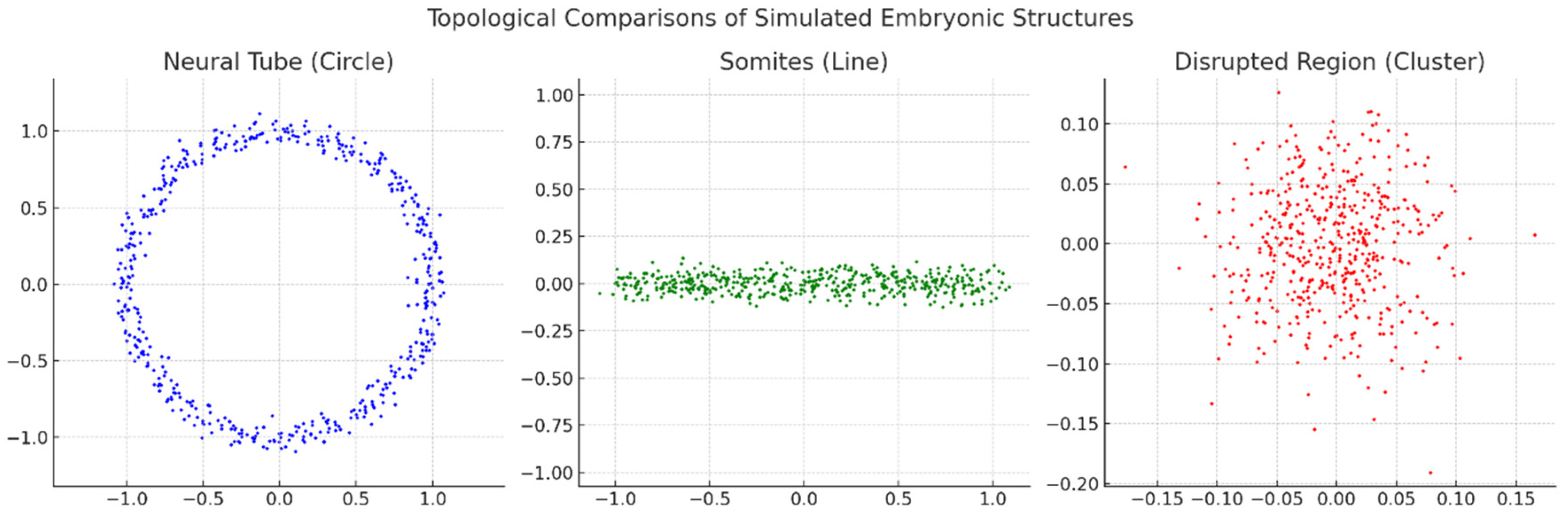
Section 3.1. Graphs Overview:

  • Neural Tube (Circle):
    The data points form a clear circular structure, representing the topology of a neural tube during embryonic development. This pattern signifies continuity and loop-like formation typical of such biological structures.
  • Somites (Line):
    The data points align in a linear configuration, symbolising somite development, which is segmental and elongated. This topological representation highlights a linear structure.
  • Disrupted Region (Cluster):
    The data points are scattered without a clear geometric or topological form, suggesting a disrupted or irregular developmental region. This randomness indicates a breakdown or abnormality in the structure.
Graph 1. These graphs use topology as a lens to explore and classify embryonic structures based on shape, symmetry, and abnormalities. The results could help identify normal versus abnormal development patterns. Blue Points: Neural Tube (Circle), Green Points: Somites (Line), Red Points: Disrupted Region (Cluster).
Preprints 140659 g001
Graph 2. This graph visually maps the hierarchy of embryonic development, showcasing how primary germ layers give rise to specialised tissues and organ systems. Blue Nodes: Ectoderm and derivatives, Green Nodes: Mesoderm and derivatives, Pink Nodes: Endoderm and derivatives.
Preprints 140659 g002

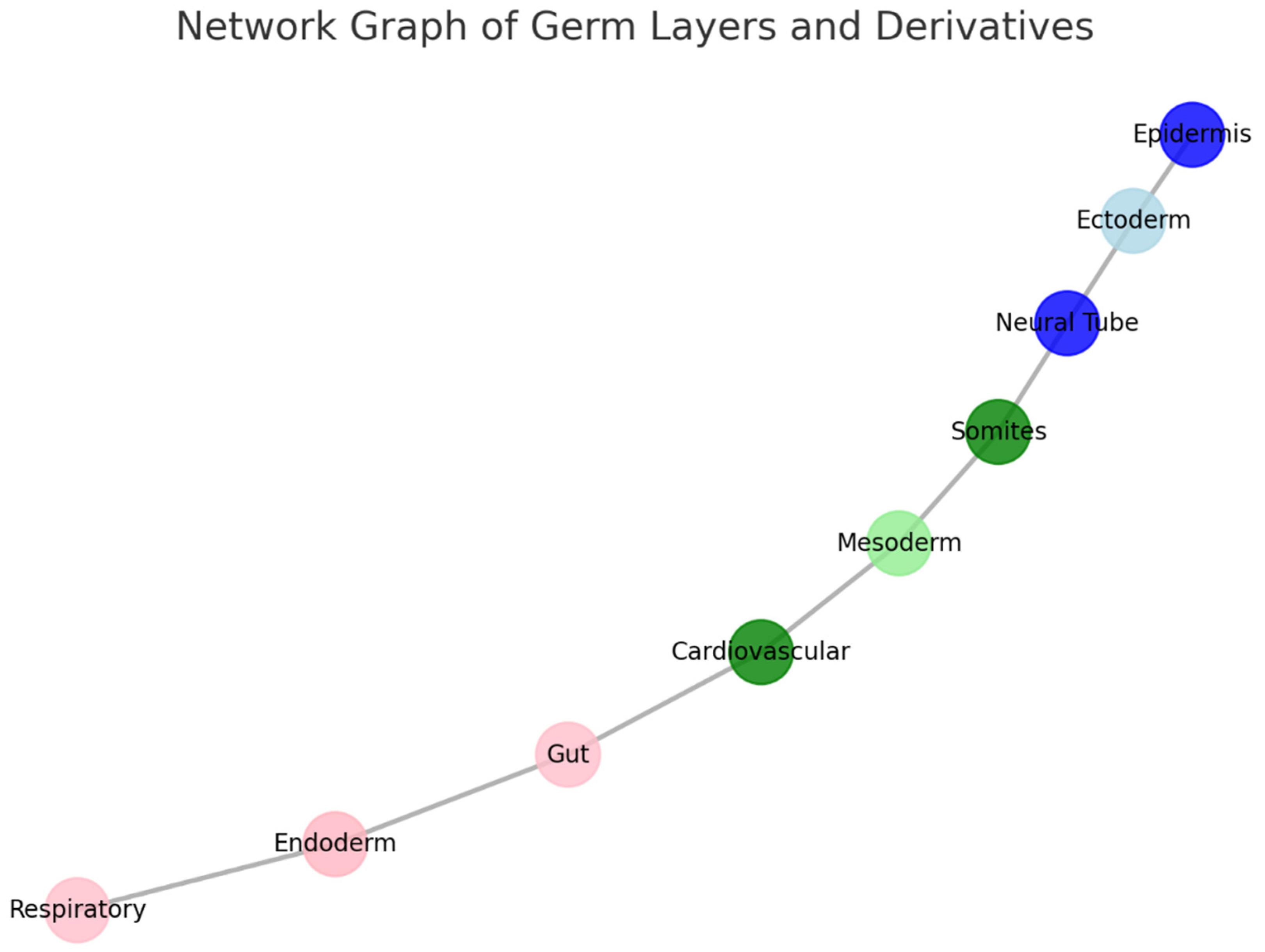
Section 3.2. Explanation:
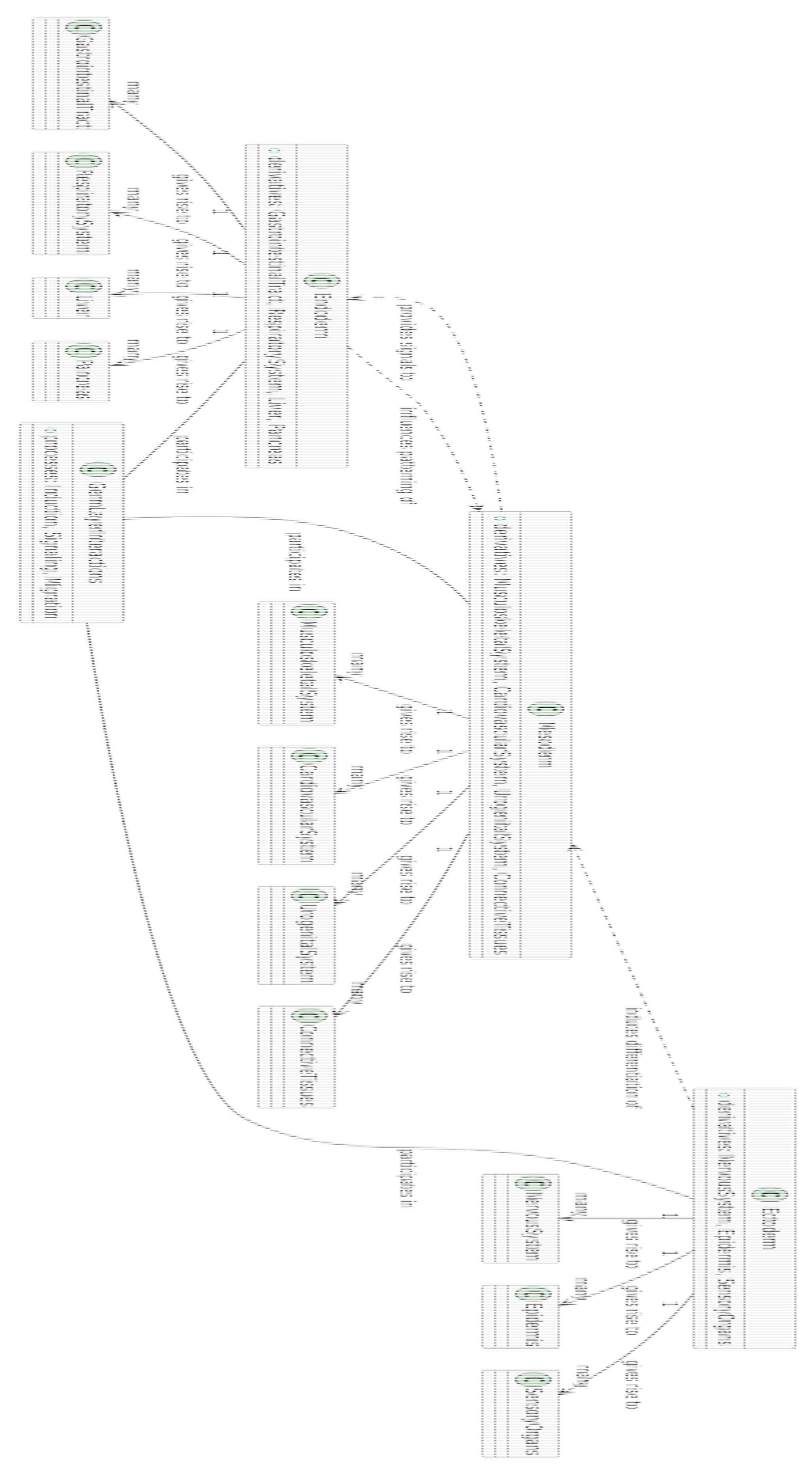
  • Nodes:
    Ectoderm (light blue) and its derivatives:
    Neural Tube (blue): Develops into the central nervous system.
    Epidermis (dark blue): Forms the outer skin layer.
    Mesoderm (green shades) and its derivatives:
    Somites (green): Give rise to skeletal muscles and vertebrae.
    Cardiovascular system (dark green): Includes the heart and blood vessels.
    Endoderm (pink shades) and its derivatives:
    Gut (red-pink): Forms the digestive tract.
    Respiratory system (light pink): Includes the lungs and airways.
  • Edges:
    The lines between nodes represent developmental pathways or relationships, tracing how germ layers differentiate into specific structures.
Graph 3. This figure shows 3D manifold representations of germ layers and their derivatives, highlighting the topological structures and potential relationships between the ectoderm, mesoderm, and endoderm.
Preprints 140659 g003

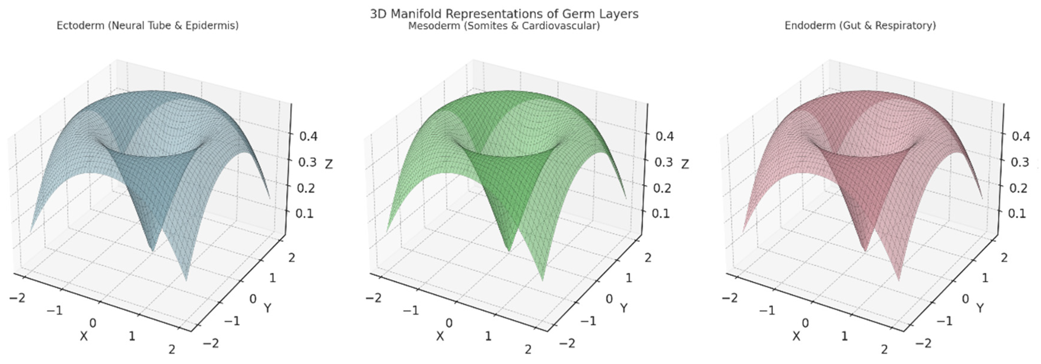
Section 3.3. Explanation of Each Plot:

  • Left Plot: Ectoderm (Neural Tube & Epidermis):
    A 3D surface representing the ectoderm and its derivatives (e.g., neural tube and epidermis).
    The structure suggests branching and separation into distinct pathways for neural and epidermal development.
    Axes:
    X,YX, YX,Y: Represent spatial or topological coordinates.
    ZZZ: Encodes aspects of differentiation or development over time.
  • Middle Plot: Mesoderm (Somites & Cardiovascular):
    Depicts the 3D topology of the mesoderm and its derivatives, including somites (segments) and the cardiovascular system.
    This manifold is smoother and broader, symbolising the more diverse range of tissues formed by the mesoderm.
    Axes:
    Similar spatial or developmental coordinates (X,Y, Z,X, Y, ZX, Y,Z).
  • Right Plot: Endoderm (Gut & Respiratory):
    Represents the endoderm's contribution to forming the gut and respiratory system.
    The surface exhibits a more funnel-shaped topology, indicating internalisation processes like tube formation for the digestive and respiratory tracts.
Key Features:
  • Topological Differences:
    The ectoderm's shape reflects branching for neural and external layers.
    The mesoderm's smoothness indicates the spread into multiple structures.
    The endoderm's inward topology corresponds to tube-like organ formation.
  • Interpretation:
    These manifolds highlight the spatial and developmental pathways, visually encoding differentiation patterns among the germ layers.
This representation aids in understanding how the germ layers' topology correlates with their developmental roles.
Graph 4. The dendrogram below maps the hierarchical and interconnected relationships between germ layers, molecular pathways, environmental factors, and developmental processes, providing an overview of normal and disrupted embryogenesis.
Preprints 140659 g004

Section 3.4. Explanation

This UML dendrogram uses various elements to represent the intricate interactions and relationships in biological systems. Rectangles with icons provide essential classifications: "C" denotes classes or components, which include key entities, processes, or pathways such as tissues, germ layers, and regulatory mechanisms. "M" represents molecular pathways, highlighting signalling cascades like the Notch pathway or SHH pathway that play pivotal roles in embryogenesis. Meanwhile, "P" stands for processes, capturing biological phenomena like segmentation, differentiation, or morphogenesis.
The arrows in the diagram indicate the nature of the relationships. Solid arrows represent direct dependencies or pathways, such as one component giving rise to another or a process controlled by a specific molecular mechanism. Dashed arrows, on the other hand, illustrate conditional influences, such as mutations affecting pathways or environmental factors altering processes. The diagram also reveals key relationships, including many-to-one dynamics, where multiple components or processes converge into a single outcome, and one-to-many dynamics, where a single process or pathway branches out into multiple downstream effects.
The content mapped in this dendrogram captures various aspects of development. It includes critical developmental pathways such as Wnt, SHH, and Notch, which play regulatory roles in embryogenesis. It also visualises the differentiation of germ layers into tissues like neural and mesodermal structures. Environmental influences, such as teratogens or nutrient deficiencies, are shown to potentially disrupt these processes. Additionally, the dendrogram highlights developmental disorders, showcasing how pathways and mutations can lead to conditions like spina bifida or holoprosencephaly.
Labels on the arrows clarify the nature of these relationships, using terms like "may regulate," "can cause," "interacts with," or "influenced by." Together, these elements create a comprehensive representation of the complex interactions driving embryonic development and potential disruptions that can lead to abnormalities.
Graph 5. Below, the dendrogram provides a comprehensive view of germ layer derivatives, their relationships, and the processes that guide embryonic development. It visually encodes the hierarchy and interactions crucial for organogenesis.
Preprints 140659 g005

Section 3.5. Explanation

The dendrogram visually represents the relationships between germ layers, their derivatives, and the biological processes involved in embryonic development. Rectangles with icons provide key information: "C" represents classes or components, which include germ layers such as the ectoderm, mesoderm, and endoderm, as well as their derivatives like specific tissues and organ systems. Meanwhile, "P" denotes processes like induction, migration, and patterning. Arrows are used to illustrate the connections between these elements, with solid arrows indicating direct relationships, such as one germ layer giving rise to specific organ systems, and dashed arrows showing influences or signalling interactions that regulate developmental processes. Labels on the arrows further clarify the nature of these interactions, describing relationships such as "gives rise to," "induces differentiation of," or "influences patterning of."
The diagram highlights key developmental pathways and relationships. For example, the ectoderm gives rise to the nervous system, including the brain and spinal cord, the epidermis, and sensory organs like the eyes and ears. It plays a critical role in neural and sensory organ development and interacts with other components to induce differentiation. The mesoderm, in contrast, contributes to the formation of the musculoskeletal system, the cardiovascular system, the urogenital system, and connective tissues. This layer is involved in diverse organ system development and participates in processes like signalling and cell migration. The endoderm primarily forms internal structures, including the gastrointestinal tract, respiratory system, liver, and pancreas. It provides essential signals that influence the patterning and differentiation of nearby tissues.
In addition to outlining these germ layer derivatives, the dendrogram emphasises the processes that guide their development. Induction and signalling are key, as germ layers interact to regulate the timing and differentiation of their derivatives. For instance, signals from the endoderm help shape the gastrointestinal and respiratory systems. Migration and differentiation processes are equally vital, as cells originating from the germ layers move to specific locations and transform into specialised tissues.
This interconnected network of germ layers, derivatives, and processes offers a comprehensive view of embryonic development, illustrating the complex interactions and pathways that drive organogenesis.

Section 4. Discussion

Section 4.1 Embryogenesis: The Interrelations between Topology, Molecular Pathways, and Developmental Disorders

Embryogenesis is the remarkable process by which a single-cell zygote transforms into a complex, multicellular organism. This transformation involves not only cellular proliferation and differentiation but also the spatial and temporal coordination of topological changes—such as folding, segmentation, and closure—that shape the embryo's structure. These morphological transformations are intricately linked with molecular signaling pathways and mechanical forces, creating a dynamic interaction that dictates normal development. Disruptions in these finely tuned processes can lead to congenital anomalies, highlighting the importance of understanding the underlying mechanisms.

Section 4.2. The Role of Topology in Embryogenesis

Topology, the mathematical study of properties preserved through deformations, finds profound applications in biology, especially during embryogenesis. The formation of complex structures in the embryo often involves topological transformations that maintain the continuity of tissues while altering their shape. A quintessential example is neural tube closure, a critical event in early development that gives rise to the central nervous system.
The neural tube forms from the neural plate, a flat layer of neuroepithelial cells. Through a series of coordinated movements and shape changes, the neural plate folds along the dorsal midline to create a tube. This process involves the elevation of neural folds and their convergence towards the midline, driven by mechanical forces such as apical constriction of neuroepithelial cells (Haigo et al., 2003). Cell adhesion molecules like N-cadherin facilitate the fusion of neural folds, ensuring the tube's continuity (Colas & Schoenwolf, 2001).
Molecular signals tightly regulate these mechanical processes. Sonic Hedgehog (SHH), secreted by the notochord and floor plate, establishes a ventral-to-dorsal gradient that patterns the neural tube, influencing cell fate decisions (Briscoe & Novitch, 2008). Bone Morphogenetic Proteins (BMPs), produced by the roof plate and surrounding ectoderm, form a dorsalizing gradient that promotes differentiation of dorsal neural cell types (Anderson et al., 2006). The balance between SHH and BMP signaling is crucial for proper neural tube development.
Failures in neural tube closure lead to neural tube defects (NTDs), such as spina bifida and anencephaly. These defects underscore the importance of maintaining topological integrity during development. Genetic mutations affecting SHH signaling components, folate metabolism enzymes, or planar cell polarity genes can disrupt neural tube closure (Greene et al., 2009). Environmental factors like folate deficiency also play a significant role, as folate is essential for DNA synthesis and methylation during rapid cell proliferation (Blom et al., 2006). This understanding has led to public health initiatives promoting folic acid supplementation, significantly reducing NTD incidence worldwide (Berry et al., 1999).

Section 4.3. Interactions and Shared Pathologies Among Germ Layers

The three primary germ layers—ectoderm, mesoderm, and endoderm—give rise to all tissues and organs in the body. Their development is highly interconnected, and disruptions can lead to overlapping pathologies. Neural crest cells (NCCs), derived from the ectoderm, exemplify this interconnection. NCCs migrate extensively and differentiate into a diverse range of cell types, contributing to structures traditionally associated with all three germ layers (Bronner & LeDouarin, 2012).
Disorders of NCC development, known as neurocristopathies, highlight the impact of germ layer interactions. Waardenburg syndrome, caused by mutations in genes such as PAX3 and MITF, results in pigmentary anomalies, sensorineural deafness, and facial dysmorphisms due to defective melanocyte and inner ear development (Read & Newton, 1997). DiGeorge syndrome, often resulting from a deletion on chromosome 22q11.2, affects NCC-derived tissues including the thymus, parathyroid glands, and parts of the heart, leading to immunodeficiency, hypocalcemia, and congenital heart defects (McDonald-McGinn et al., 2015).
Craniofacial development further illustrates germ layer interactions. Structures like the palate and upper lip form through the fusion of facial prominences derived from both ectodermal and mesenchymal tissues, with significant contributions from NCCs. Failure of these structures to fuse properly results in cleft lip and palate, common congenital anomalies with both genetic and environmental etiologies (Dixon et al., 2011). Genes such as IRF6 and environmental factors like maternal smoking influence the risk, emphasizing the complexity of germ layer interactions in development.
Neurocutaneous syndromes also demonstrate shared pathologies across germ layers. Neurofibromatosis type 1 (NF1), caused by mutations in the NF1 gene encoding neurofibromin, affects both the nervous system and the skin. Patients develop benign tumors along nerves (neurofibromas) and exhibit skin pigment changes like café-au-lait spots (Gutmann et al., 2017). This reflects the shared ectodermal origin of these tissues and the role of neurofibromin in regulating cell proliferation and differentiation.

Section 4.4. Molecular Pathways as Orchestrators of Topology

Molecular signaling pathways act as the blueprint guiding the topological transformations during embryogenesis. SHH signaling is a master regulator influencing multiple aspects of development. In addition to its role in neural tube patterning, SHH establishes the anterior-posterior axis in limb buds, determining digit identity (Riddle et al., 1993). Disruptions in SHH signaling can lead to holoprosencephaly, characterized by incomplete separation of the cerebral hemispheres, and limb abnormalities like polydactyly (Roessler & Muenke, 2010).
Bone Morphogenetic Proteins (BMPs) are critical for tissue differentiation and morphogenesis. They regulate not only neural tube closure but also skeletal development by influencing osteoblast and chondrocyte differentiation (Zhang & Bradley, 1996). Altered BMP signaling can result in craniosynostosis, where premature fusion of cranial sutures affects skull shape and brain development (Wilkie et al., 2007). BMP antagonists like Noggin and Chordin modulate BMP activity, highlighting the importance of precise regulation in development (McMahon et al., 1998).
The Wnt and Notch signaling pathways are essential for somite segmentation, which gives rise to the vertebral column and skeletal muscles. These pathways interact in a segmentation clock mechanism, with oscillations in gene expression coordinating the periodic formation of somites (Oates et al., 2012). Mutations affecting components of these pathways can lead to congenital vertebral anomalies and disorders like spondylocostal dysostosis, characterized by abnormal vertebral segmentation and rib anomalies (Sparrow et al., 2012).
Cross-talk between these molecular pathways ensures the coordinated development of tissues. For example, interactions between SHH and BMP signaling regulate dorsoventral patterning in the neural tube, while Wnt signaling modulates both SHH and BMP pathways during limb development. This intricate network of signaling underscores the complexity of embryogenesis and the potential for disruptions to cause developmental disorders.

Section 4.5. Clinical Implications and Future Perspectives

Understanding the mechanisms of embryogenesis has significant clinical implications. Preventive strategies have emerged from insights into developmental biology. The recognition of folate's role in neural tube closure led to folic acid supplementation recommendations for women of childbearing age, reducing the incidence of NTDs (Williams et al., 2015). Public health initiatives continue to emphasize maternal nutrition and avoidance of teratogens during pregnancy.
Advances in molecular biology have opened avenues for targeted therapies. Modulating SHH signaling is being explored for treating conditions like holoprosencephaly and certain cancers, though balancing therapeutic benefits with potential side effects is critical (Muenke et al., 2004). BMP inhibitors are under investigation for treating disorders involving excessive bone growth, such as fibrodysplasia ossificans progressiva (Shore et al., 2006). Gene therapy and gene editing technologies like CRISPR/Cas9 hold promise for correcting genetic defects, but ethical considerations and safety concerns remain paramount (Doudna & Charpentier, 2014).
Regenerative medicine and stem cell therapies offer potential for repairing or replacing damaged tissues. Induced pluripotent stem cells (iPSCs) provide a platform for modeling developmental disorders and testing therapeutic interventions. Combining these approaches with tissue engineering could lead to novel treatments for congenital anomalies.
Computational modeling enhances our understanding of embryogenesis by simulating complex biological processes. Finite Element Analysis (FEA) models mechanical forces during morphogenesis, providing insights into how changes in cellular properties influence development (Brodland & Veldhuis, 2006). Integrating computational tools like persistent homology with high-resolution imaging allows for quantitative analysis of embryonic structures, potentially aiding in early detection of developmental abnormalities (Edelsbrunner & Harer, 2010). Future research aims to combine these models with genetic and molecular data to create comprehensive maps of embryogenesis.

Section 5. Conclusion

Embryogenesis is a complex and finely tuned process involving mutual interrelations of topology, molecular pathways, and mechanical forces. Understanding this intricate choreography is essential for unraveling the mechanisms behind normal development and congenital anomalies. By integrating theoretical models, experimental data, and computational tools, we gain deeper insights into embryogenesis, leading to improved preventive strategies and therapies for developmental disorders. Continued interdisciplinary research promises to unveil new dimensions of developmental biology, ultimately enhancing human health and well-being.
*The Author claims there are no conflicts of interests

References

  1. Anderson, R. M., Lawrence, A. R., Stottmann, R. W., Bachiller, D., & Klingensmith, J. (2006). Chordin and noggin promote organizing centers of forebrain development in the mouse. Development, 133(21), 4935–4947. [CrossRef]
  2. Berry, R. J., Li, Z., Erickson, J. D., et al. (1999). Prevention of neural-tube defects with folic acid in China. New England Journal of Medicine, 341(20), 1485–1490. [CrossRef]
  3. Blom, H. J., Shaw, G. M., den Heijer, M., & Finnell, R. H. (2006). Neural tube defects and folate: case far from closed. Nature Reviews Neuroscience, 7(9), 724–731. [CrossRef]
  4. Briscoe, J., & Novitch, B. G. (2008). Regulatory pathways linking progenitor patterning, cell fates, and neurogenesis in the ventral neural tube. Philosophical Transactions of the Royal Society B: Biological Sciences, 363(1489), 57–70. [CrossRef]
  5. Brodland, G. W., & Veldhuis, J. H. (2006). Computer simulations of convergent extension: Cell rearrangements and tissue mechanical properties. Developmental Biology, 303(1), 256–270.
  6. Bronner, M. E., & LeDouarin, N. M. (2012). Development and evolution of the neural crest: an overview. Developmental Biology, 366(1), 2–9. [CrossRef]
  7. Colas, J. F., & Schoenwolf, G. C. (2001). Towards a cellular and molecular understanding of neurulation. Developmental Dynamics, 221(2), 117–145. [CrossRef]
  8. Dixon, M. J., Marazita, M. L., Beaty, T. H., & Murray, J. C. (2011). Cleft lip and palate: understanding genetic and environmental influences. Nature Reviews Genetics, 12(3), 167–178. [CrossRef]
  9. Doudna, J. A., & Charpentier, E. (2014). The new frontier of genome engineering with CRISPR-Cas9. Science, 346(6213), 1258096. [CrossRef]
  10. Edelsbrunner, H., & Harer, J. (2010). Computational Topology: An Introduction. American Mathematical Society.
  11. Greene, N. D., & Copp, A. J. (2009). Development of the vertebrate central nervous system: formation of the neural tube. Prenatal Diagnosis, 29(4), 303–311. [CrossRef]
  12. Gutmann, D. H., Ferner, R. E., Listernick, R. H., Korf, B. R., Wolters, P. L., & Johnson, K. J. (2017). Neurofibromatosis type 1. Nature Reviews Disease Primers, 3, 17004. [CrossRef]
  13. Haigo, S. L., Hildebrand, J. D., Harland, R. M., & Wallingford, J. B. (2003). Shroom induces apical constriction and is required for hingepoint formation during neural tube closure. Current Biology, 13(24), 2125–2137. [CrossRef]
  14. McDonald-McGinn, D. M., Sullivan, K. E., Marino, B., et al. (2015). 22q11.2 deletion syndrome. Nature Reviews Disease Primers, 1, 15071.
  15. McMahon, J. A., Takada, S., Zimmerman, L. B., et al. (1998). Noggin-mediated antagonism of BMP signaling is required for growth and patterning of the neural tube and somite. Genes & Development, 12(10), 1438–1452. [CrossRef]
  16. Muenke, M., Beachy, P. A., & Rosenthal, A. (2004). Therapeutic implications of possible genetic and teratogenic interactions in holoprosencephaly. Current Opinion in Pediatrics, 16(6), 614–621.
  17. Oates, A. C., Morelli, L. G., & Ares, S. (2012). Patterning embryos with oscillations: structure, function and dynamics of the vertebrate segmentation clock. Development, 139(4), 625–639. [CrossRef]
  18. Read, A. P., & Newton, V. E. (1997). Waardenburg syndrome. Journal of Medical Genetics, 34(8), 656–665. [CrossRef]
  19. Riddle, R. D., Johnson, R. L., Laufer, E., & Tabin, C. (1993). Sonic hedgehog mediates the polarizing activity of the ZPA. Cell, 75(7), 1401–1416. [CrossRef]
  20. Roessler, E., & Muenke, M. (2010). The molecular genetics of holoprosencephaly. American Journal of Medical Genetics Part C: Seminars in Medical Genetics, 154C(1), 52–61. [CrossRef]
  21. Shore, E. M., Xu, M., Feldman, G. J., et al. (2006). A recurrent mutation in the BMP type I receptor ACVR1 causes inherited and sporadic fibrodysplasia ossificans progressiva. Nature Genetics, 38(5), 525–527. [CrossRef]
  22. Sparrow, D. B., Chapman, G., & Dunwoodie, S. L. (2012). The mouse notches up another success: understanding the causes of human vertebral malformation. Mammalian Genome, 23(1-2), 27–40. [CrossRef]
  23. Williams, J., Mai, C. T., Mulinare, J., et al. (2015). Updated estimates of neural tube defects prevented by mandatory folic acid fortification—United States, 1995–2011. Morbidity and Mortality Weekly Report, 64(1), 1–5.
  24. Wilkie, A. O., & Morriss-Kay, G. M. (2001). Genetics of craniofacial development and malformation. Nature Reviews Genetics, 2(6), 458–468. [CrossRef]
  25. Zhang, H., & Bradley, A. (1996). Mice deficient for BMP2 are nonviable and have defects in amnion/chorion and cardiac development. Development, 122(10), 2977–2986.
  26. Copp, A. J., Greene, N. D. E., & Murdoch, J. N. (2003). The genetic basis of mammalian neurulation. Nature Reviews Genetics, 4(10), 784–793. [CrossRef]
  27. Greene, N. D. E., & Copp, A. J. (2014). Neural tube defects. Annual Review of Neuroscience, 37, 221–242. [CrossRef]
  28. Dixon, M. J., Marazita, M. L., Beaty, T. H., & Murray, J. C. (2011). Cleft lip and palate: understanding genetic and environmental influences. Nature Reviews Genetics, 12(3), 167–178. [CrossRef]
  29. Lan, Y., Xu, J., & Jiang, R. (2015). Neural crest cells regulate FGF-dependent epithelial-mesenchymal interactions during palatal development. Science Signaling, 8(404), ra89. [CrossRef]
  30. Oates, A. C., Morelli, L. G., & Ares, S. (2012). Patterning embryos with oscillations: Structure, function and dynamics of the vertebrate segmentation clock. Development, 139(4), 625–639. [CrossRef]
  31. Clugston, R. D., & Klatt, J. (2010). Embryogenesis of congenital diaphragmatic hernia. Seminars in Pediatric Surgery, 19(2), 82–90. [CrossRef]
  32. Trainor, P. A. (2010). Neural crest cells: Evolution, development, and disease. Developmental Biology, 344(1), 3–6. [CrossRef]
Disclaimer/Publisher’s Note: The statements, opinions and data contained in all publications are solely those of the individual author(s) and contributor(s) and not of MDPI and/or the editor(s). MDPI and/or the editor(s) disclaim responsibility for any injury to people or property resulting from any ideas, methods, instructions or products referred to in the content.
Copyright: This open access article is published under a Creative Commons CC BY 4.0 license, which permit the free download, distribution, and reuse, provided that the author and preprint are cited in any reuse.
Prerpints.org logo

Preprints.org is a free preprint server supported by MDPI in Basel, Switzerland.

Subscribe

Disclaimer

Terms of Use

Privacy Policy

Privacy Settings

© 2025 MDPI (Basel, Switzerland) unless otherwise stated