
Submitted:
26 September 2024
Posted:
27 September 2024
You are already at the latest version
Abstract
Two new harziane diterpenes (1-2), five undescribed cyclonerane sesquiterpenes (3-7) and three known compounds, 11-cycloneren-3,7,10-triol (8), harziandione (9), and dehydroacetic acid (10) were isolated from Trichoderma hamatum b-3. Their structures were elucidated via comprehensive inspection of spectral evidences in HRESIMS, 1D and 2D NMR, the absolute configuration of 1-8 was confirmed by NMR, ECD calculation as well as Mosher's method. In vitro fungicidal activity showed that some compounds showed great inhibitory activity against pathogenic fungi including Fusarium graminearum, Sclerotinia sclerotiorum, Botrytis cinerea, and Rhizoctonia solani, among which compound 10 showed 100% inhibition of S. sclerotiorum and B. cinerea. In vivo activity test showed that compound 10 was 65.8% effective against B. cinerea and has the potential to be developed as a biocontrol pesticide.
Keywords:
Trichoderma
; diterpene
; sesquiterpene
; fungicidal activity
1. Introduction
The continuous growth of the global population exerts tremendous pressure on agricultural production systems. Concurrently, the food and economic losses caused by plant pathogens in agriculture have raised serious concerns [1,2]. Chemical control can mitigate plant diseases and enhance grain yield, making it one of the most effective methods for prevention and control. However, the frequent use of chemical pesticides can result in pesticide residues in crops; contaminate soil and water sources; as well as pose risks to human health [3,4]. In contrast, biological control involves managing plant diseases through microorganisms and their metabolites, offering a safer, more effective, and environmentally friendly alternative to chemical control [5]. Trichoderma strains are widely utilized in the biological control of plant diseases within agriculture, employing diverse mechanisms such as competition, antibiosis, induced systemic resistance and mycoparasitism [6,7,8,9].
Antibiosis is a primary biological control mechanism employed by Trichoderma, which involves the production of secondary metabolites that inhibit the growth of pathogenic fungi [10,11]. Trichoderma strains are known to produce a diverse array of natural products, including epipolythiodioxopiperazines [12], peptaibols [13], pyrones [14], butenolides [15], koninginins [16], steroids [17], lactones [18], and trichothecenes [19,20]. Our research team has been dedicated to the study of fungal secondary metabolites with bioactive properties over an extend period, resulting in the isolation of several previously undescribed compounds from fungal strains such as Chaetomium elatum [21], and Nigrospora sphaerica [22]. In this study, we investigated the secondary metabolites of Trichoderma hamatum, leading to the isolation of two new harziane diterpenes (1-2) and five cyclonerane sesquiterpenes (3-7). The structures of these compounds were determined by HRESIMS, NMR, ECD, Mosher's method [23] combined with chemical calculation. Compounds 1-10 were tested for their in vitro fungicidal activity against six agriculture pathogenic fungi. Compound 10 was then tested for in vivo fungicidal activities against some pathogenic fungi. Herein, we report the details of isolation, elucidation and fungicidal activity of above-mentioned secondary metabolites.
2. Materials and Methods
2.1. General Experimental Procedures
Optical rotation was measured on a Perkin-Elmer 241 Polarimeter (Perkin Elmer, Inc., Waltham, MA, USA). UV, CD and ICD spectra were measured on a Chirascan circular dichroism (Applied Photophysics Ltd., Leatherhead, UK). IR spectrum was acquired from an Agilent Cary 600 FT-IR infrared spectrometer (Agilent Technologies, Santa Clara, USA). Mass spectrum was obtained on an Ultra-performance liquid chromatography coupled with Q Exactive Quadrupole-Electrostatic field Orbital trap high resolution mass spectrometer (Thermo Fisher Scientific, Bremen, Germany). NMR spectra were measured with a Bruker Ascend 600 MHz (Burker, Karlsruhe, Germany) with TMS as internal standard. Fractionation was conducted on a Column chromatography silica gel (200-300 mesh). Sample fractionated by dynamic axial compression column (Hanbon Sci.& Tech, Huaian, China). Purification was performed with a NP7000 preparative high performance liquid chromatograph (Hanbon Sci.& Tech, Huaian, China) with a kromasil C18 5 μm semi-preparative column (10×250 mm). Methanol, acetone, ethyl acetate are analytical pure and dimethyl sulfoxide (chromatographic pure) were purchased from Chengdu Kelong Chemical Co., Ltd. Dichloromethane (analytically pure), methanol (chromatography preparative pure) and acetonitrile (chromatography preparative pure) were purchased from Chengdu Jinshan Chemical Reagent Co., Ltd. Deuterated chloroform was purchased from Adamas Reagent Co., Ltd. N, N-dimethylformamide (analytically pure) was purchased from Tianjin Fuyu Fine Chemical Co. Ltd. Pyrimethanil and dimethachlon (analytically pure) were purchased from Alta Scientific Co., Ltd.
2.2. Fungal Material and Fermentation
The strain (GenBank accession no. OR553890, sequence no. NR134371.1) was isolated from the rhizomes of Bergenia purpurascens collected from Mount Emei and identified to be T. hamatum by Shanghai Sangong Bioengineering Co., Ltd. through morphological analysis and BLAST comparison.
Fungus 2 medium (maltose 20 g; monosodium glutamate 10 g; dextrose 10 g; yeast paste 3 g; corn syrup 1 g; mannitol 20 g; KH2PO4 0.5 g; MgSO4.7H2O 0.3 g; CaCO3 20 g; 5-azacytidine 10 mg; distilled water 1 L) was used for the cultivation of T. hamatum b-3[24]. The experimental strains were inoculated onto potato dextrose agar (PDA) medium for duration of 5 days. Subsequently, five pieces (0.5×0.5 cm2) of mycelial agar plugs were transferred into sterilized and cooled Fungi No. 2 liquid medium contained in 500 mL conical flasks, with 200 mL medium per flask. These flasks were then placed in a constant temperature shaker set at 28 ℃ and 120 rpm/min for 7 days to obtain the T. hamatum seed cultures. Following this, the seed cultures were inoculated into the sterilized Fungus 2 medium for expansion cultures, with 2.5 mL of seed cultures added into each 100 mL of medium. The inoculated medium was cultured in a constant temperature shaker at 28 ℃ and 120 rpm/min for 14 days to produce the fermentation broth of T. hamatum b-3 resulting in a total culture volume of 564.4 L.
2.3. Extraction and Isolation
The cultured fermentation broth was centrifuged at 4000 r/min in a high-speed centrifuge to separate the fermentation broth. The broth was then treated with an equal volumn of ethyl acetate for 24 h. Subsequently, the extraction process was repeated three times using an equal volume of ethyl acetate. The ethyl acetate layer was collected and concentrated under reduced pressure, yielding 126 g of ethyl acetate extract.
The ethyl acetate extract was separated by silica gel CC, eluted with petroleum ether and acetone (30: 1~1: 1, V/V), yield seven fractions (Fr.1~Fr.7). Fr.1 was separated through column chromatography on dynamic axial compression column (MeOH/H2O, 90: 10) and semi-pHPLC (MeOH/H2O, 68: 32) to obtain 9 (11.5, tR 20.3 min). Fr.2 was separated via RP-18 CC (MeOH/H2O, 95:5) to afford three fractions (Fr.2.1~ Fr.2.3). Fr.2.1 was then separated through semi-pHPLC (MeOH/H2O, 50: 50 to 100:0) to obtain eleven fractions (Fr.2.1.1~ Fr.2.1.11), Fr.2.1.5 was then separated through semi-pHPLC (MeOH/H2O, 40: 60) to yield 10 (87 mg, tR 25.8 min). Fr.2.1.6 was subjected to silica gel CC and semi-pHPLC (MeOH/H2O, 70: 30) to yield 1 (1.8 mg, tR 28.0 min). Fr.2.1.8 was separated via Sephadex LH-20 CC (CH2Cl2/MeOH, 70: 30) and purified by semi-pHPLC (MeCN/H2O, 20: 80) to produce 8 (13.5 mg,tR 30.3 min) and semi-pHPLC (MeCN/H2O, 24: 76) to produce 4 (1.8 mg, tR 19.7 min) and 5 (9.3 mg, tR 26.6 min). Fr.3 was separated through dynamic axial compression column chromatography (MeOH/H2O, 50:50 to 50:100) to give six fractions (Fr.3.1~ Fr.3.6), Fr.3.4 was further purified by CC on Sephadex LH-20 (MeOH) as well as semi-pHPLC (MeCN/H2O, 57:43) to afford 3 (1.3 mg, tR 8.3 min). Fr.3.4.1.10 was further purified by semi-pHPLC (MeCN/H2O, 35: 65) to obtained 6 (1.4 mg, tR 35.4 min). Fr.5 was separated via dynamic axial compression column chromatography (MeOH/H2O, 60: 40 to 100: 0) to give five fractions (Fr.5.1~ Fr.5.5), Fr.5.2 was then purified by CC on Sephadex LH-20 (CH2Cl2/MeOH, 70:30) and semi-pHPLC (MeCN/H2O, 40:60) to yield 7 (5.7 mg, tR 19.4 min).
Compound 1: Colorless oil; = +81.8 (c = 0.011, MeOH); UV (MeOH) λmax 248 (0.58); IR (KBr): 3449, 2920, 2851, 1740, 1384, 1234, 1026 cm-1; CD (c 2.23 mM, MeOH) λmax (Δε) 238 (-1.21), 295 (0.53), 342 (0.67) nm; HRESIMS m/z 359.2212 [M+H]+ (calcd. For C22H31O4+, 359.2217); For 1H-NMR, 13C-NMR data see Table 1 and Table 2.
Compound 2: Colorless oil; = +7.1 (c = 0.028, MeOH); UV (MeOH) λmax 255 (0.48); IR (KBr): 3439, 2936, 1730, 1643, 1386, 1199 cm-1; CD (c 2.51 mM, MeOH) λmax (Δε) 222 (-0.31), 251 (-0.73), 299 (0.11), 343 (0.53) nm; HRESIMS m/z 319.2268 [M+H]+ (calcd. For C20H31O3+, 319.2268); For 1H-NMR, 13C-NMR data see Table 1 and Table 2.
Compound 3: Colorless oil; = -8.0 (c = 0.025, MeOH); UV (MeOH) λmax 209 (4.32); IR (KBr): 3436, 2958, 2926, 2850, 1666, 1622, 1386 cm-1; CD (c 3.05 mM, MeOH) λmax (Δε) 224 (0.33), 321 (-0.12) nm; HRESIMS m/z 263.1624 [M+Na]+ (calcd. For C14H24NaO3+, 263.1618); For 1H-NMR, 13C-NMR data see Table 3 and Table 4.
Compound 4: Colorless oil; = -24.0 (c = 0.05, MeOH); IR (KBr): 3424, 2962, 2875, 1716, 1647, 1455, 1377, 918 cm-1; HRESIMS m/z 279.1940 [M+Na]+ (calcd. For C15H28NaO3+, 279.1931); For 1H-NMR, 13C-NMR data see Table 3 and Table 4.
Compound 5: Colorless oil; [α]20 D -19.7 (c 0.20, MeOH); UV (MeOH) λmax 213 (0.49); IR (KBr): 3402, 2966, 2931, 1458, 1377, 1156, 920 cm-1; CD (c 2.94 mM, MeOH) λmax (Δε) 216 (-0.05) nm; HRESIMS m/z 271.1910[M-H]- (calcd. For C15H27O4-, 271.1914); For 1H-NMR, 13C-NMR data see Table 3 and Table 4.
Compound 6: colorless oil; = -3.2 (c = 0.06, MeOH); IR (KBr): 3453, 2964, 2937, 1737, 1459, 1383, 1211, 1169 cm-1; CD (c 1.62 mM, MeOH) λmax (Δε) 205 (-0.21), 243 (-0.04), 307 (0.3) nm; HRESIMS m/z 393.2249 [M+Na]+ (calcd. For C20H34NaO6+, 393.2248); For 1H-NMR, 13C-NMR data see Table 1 and Table 2.
2.4. NMR and ECD Calculation Methods
The chemical calculations of compounds were conducted using Gaussian 16 1. Initially, a conformational analysis was performed with Conflex 8 to generate conformations via Boltzmann Jump [25]. All geometric configurations with relative energies between 0 and 5.0 kcal/mol were optimized at the B3LYP/6-31G level in the gas phase, as well as at ωB97XD/DGDZVP level in methanol. Room-temperature equilibrium populations were determined based on the Boltzmann distribution law [26]. Shielding tensor calculations were conducted at the PCM/mPW1PW91/6-31+G (d, p) level (with Boltzmann distribution ≥1%) employing the GIAO method [27]. The isotropic values of TMS were calculated at the same level and used as a reference. The DP4+ parameters were computed using the Excel file provided by Sarotti [28]. ECD calculations were performed using TD-DFT at the CAM-B3LYP/DGDZVP level in methanol. The ECD spectra were generated by considering the Boltzmann distribution of each geometric conformation. Subsequently, SpecDis 1.71 was utilized to combine the individual CD spectra with a Boltzmann statistical weighting, resulting in a Gaussian curve (σ = 0.16-0.4 eV), which was then compared with experimental data.
2.5. Fungicidal Activity Assay of Compounds 1-10 In Vitro
Pathogenic fungi, including Alternaria solani, Fusarium graminearum, Phytophthora capsici, Sclerotinia sclerotiorum, Botrytis cinerea, and Rhizoctonia solani, were inoculated onto petri dishes containing a compound solution at a concentration of 50 μg/mL. The dishes were then incubated in a biochemical incubator at 25 °C in the dark. The assessment of bactericidal activity was conducted following a three days incubation period, with each experimental group replicated three times. The control group was treated with sterile water. The results of the activity assessment were quantified on a percentage scale ranging from 0 to 100, where 0 indicates no activity and 100 signifies complete eradication [29].
2.6. Fungicidal Activity Assay of Compound 10 In Vivo
The commercial fungicides pyrimethanil and dimethachlon were utilized as positive controls in this study. Both compounds, along with the controls, were dissolved in N, N-dimethylformamide and subsequently diluted to a concentration of 200 µg/mL. Cucumber plants were then sprayed with these solutions and allowed to air dry for approximately 2 h. Following this drying period, the undersurfaces of the treated cucumber leaves were sprayed with a pathogen spore suspension containing approximately 1×10⁴ spores/mL. The plants were then placed in an incubator maintained at 20 °C with humidity levels exceeding 90% for a 5 day infection period. After this initial incubation, the plants were transferred to a greenhouse for an additional 5 days before being assessed for disease control scores [29].
3. Results
3.1. Structural Identification of Compounds
Compound 1 was isolated as a colorless oil, with its molecular formula determined to be C22H30O4 by HRESIMS, revealing eight degrees of unsaturation. The FT-IR absorption bands indicate the presence of hydroxyl (3449 cm−1), methyl (2920 and 2851 cm−1), carbonyl (1740 cm−1) groups. The 1H-NMR, 13C-NMR (Table 1 and Table 2) and HSQC spectra reveal the presence of three carbonyl groups [δC: 214.2 (C-3), 196.5 (C-11), 170.9 (C-21)], a set of conjugated double bonds [δC: 153.0 (C-10), 141.1 (C-9)], six aliphatic methylene groups [δH: 5.12 (H-20a), 4.76 (H-20b), 2.90 (H-4a), 2.66 (H-12a), 2.51 (H-12b), 2.29 (H-8), 2.09 (H-4b), 2.05 (H-15a), 1.97 (H-7a), 1.54 (H-15b), 1.40 (H-7b); δC: 63.3 (C-20), 60.2 (C-12), 42.7 (C-4), 30.3 (C-7), 26.7(C-15), 23.8 (C-8)], three hypomethyl groups [δH: 2.90 (H-5), 2.52 (H-14), 2.29 (H-2); δC: 59.4 (C-2), 52.9 (C-14), 30.1 (C-5)], five methyl groups [δH: 2.10 (H-22), 1.54 (H-19), 1.13 (H-18), 1.01 (H-17), 1.00 (H-16); δC: 25.2 (C-16), 23.4 (C-17), 21.1 (C-18), 21.0 (C-22)], 20.6 (C-19)] and three quaternary carbons [δC: 51.8 (C-6), 49.7 (C-1), 40.4 (C-13)]. The 1H-1H COSY correlations of H-2/H-15/H-14; H-4/H-5/H-18; and H-7/H-8 along with HMBC correlations of H-2, H-4, H-15/C-3; H-18/C-4, C-5, C-6; H-16, H-17/C-1, C-2, C-6; H-7/C-5; H-19/C-10, C-12, C-13, C-14; H-20/C-8, C-9, C-10, C-21 and of H-22/C-21, elucidated the plannar structure of 1 as shown in Figure 1.
The relative configuration of compound 1 was determined to be 2S*, 5R*, 6R*, 13S*, 14S* based on the NOESY correlations of H-16/H-14, H-2 and H-5/H-19. The absolute configuration of 1 was confirmed to be 2S, 5R, 6R, 13S, 14S by subsequent ECD calculations.
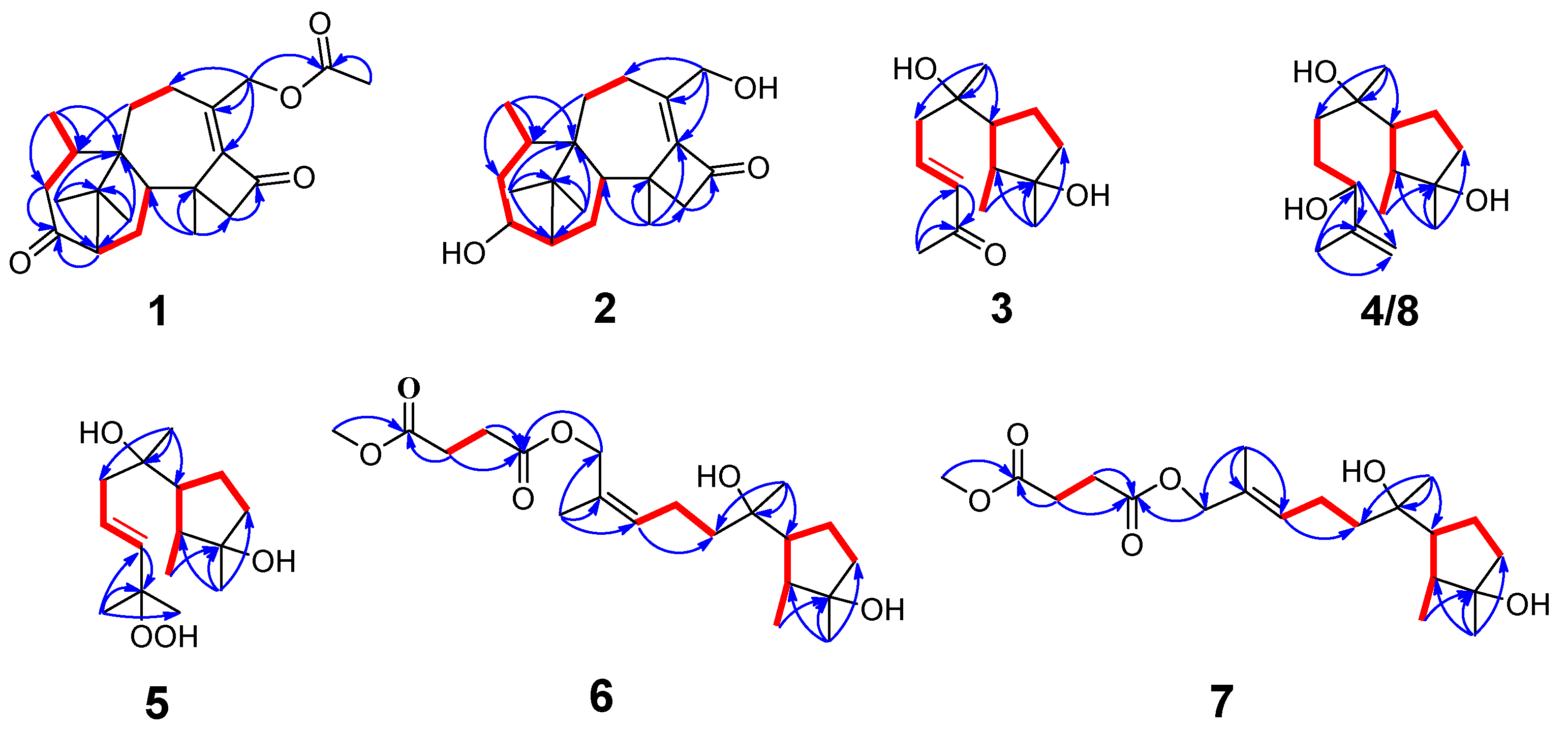
Figure 2.
Key HMBC (bold lines) and 1H–1H COSY (arrows) correlations of compound 1–8.
Compound 2 was purified as colorless oil, and its molecular formula was deduced to be C20H30O3 by (+)-HRESIMS m/z 319.2268 [M+H]+ (calcd. For C20H31O3+, 319.2268), implying six degrees of unsaturation. The IR spectrum of 2 showed absorption bands for hydroxyl (3439 cm−1), methyl (2936 cm−1) and carbonyl (1730 cm−1). The 1H-NMR, 13C-NMR (Table 1 and Table 2) and HSQC spectra indicate that compound 2 is a harziane diterpenes type compound like compound 1, with the difference that the substituents of compound 2 at C-3 and C-20 are both hydroxyl groups. The entire structure was confirmed by the 1H-1H COSY correlations of H-14/H-15/H-2/H-3/H-4/H-5/H-18; H-7/H-8 and the HMBC correlations of H-7/C-5; H-12/C-11; H-16, H-17/C-1, C-2, C-6; H-18/C-4, C-5, C-6; H-19/C-10, C-12, C-13, C-14 as well as H-20/C-8, C-9, C-10. The relative configuration of compound 2 was assigned to be 2S*, 3S*, 5R*, 6R*, 13S*, 14S* by the NOESY correlation of H-15b/H-19, H-3; H-19/H-5. The experimental and calculated ECD spectra did match well, suggesting 2S, 3S, 5R, 6R, 13S, 14S was the correct absolute configuration (Figure 4).
Compound 3 was isolated as colorless oil, and its molecular formula was deduced to be C14H24O3 by (+)-HRESIMS m/z 263.1624 [M+Na]+ (calcd. For C14H24NaO3+, 263.1618), implying three degrees of unsaturation. The FT-IR absorption band suggested the presence of hydroxy group (3436 cm−1), methyl group (2958 cm−1, 2926 cm−1 and 2850 cm−1), olefinic group (1622 cm−1) and carbonyl group (1666 cm−1). The 1H-NMR, 13C-NMR (Table 3 and Table 4) and HSQC spectra indicate the presence of one carbonyl signal [δC: 198.5 (C-11)], a group of conjugated olefin signals [δH: 6.88 (H-9), 6.13 (H-10); δC: 144.1 (C-9), 134.2 (C-10)], three aliphatic methylene signals [δH: 2.45 (H-8a), 2.36 (H-8b), 1.91 (H-5a), 1.72 (H-4a), 1.58 (H-4b, 5b); δC: 43.8 (C-8), 40.4 (C-4), 24.6 (C-5)], two hypomethyl signals [δH: 1.86 (H-6), 1.62 (H-2); δC: 54.9 (C-6), 44.6 (C-2)],two oxidized quaternary carbon signals [δC: 81.4 (C-3), 75.0 (C-7)] and four methyl signals [δH: 2.27 (H-12), 1.27 (H-13), 1.19 (H-14), 1.06 (H-1); δC: 27.2 (C-12), 26.2 (C-13), 25.9 (C-14) , 14.6 (C-1)]. The 1H-1H COSY correlations of H-1/H-2/H-6/H-5/H-4; H-8/H-9/H-10 as well as HMBC correlations of H-1/C-3, H-13/C-2, C-3, C-4; H-14/C-6, C-7, C-8; H-10/C-11 and of H-12/C-10, C-11 generated the planar structure of 3. The configuration of the double bond at C-9 was assigned as trans by the NOESY correlations signal of H-8/H-10, and the large coupling constant between H-9 and H-10. The NOESY correlations of H-2/H-13 and H-1/H-6 indicates the relative configuration of C-2, C-3, C-6.
The absolute configuration of C-7 could not be determined by ECD. To solve this difficult stereoscopic problem, the NMR data of 3a (2S*, 3R*, 6R*, 7R*) and 3b (2S*, 3R*, 6R*, 7S*) were further calculated at the PCM/mPW1PW91/6–31+G (d, p) level using the GIAO method [26]. The calculated 13C NMR chemical shifts of 3a showed a better agreement with the experimental values of compound 3 with a higher correlation coefficient [R2 for 3a: 0.9958; R2 for 3b: 0.9956, Figure S 79]. In addition, DP4+ probability analysis [27] based on both 1H and 13C NMR data predicated 3a as the correct relative structure with 96.49 % probability (Tables S 13). The experimental and calculated ECD spectra for 2S, 3R, 6R, 7R in MeOH did match well, indicating that 2S, 3R, 6R, 7R was the correct absolute configuration (Figure 4).
Compound 4 was isolated as colorless oil, its molecular formula was determined as C15H28O3 by (+)-HRESIMS m/z 279.1940 [M+Na]+ (calcd. For C15H28NaO3+, 279.1931), implying two degrees of unsaturation. The FT-IR absorption band indicate that compound 4 contains hydroxyl group (3424 cm−1), methyl group (2962 and 2875 cm−1), olefinic groups (1647 cm−1). The planar structure of 4 was deduced to be the same as that of 11-cycloneren-3,7,10- triol [30] by comparison of its 1H- and 13C-NMR data (Table 3 and Table 4) with those for 4, which was supported by HMBC correlations of and 1H-1H COSY correlations (Figure 2).
The relative configurations of the cyclopentane in compound 4 were determined to be 2S*, 3R*, 6R* by the NOESY-related signals of H-1/H-6 as well as H-2/H-13. Since the amount of compound 4 was not sufficient for the Mosher's reaction, we determined its absolute configuration by calculating NMR and ECD. The relative configuration of the compound 4 was further determined to be 2S*, 3R*, 6R*, 7R*, 10R* based on DP4+ probability (100% for 2S*, 3R*, 6R*, 7R*, 10R*, Table S 26) analysis of 1H-NMR and 13C-NMR data. The absolute configuration of compound 4 was determined to be 2S, 3R, 6R, 7R, 10R based on the comparison of calculated and experimental ECD spectra (Figure 4).
Compound 5 was separated as colorless oil, with the molecular formula C15H28O4 established by the (-)-HRESIMS data at m/z 271.1910 [M-H]- (calcd. For C15H27O4- 271.1914), indicating two degrees of unsaturation. The FT-IR absorption band indicate that compound 5 contains hydroxyl group (3402 cm−1) and methyl group (2966 and 2931 cm−1). The NMR data of 5 (Table 3 and Table 4) closely resembled those of 9-cycloneren-3, 7, 11-triol [30], with the main difference is that -OH at the C-10 is substituted with -OOH. This was demonstrated by the HRESIMS data and the HMBC correlations of H-10/H-11; H-12/H-10, H-11, H-15. Other HMBC and 1H-1H COSY correlations confirm the entire structure of the compound 5. The trans relative configuration of C-9 and C-10 was established according to the large coupling constant of H-9 and H-10 and the NOESY correlations of H-10/H-8 (Figure 3). The relative configuration of 5 was determined to be 2S*, 3R*, 6R* by NOESY cross-peaks of H-2/H-13 and of H-1/H-6.
Additionally, the relative configuration of C-7 was deduced as 7R* by NMR calculations with the DP4+ probability of 100% (Table S 35). The absolute configuration of compound 5 was determined to be 2S, 3R, 6R, 7R by comparing the calculated ECD and experimental ECD spectra (Figure 4).
Compound 6 was isolated as colorless oil, its molecular formula was deduced to be C20H34O6 by (+)-HRESIMS m/z 393.2249 [M+Na]+ (calcd. For C20H34NaO6+, 393.2248), with four degrees of unsaturation. The FT-IR absorption band suggested the presence of hydroxy group (3453 cm−1), methyl group (2964 and 2937 cm−1) and carbonyl group (1737 cm−1). The 1H-NMR, 13C-NMR(Table 1 and Table 2)and HSQC spectra indicate the presence of two carbonyl signal [δC: 172.9 (C-16), 172.4 (C-13)], a group of conjugated olefin signals [δH: 5.41 (H-10); δC: 131.2 (C-10), 129.9 (C-11)], six aliphatic methylene signals [δH: 2.64 (H-14, H-15), 2.19 (H-9a), 2.12 (H-9b), 1.85 (H-5a), 1.68 (H-4a), 1.56 (H-4b), 1.55 (H-5b), 1.49 (H-8);δC: 40.5 (C-4, C-8), 29.3 (C-15), 29.1 (C-14), 24.5 (C-5), 22.5 (C-9)], an oxidized methylene signal [δH: 4.66 (H-12a), 4.60 (H-12b); δC: 63.6 (C-12)], two hypomethyl signals [δH: 1.84 (H-6), 1.60 (H-2); δC: 54.6 (C-6), 44.4 (C-2)], two oxidized quaternary carbon signals [δC: 81.4 (C-3), 74.8 (C-7)] and four methyl signals [δH: 1.74 (H-20), 1.26 (H-18), 1.15 (H-19), 1.04 (H-1); δC: 26.2 (C-18), 25.0 (C-19), 21.6 (C-20), 14.7 (C-1)] and a methoxy signal [δH: 3.69 (H-17); 52.0 (C-17)].
The 1H-1H COSY correlations of H-1/H-2/H-6/H-5/H-4; H-8/H-9/H-10; H-14/H-15 as well as HMBC correlations of H-1/C-3; H-18/C-2, C-3, C-4; H-19/C-6, C-7, C-8; H-10/C-8; H-20/C-10, C-11, C-12; H-12/C-13; H-14/C-13; H-15/C-13, C-16 and of H-17/C-16 generated the primary structure of 6. The configuration of the double bond at C-10 was assigned as cis by the NOESY correlations signal between H-9/H-12 as well as H-10/H-20. The correlations between H-2 and H-18, H-1 and H-6 suggests that the relative configurations of C-2, C-3 and C-6 are 2S, 3R, 6R.
The relative configuration of the C-7 is 7R*, as shown by the theoretical NMR calculation using the GIAO's method combined with a DP4+ probability analysis (Table S 44). The absolute configuration of 6 was confirmed by the similarity between the calculated ECD curve of 2S, 3R, 6R, 7R-6 and its experimental ECD spectrum (Figure 4). Thus, the structure of 6 is as shown in Figure 1.
Compound 7 was obtained as colorless oil, The molecular formula C20H34O6 was given by analysis of (+)-HRESIMS m/z 393.2252 [M+Na]+ (calcd. For C20H34NaO6+, 393.2248), suggesting four degrees of unsaturation. The FT-IR absorption band shows the presence of hydroxyl (3456 cm-1), methyl (2968 and 2936 cm-1) and carbonyl (1738 cm-1). Its 1H- and 13C-NMR (Table 1 and Table 2) as well as HREIMS data closely resembled those of 7, and 1H-1H COSY and HMBC correlations (Figure2) also suggested 8 has a similar planar structure to 7. The difference between compound 8 and 7 is that the geometry of double bond at C-10 is trans which was inferred from the NOESY correlations of H-9/H-20 as well as H-10/H-12. The relative configuration of C-2, C-3, C-6 was determined to be 2S, 3R, 6R based on the correlation signals of H-2/H-18 as well as H-1/H-6 in the NOESY spectra (Figure 3).
The relative configuration of C-7 was determined by NMR calculations combined with DP4+ probability analysis (100% for 2S*, 3R*, 6R*, 7R*, Table S 43). By contrast of the experimental and calculated ECD data, the absolute configuration was validated.
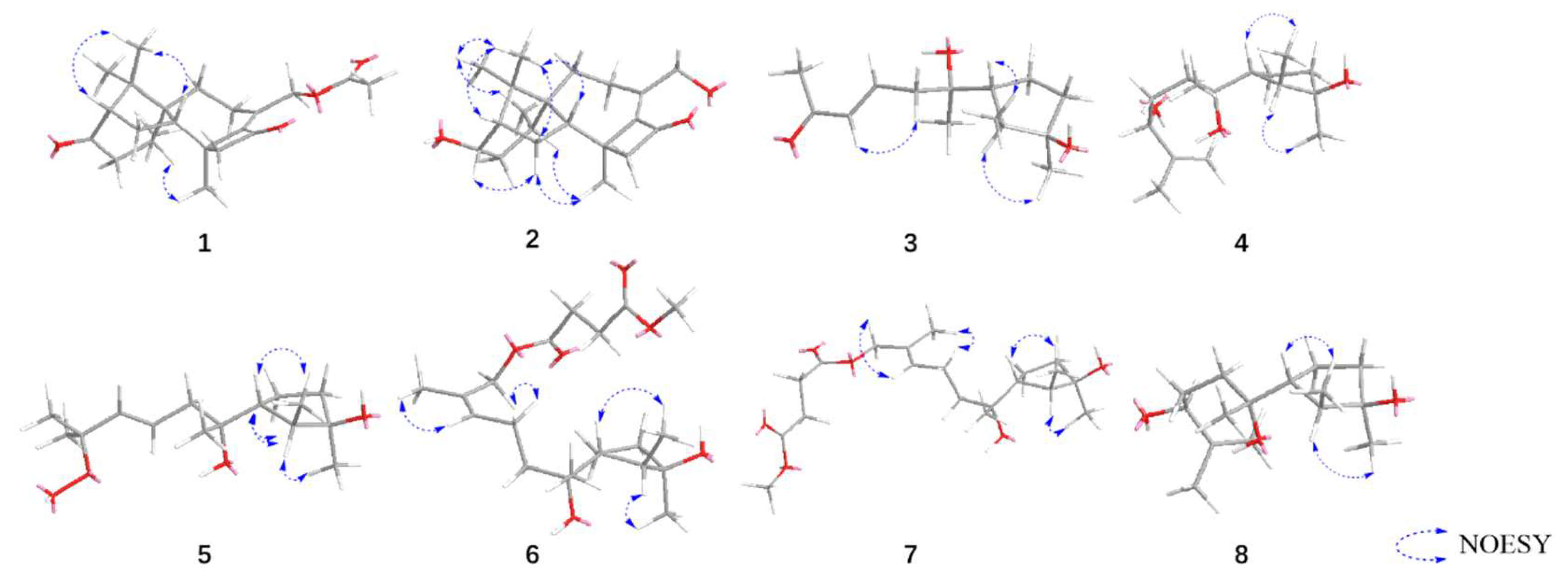
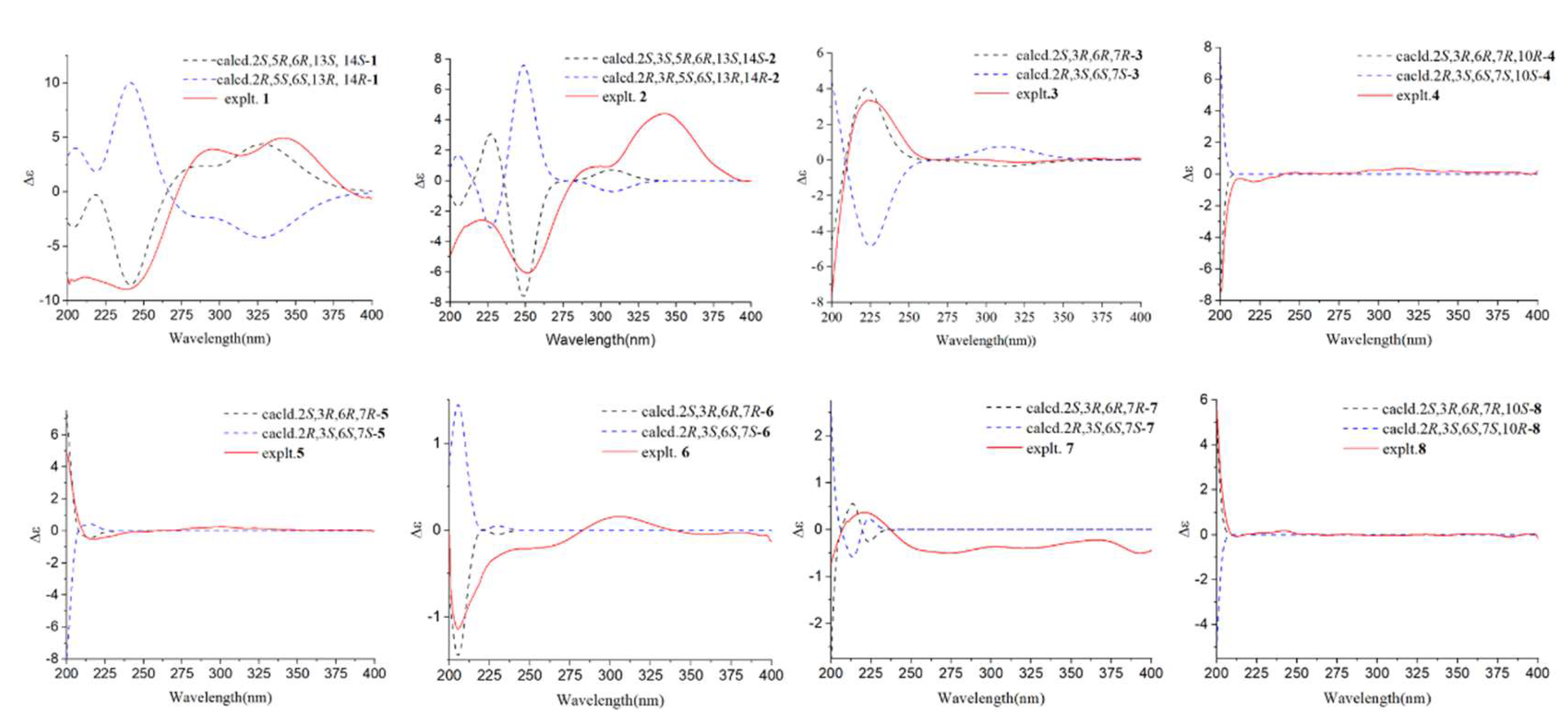
Figure 3.
Key NOESY correlations of compound 1-8.
Figure 4.
Calculated ECD spectra and experimental ECD curves of compound 1–8 in MeOH.
Compound 8 was obtained as colorless oil, its molecular formula was determined as C15H28O3 by (+)-HRESIMS m/z 279.1941 [M+Na]+ (calcd. For C15H28NaO3 +, 279.1931), implying two degrees of unsaturation. The FT-IR absorption band indicate that compound 8 contains hydroxyl group (3408 cm−1), methyl group (2960 and 2873cm−1), olefinic groups (1647 cm−1). The 1H-NMR, 13C-NMR (Table 3 and Table 4) and HSQC spectra exhibited the presence of a group of olefinic signals [δH: 4.97 (H-12a), 4.86 (H-12b); δC: 147.7 (C-11), 111.0 (C-12)], four methylene groups [δH: 1.87 (H-5a), 1.71 (H-9a), 1.69 (H-4a), 1.61 (H-9b), 1.57 (H-8a, H-4b), 1.56 (H-5b), 1.50 (H-8b); δC: 40.5 (C-4), 35.8 (C-8), 29.1 (C-9), 24.5 (C-5)], two hypomethyl groups [δH: 1.87 (H-6), 1.59 (H-2); δC: 54.8 (C-6), 44.5 (C-2)], one oxidized hypomethyl group [δH: 4.09 (H-10); δC: 76.0 (C-10)], two oxidized quaternary carbon signals [δC: 81.4 (C-3), 74.8 (C-7)] and four methyl signals [δH: 1.72 (H-15), 1.26 (H-13) 1.16 (H-14), 1.04 (H-1); δC: 26.2 (C-13), 25.2 (C-14) , 18.2 (C-15), 14.6 (C-1)].
The 1H-1H COSY correlations of H-1/H-2/H-6/H-5/H-4; H-8/H-9/H-10 along with the HMBC correlations signals of H-1/C-3; H-13/C-2, C-3, C-4; H-14/C-6, C-7, C-8; H-10/C-11, C-12 and of H-15/C-10, C-11, C-12 demonstrated that compound 8 is 11-cycloneren-3, 7, 10-triol [24].According to previous research[33], the chirality of 11-cycloneren-3,7,10-triol at C-10 was undetermined, so the Mosher's method was used to determine its conformation. Compound 8 was esterified with (S)-MTPA-Cl and (R)-MTPA-Cl, respectively, to give Mosher ester derivatives. The ΔδS-R of the 1H-NMR data of each proton adjacent to C-10 in the (S)-MTPA ester and (R)-MTPA ester products were compared (Fig. 50), and the final absolute configuration of C-10 was determined to be 10S. The ECD of compound 8 was calculated, and the calculated ECD curves of 2S, 3R, 6R, 7R, 10S-8 were found to be in good agreement with the experimental ECD curves, thus the absolute configuration of compound 8 was determined.
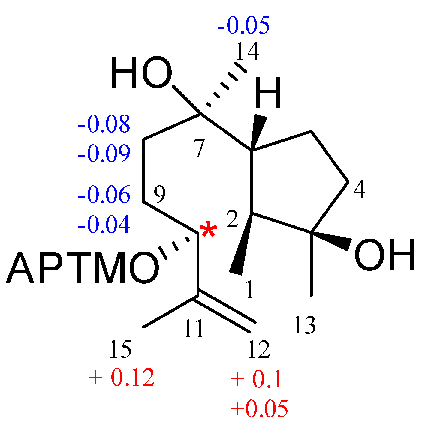
Figure 5.
ΔδS-R value (ppm) of MTPA ester of 8.
3.2. Fungicidal Activities of Compounds 1-10 In Vitro
We tested the fungicidal activity of compounds 1-10 against six crop pathogens at a concentration of 50 µg/mL. Fungicidal tests showed that compounds 1-10 all showed good inhibition of S. sclerotiorum at a concentration of 50 µg/mL. In addition, compound 3 and 10 showed significant inhibition against B. cinerea with inhibition rates of 81.6% and 100%, respectively.
3.3. Fungicidal Activities of Compound 10 In Vivo
In the in vivo activity test, compound 10 was effective against two pathogens, and it showed significant inhibitory activity against B. cinerea, with a control effect of 65.8%.


Table 6.
Activities of compound 10 against Botrytis cinerea and Sclerotinia sclerotiorum in vivo.
| Preventative efficiency (%) | ||
| pathogenic fungi | Botrytis cinerea | Sclerotinia sclerotiorum |
| 10 | 65.8 % | 49.1 % |
| PC | 88.6 % | 100 % |
Figure 6.
Activities of compound 10 against B. cinerea and S. sclerotiorum in vivo.
4. Discussion
A variety of Trichoderma strains have been utilized worldwide as effective biocontrol agents, including T. harzianum, T. hamatum, T. asperellum, T. atroviride, T. koningii, and T. viride [31,32,33,34,35]. One of the mechanisms by which Trichoderma exerts biological control is through the production of secondary metabolites [36,37]. In the present study, we isolated and purified secondary metabolites extracted using ethyl acetate from T. hamatum and identified the structures of the previously undescribed compounds 1-7, as well as the known compound 8, utilizing HRESIMS, NMR, UV, IR, circular dichroism, and Mosher’s method in conjunction with computational chemistry. Compounds 1, 2, and 9 are harziane diterpenes characterized by a unique 6-5-4-7 tetracyclic carbon skeleton; compounds 3-8 are cyclonerane sesquiterpenes, among which compound 5 represents the first -OOH-substituted cyclonerane sesquiterpene to be discovered, while compound 10 was identified as dehydroacetic acid.
Dehydroacetic acid (10) was first isolated from Solandra nitida in 1866 and has been utilized as a food preservative due to its efficacy in inhibiting the growth of molds, yeasts, and bacteria. Its derivatives have been investigated for the development of effective antimicrobial agents against various bacteria and fungi[38]; however, research on its antagonistic effects against agricultural pathogens remains relatively limited. In this study, the fungicidal activities of compounds 1-9 and dehydroacetic acid (10) against six agricultural pathogens were assessed. The results indicated that some of these compounds exhibited promising fungicidal activities against pathogenic fungi. Notably, dehydroacetic acid (10) significantly inhibited the growth of both B. cinerea and R. solani. Furthermore, in vivo examinations of the effects of dehydroacetic acid (10) on the control of B. cinerea and S. sclerotiorum suggested its potential for development as an agricultural antibiotic. Additionally, the substantial isolation of dehydroacetic acid may partially account for the antibiosis activity observed in T. hamatum.
This work not only enriched the active material base of T. hamatum but also advanced the application of its secondary metabolites in controlling agricultural pathogenic fungi, thereby providing a valuable reference for the development of fungal biocontrol strategies.
5. Patents
Chengdu University of Traditional Chinese Medicine has filed patents ZL202311414683.0 and ZL202311251991.6 concerning the in vitro antifungal activity of compounds 2 and 3.
Supplementary Materials
The following supporting information can be downloaded at the website of this paper posted on Preprints.org, Figure S1-S9:HRESIMS, IR and NMR spectra for compound 1; Figure S10-S18: HRESIMS, IR and NMR spectra for compound 2; Figure S19-S27: HRESIMS, IR and NMR spectra for compound 3; Figure S28-S36: HRESIMS, IR and NMR spectra for compound 4; Figure S37-S45: HRESIMS, IR and NMR spectra for compound 5; Figure S46-S54: HRESIMS, IR and NMR spectra for compound 6; Figure S55-S63: HRESIMS, IR and NMR spectra for compound 7; Figure S64-S72: HRESIMS, IR and NMR spectra for compound 8; Figure S73-S110: Calculated NMR and calculated ECD supplementary figure for compound 1-8; Table S1-S61: Calculated NMR and calculated ECD supplementary tables for compound 1-8.
Author Contributions
Conceptualization, D. G, G.W. and Q.B.; methodology, Q.B. and Q.Z..; software, L.H.; validation, D.G., G.W. and L.C..; investigation, L.H., M.L., Y.H. and L.C.; resources, G.W.; data curation, L.H., M.L., Y.H. and L.C.; writing—original draft preparation, L.H.; writing—review and editing, D.G. and Y.G.; visualization, L.H.; supervision, G.W. and D.G.; funding acquisition, D.G. All authors have read and agreed to the published version of the manuscript.
Funding
This research was funded by Sichuan Science and Technology Program, grant number 2021YFN0134.
Data Availability Statement
The authors will make all raw data supporting the conclusions of this article available to any qualified researcher, without undue reservation.
Acknowledgments
On the auspicious occasion of her 95th birthday, this paper is dedicated to Professor Youyou Tu, the esteemed recipient of the 2015 Nobel Prize in Physiology or Medicine, in recognition of her groundbreaking discovery of Artemisinin, which has saved millions of lives worldwide.
Conflicts of Interest
The authors declare no conflicts of interest.
References
- Salazar, B.; Ortiz, A.; Keswani, C.; Minkina, T.; Mandzhieva, S.; Pratap, S. S.; Rekadwad, B.; Borriss, R.; Jain A.; Singh HB.; Sansinenea E. Bacillus spp. as Bio-factories for Antifungal Secondary Metabolites: Innovation Beyond Whole Organism Formulations. Microb Ecol, 2023; 86, 1-24. DOI: 10.1007/s00248-022-02044-2. [CrossRef]
- Syed Ab Rahman, S.F.; Singh, E.; Pieterse, C.M.J.; Schenk, P.M. Emerging microbial biocontrol strategies for plant pathogens. Plant Sci, 2018, 267, 102-111. DOI: 10.1016/j.plantsci.2017.11.012. [CrossRef]
- Saldaña-Mendoza, S.A.; Pacios-Michelena, S.; Palacios-Ponce, A.S.; Chávez-González, M.L.; Aguilar, C.N. Trichoderma as a biological control agent: mechanisms of action, benefits for crops and development of formulations. World J Microbiol Biotechnol, 2023; 39, 269. DOI: 10.1007/s11274-023-03695-0. [CrossRef]
- Li, Z.; Jennings, A. Worldwide Regulations of Standard Values of Pesticides for Human Health Risk Control: A Review. Int J Environ Res Public Health, 2017, 14 (7), 826. DOI: 10.3390/ijerph14070826. [CrossRef]
- Harman, G.; Khadka, R.; Doni, F.; Uphoff, N. Benefits to Plant Health and Productivity from Enhancing Plant Microbial Symbionts. Front Plant Sci, 2021, 11, 610065. DOI: 10.3389/fpls.2020.610065. [CrossRef]
- Zhao, D.; Shen, D.; Fan, H.; Zhu, X.; Wang, Y.; Duan, Y.; Chen, L. Virulent and attenuated strains of Trichoderma citrinoviride mediated resistance and biological control mechanism in tomato. Front Plant Sci, 2023, 14, 1179605. DOI: 10.3389/fpls.2023.1179605. [CrossRef]
- Martinez, Y.; Ribera, J.; Schwarze F.W.M.R.; De France, K. Biotechnological development of Trichoderma-based formulations for biological control. Appl Microbiol Biotechnol, 2023, 107 (18), 5595-5612. DOI: 10.1007/s00253-023-12687-x. [CrossRef]
- Tyśkiewicz, R.; Nowak, A.; Ozimek, E.; Jaroszuk-Ściseł, J. Trichoderma: The Current Status of Its Application in Agriculture for the Biocontrol of Fungal Phytopathogens and Stimulation of Plant Growth. Int J Mol Sci, 2022, 23 (4), 2329. DOI: 10.3390/ijms23042329. [CrossRef]
- Yao, X.; Guo, H.; Zhang, K.; Zhao, M.; Ruan, J.; Chen, J. Trichoderma and its role in biological control of plant fungal and nematode disease. Front Microbiol, 2023, 14, 1160551. DOI: 10.3389/fmicb.2023.1160551. [CrossRef]
- Bolzonello, A.; Morbiato, L.; Tundo, S.; Sella, L.; Baccelli I.; Echeverrigaray S.; Musetti R.; De Zotti M.; Favaron F.; Peptide Analogs of a Trichoderma Peptaibol Effectively Control Downy Mildew in the Vineyard. Plant Dis, 2023, 107 (9), 2643-2652. DOI: 10.1094/PDIS-09-22-2064-RE. [CrossRef]
- El-Hasan, A.; Walker, F.; Klaiber, I.; Schöne, J. P.; fannstiel, J.; Voegele, R.T. New Approaches to Manage Asian Soybean Rust (Phakopsora pachyrhizi) Using Trichoderma spp. or Their Antifungal Secondary Metabolites. Metabolites, 2022, 12 (6), 507. DOI: https://doi.org/10.3390/metabo12060507. [CrossRef]
- Scharf, D.H.; Brakhage, A.A.; Mukherjee, P.K. Gliotoxin--bane or boon? Environ Microbiol, 2016, 18 (4), 1096-1109. DOI: 10.1111/1462-2920.13080. [CrossRef]
- Lin, X.; Tang, Z.; Gan, Y.; Li, Z.; Luo, X.; Gao, C.; Zhao, L.; Chai, L.; Liu, Y. 18-Residue Peptaibols Produced by the Sponge-Derived Trichoderma sp. GXIMD 01001. J Nat Prod, 2023, 86 (4), 994-1002. DOI: 10.1021/acs.jnatprod.3c00014. [CrossRef]
- Torres-Ortega, R.; Guillén-Alonso, H.; Alcalde-Vázquez, R.; Ramírez-Chávez, E.; Molina-Torres, J.; Winkler, R. In Vivo Low-Temperature Plasma Ionization Mass Spectrometry (LTP-MS) Reveals Regulation of 6-Pentyl-2H-Pyran-2-One (6-PP) as a Physiological Variable during Plant-Fungal Interaction. Metabolites, 2022, 12 (12), 1231. DOI: 10.3390/metabo12121231. [CrossRef]
- Li, C.P.; Shi, Z.Z.; Fang, S.T.; Song, Y.P.; Ji, N.Y. Lipids and Terpenoids from the Deep-Sea Fungus Trichoderma lixii R22 and Their Antagonism against Two Wheat Pathogens. Molecules, 2023, 28 (17), 6220. DOI: 10.3390/molecules28176220. [CrossRef]
- Hu, M.; Li, Q.L.; Yang, Y.B.; Liu, K.; Miao, C.P.; Zhao, L.X.; Ding, Z.T. Koninginins R-S from the endophytic fungus Trichoderma koningiopsis. Nat Prod Res, 2017, 31 (7), 835-839. DOI: 10.1080/14786419.2016.1250086. [CrossRef]
- Ahluwalia, V.; Kumar, J.; Rana, V.S.; Sati, O.P.; Walia, S. Comparative evaluation of two Trichoderma harzianum strains for major secondary metabolite production and antifungal activity. Nat Prod Res, 2015, 29 (10), 914-920. DOI: 10.1080/14786419.2014.958739. [CrossRef]
- Khan, R.A.A.; Najeeb, S.; Hussain, S.; Xie, B.; Li, Y. Bioactive Secondary Metabolites from Trichoderma spp. against Phytopathogenic Fungi. Microorganisms, 2020, 8 (6), 817. DOI: 10.3390/microorganisms8060817. [CrossRef]
- Shi, Z.Z.; Liu, X.H.; Li, X.N.; Ji, N.Y. Antifungal and Antimicroalgal Trichothecene Sesquiterpenes from the Marine Algicolous Fungus Trichoderma brevicompactum A-DL-9-2. J Agric Food Chem, 2020, 68 (52), 15440-15448. DOI: 10.1021/acs.jafc.0c05586. [CrossRef]
- Guo, Q.; Shi, L.; Wang, X.; Li, D.; Yin, Z.; Zhang, J.; Ding, G.; Chen, L. Structures and Biological Activities of Secondary Metabolites from the Trichoderma genus (Covering 2018-2022). J Agric Food Chem, 2023, 71 (37), 13612-13632. DOI: 10.1021/acs.jafc.3c04540. [CrossRef]
- Ju, F.; Kuang, Q.X.; Li, Q.Z.; Huang, L.J.; Guo, W.X.; Gong, L.Q.; Dai, Y.F.; Wang, L.; Gu, Y.C.; Wang, D.; Deng, Y.; Guo, DL. Aureonitol Analogues and Orsellinic Acid Esters Isolated from Chaetomium elatum and Their Antineuroinflammatory Activity. J Nat Prod, 2021, 84 (12), 3044-3054. DOI: 10.1021/acs.jnatprod.1c00783. [CrossRef]
- Kuang, Q.X.; Luo, Y.; Lei, L.R.; Guo, W.X.; Li, X.A.; Wang, Y.M.; Huo, X.Y.; Liu, M.D.; Zhang, Q.; Feng D.; Huang, L.J.; Wang, D.; Gu, Y.C.; Deng, Y.; Guo, D.L. Hydroanthraquinones from Nigrospora sphaerica and Their Anti-inflammatory Activity Uncovered by Transcriptome Analysis. J Nat Prod, 2022, 85 (6), 1474-1485. DOI: 10.1021/acs.jnatprod.1c01141. [CrossRef]
- Hoye, T. R.; Jeffrey, C. S.; Shao, F. Mosher ester analysis for the determination of absolute configuration of stereogenic (chiral) carbinol carbons. Nat Protoc, 2007, 2 (10), 2451-2458. DOI: 10.1038/nprot.2007.354. [CrossRef]
- Zou, G.; Yang, W.; Chen, T.; Liu, Z.; Chen, Y.; Li, T.; Said, G.; Sun, B.; Wang, B.; She, Z. Griseofulvin enantiomers and bromine-containing griseofulvin derivatives with antifungal activity produced by the mangrove endophytic fungus Nigrospora sp. QQYB1. Mar Life Sci Technol, 2023, 6 (1), 102-114. DOI: 10.1007/s42995-023-00210-0. [CrossRef]
- Goto, H.; Osawa, E. An efficient algorithm for searching low-energy conformers of cyclic and acyclic molecules. J. Chem. Soc., Perkin Trans, 1993, 2, 187−198. DOI: 10.1039/P29930000187. [CrossRef]
- Frisch, M.J.; Trucks, G.W.; Schlegel, H.B.; Scuseria, G.E.; Robb, M.A.; Cheeseman, J.R.; Scalmani, G.; Barone, V.; Petersson, G.A.; Nakatsuji, H.; Li, X.; et al. 2016, Gaussian 16, Revision B.01, Gaussian, Inc., Wallingford CT.
- Li, Y.H.; Mándi, A.; Li, H.L.; Li, X.M.; Li, X.; Meng, L.H.; Yang, S.Q.; Shi, X.S.; Kurtán, T.; Wang, B.G. Isolation and characterization of three pairs of verrucosidin epimers from the marine sediment-derived fungus Penicillium cyclopium and configuration revision of penicyrone A and related analogues. Mar Life Sci Technol, 2023, 5 (2), 223-231. DOI: 10.1007/s42995-023-00173-2. [CrossRef]
- Grimblat, N.; Zanardi, M.M.; Sarotti, A.M. Beyond DP4: an Improved Probability for the Stereochemical Assignment of Isomeric Compounds using Quantum Chemical Calculations of NMR Shifts. J Org Chem, 2015, 80 (24), 12526-12534. DOI: 10.1021/acs.joc.5b02396. [CrossRef]
- Bian, Q.; Zhao, R.Q.; Peng, X.J.; Gao, L.J.; Zhou, G.N.; Yu, S.J.; Zhao, W.G. Design, Synthesis, and Fungicidal Activities of Novel Piperidyl Thiazole Derivatives Containing Oxime Ether or Oxime Ester Moieties, J Agric Food Chem, 2021, 69 (13), 3848-3858. DOI: 10.1021/acs.jafc.0c07581. [CrossRef]
- Song, Y.P.; Liu, X.H.; Shi, Z.Z.; Miao, F.P.; Fang, S.T.; Ji, N.Y. Bisabolane, cyclonerane, and harziane derivatives from the marine-alga-endophytic fungus Trichoderma asperellum cf44-2. Phytochemistry, 2018, 152, 45-52. DOI: 10.1016/j.phytochem.2018.04.017. [CrossRef]
- González-Menéndez, V.; Pérez-Bonilla, M.; Pérez-Victoria, I.; et al. Multicomponent Analysis of the Differential Induction of Secondary Metabolite Profiles in Fungal Endophytes. Molecules, 2016, 21 (2), 234. DOI: 10.3390/molecules21020234. [CrossRef]
- Guo, R.; Li, G.; Zhang, Z.; et al. Structures and Biological Activities of Secondary Metabolites from Trichoderma harzianum. Mar Drugs, 2022, 20 (11), 701. DOI: 10.3390/md20110701. [CrossRef]
- Guzmán-Guzmán, P.; Kumar, A.; de Los Santos-Villalobos, S.; et al. Trichoderma Species: Our Best Fungal Allies in the Biocontrol of Plant Diseases-A Review. Plants (Basel), 2023, 12 (3), 432. DOI: 10.3390/plants12030432. [CrossRef]
- Carrero-Carrón, I.; Trapero-Casas, J.L.; Olivares-García, C.; et al. Trichoderma asperellum is effective for biocontrol of Verticillium wilt in olive caused by the defoliating pathotype of Verticillium dahliae. Crop Prot, 2016, 88, 45-52. DOI: 10.1016/j.cropro.2016.05.009. [CrossRef]
- Guo R, Ji.S.; Wang, Z.; et al. Trichoderma asperellum xylanases promote growth and induce resistance in poplar. Microbiol Res, 2021, 248, 126767. DOI: 10.1016/j.micres.2021.126767. [CrossRef]
- Bhardwaj, N.; Kumar, J. Characterization of volatile secondary metabolites from Trichoderma asperellum. JANS, 2017, 9, 954-959. DOI: 10.31018/jans.v9i2.1303. [CrossRef]
- Alfiky, A.; Weisskopf, L. Deciphering Trichoderma-Plant-Pathogen Interactions for Better Development of Biocontrol Applications. J. Fungi, 2021, 7 (1), 61. DOI: 10.3390/jof7010061. [CrossRef]
- Grover, A.; Kumar, A.; Tittal, R.K.; et al. Dehydroacetic acid a privileged medicinal scaffold: A concise review. Arch pharm, 2023, 357 (2), e2300512. DOI: 10.1002/ardp.202300512. [CrossRef]
Figure 1.
Chemical structures of compound 1-10.
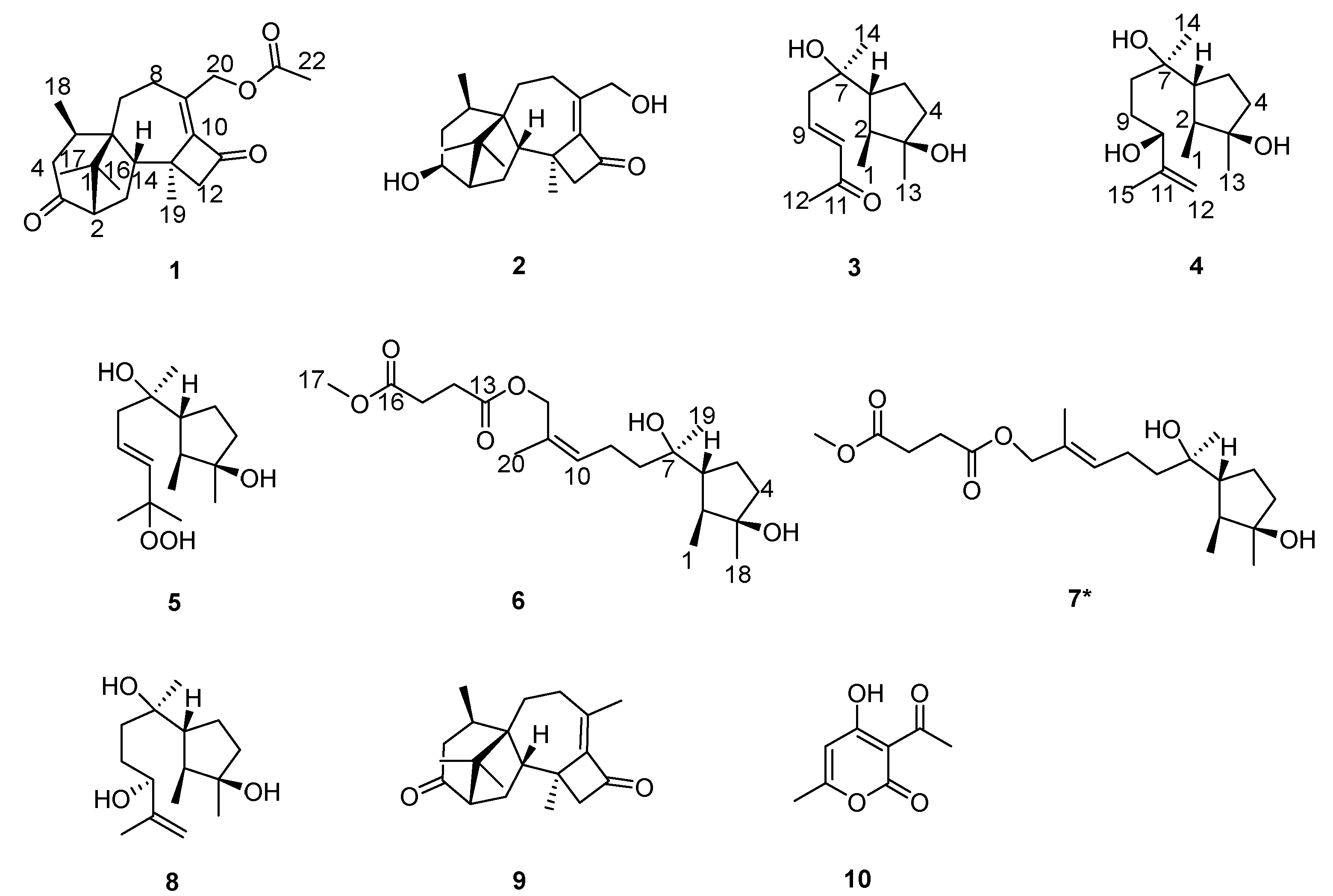
Table 1.
NMR data for compound 1-2, 6-7 (600 MHz, CDCl3).
| pos | 1 | 2 | 6 | 7 |
| δC, type | δC, type | δC, type | δC, type | |
| 1 | 49.7, C | 45.8, C | 14.7, CH3 | 14.7, CH3 |
| 2 | 59.4, CH | 49.6, CH | 44.4, CH | 44.4, CH |
| 3 | 214.2, C | 74.4, CH | 81.4, C | 81.5, C |
| 4 | 42.7, CH2 | 34.3, CH2 | 40.5, CH2 | 40.5, CH2 |
| 5 | 30.1, CH | 28.2, CH | 24.5, CH2 | 24.5, CH2 |
| 6 | 51.8, C | 50.4, C | 54.6, CH | 54.4, CH |
| 7 | 30.3, CH2 | 30.4, CH2 | 74.8, C | 74.9, C |
| 8 | 23.8, CH2 | 24.6, CH2 | 40.5, CH2 | 40.0, CH2 |
| 9 | 141.1, C | 154.0, C | 22.5, CH2 | 22.5, CH2 |
| 10 | 153.0, C | 148.9, C | 131.2, CH | 129.9, CH |
| 11 | 196.5, C | 200.2, C | 129.9, C | 130.2, C |
| 12 | 60.2, CH2 | 58.7, CH2 | 63.6, CH2 | 70.6, CH2 |
| 13 | 40.4, C | 40.4, C | 172.4, C | 172.3, C |
| 14 | 52.9, CH | 51.4, CH | 29.1, CH2 | 29.1, CH2 |
| 15 | 26.7, CH2 | 27.6, CH2 | 29.3, CH2 | 29.3, CH2 |
| 16 | 25.2, CH3 | 26.8, CH3 | 172.9, C | 172.9, C |
| 17 | 23.4, CH3 | 23.6, CH3 | 52.0, CH3 | 52.0, CH3 |
| 18 | 21.1, CH3 | 21.4, CH3 | 26.2, CH3 | 26.2, CH3 |
| 19 | 20.6, CH3 | 21.6, CH3 | 25.0, CH3 | 25.1, CH3 |
| 20 | 63.3, CH2 | 67.3, CH2 | 21.6, CH3 | 21.6, CH3 |
| 21 | 170.9, C | |||
| 22 | 21.0, CH3 |
Table 2.
1H NMR data for compound 1-2, 6-7 (150 MHz, CDCl3).
| pos | 1 | 2 | 6 | 7 |
| δH (J in Hz) | δH (J in Hz) | δH (J in Hz) | δH (J in Hz) | |
| 1 | 1.04, d (6.8) | 1.05, d (6.8) | ||
| 2 | 2.29, m | 1.84, dd (8.2, 3.7) | 1.60, m | 1.61, m |
| 3 | 3.98, dd (3.6, 6.6) | |||
| 4a 4b |
2.90, m 2.09, m |
2.42, d (16.9) 1.50, d (15.3) |
1.68, m 1.56, m |
1.69, m, 1H 1.57, m |
| 5a 5b |
2.90, m | 2.45, m | 1.85, m 1.55, m |
1.86, m 1.55, m |
| 6 | 1.84, m | 1.85, m | ||
| 7a 7b |
1.97, m 1.40, m |
1.97, m 1.25, m |
||
| 8a 8b |
2.29, m |
2.40, m 2.00, m |
1.49, m |
1.51, t (8.4) |
| 9a 9b |
2.19, m 2.12, m |
2.12, m |
||
| 10 | 5.41, t (6.7) | 5.47, td (7.2, 1.4) | ||
| 12a 12b |
2.66, d (16.6) 2.51, d (16.4) |
2.57, d (16.7) 2.46, d (16.9) |
4.66, d (11.9) 4.60, d (11.9) |
4.48, s |
| 14 | 2.52, m | 2.14, dd (11.3, 8.9) | 2.64, m | 2.65, m |
| 15a 15b |
2.05, m 1.54, m |
1.90, m 1.09, dd (14.0, 9.3) |
2.64, m |
2.65, m |
| 16 | 1.00, s | 0.87, s | ||
| 17 | 1.01, s | 1.33, s | 3.69, s | 3.69, s |
| 18 | 1.13, d (7.2) | 1.18, d (7.6) | 1.26, s | 1.26, s |
| 19 | 1.54, s | 1.51, s | 1.15, s | 1.17, s |
| 20a 20b |
5.12, d (12.8) 4.76, d (12.9) |
4.40, d (18) 4.20, d (18.2) |
1.74, d (1.5) |
1.66, d (1.5) |
| 22 | 2.10, s |
Table 3.
NMR data for compound 3-5 and 8 (600 MHz, CDCl3).
| pos | 3 | 4 | 5 | 8 |
| δC, type | δC, type | δC, type | δC, type | |
| 1 | 14.6, CH3 | 14.7, CH3 | 14.5, CH3 | 14.6, CH3 |
| 2 | 44.6, CH | 44.5, CH | 44.6, CH | 44.5, CH |
| 3 | 81.4, C | 81.5, C | 81.6, C | 81.4, C |
| 4 | 40.4, CH2 | 40.5, CH2 | 40.4, CH2 | 40.5, CH2 |
| 5 | 24.6, CH2 | 24.5, CH2 | 24.5, CH2 | 24.5, CH2 |
| 6 | 54.9, CH | 54.8, CH | 54.4, CH | 54.8, CH |
| 7 | 75.0, C | 74.7, C | 74.8, C | 74.8, C |
| 8 | 43.8, CH2 | 36.6, CH2 | 43.6, CH2 | 35.8, CH2 |
| 9 | 144.1, CH | 29.4, CH2 | 126.7, CH2 | 29.1, CH2 |
| 10 | 134.2, CH | 76.7, CH | 138.1, CH | 76.0, CH |
| 11 | 198.5, C | 147.7, C | 82.0, C | 147.7, C |
| 12 | 27.2, CH3 | 111.2, CH2 | 24.6, CH3 | 111.0, CH2 |
| 13 | 26.2, CH3 | 26.2, CH3 | 26.1, CH3 | 26.2, CH3 |
| 14 | 25.9, CH3 | 25.2, CH3 | 25.2, CH3 | 25.2, CH3 |
| 15 | 17.8, CH3 | 24.2, CH3 | 18.2, CH3 |
Table 4.
1H NMR data for compound 3-5 and 8 (150 MHz, CDCl3).
| pos | 3 | 4 | 5 | 8 |
| δH (J in Hz) | δH (J in Hz) | δH (J in Hz) | δH (J in Hz) | |
| 1 | 1.06, d (6.8) | 1.05, d (= 6.7) | 1.04, d (6.7) | 1.04, d (6.9) |
| 2 | 1.62, m | 1.62, m | 1.60, m | 1.59, m |
| 4a 4b |
1.72, m 1.58, m |
1.69, m 1.57, m |
1.68, m 1.56, m |
1.69, m 1.57, m |
| 5a 5b |
1.91, m 1.58, m |
1.86, m 1.56, m |
1.86, m 1.57, m |
1.87, m 1.56, m |
| 6 | 1.86, m | 1.86, m | 1.86, m | 1.87, m |
| 8a 8b |
2.45, dd (14.8, 7.6) 2.36, dd (13.9, 8.2) |
1.60, m 1.48, m |
2.23, m |
1.57, m 1.50, m |
| 9a 9b |
6.88, m |
1.65, m |
5.74, dt (15.0, 7.4) |
1.71, m 1.61, m |
| 10 | 6.13, d (16.0) | 4.05, dd (7.2, 5.3) | 5.63, d (15.8) | 4.09, dd (7.6, 4.6) |
| 12a 12b |
2.27, s |
4.95, s 4.84, s |
1.32, s |
4.97, s 4.86, s |
| 13 | 1.27, s | 1.26, s | 1.25, s | 1.26, s |
| 14 | 1.19, s | 1.16, s | 1.14, s | 1.16, s |
| 15 | 1.74, s | 1.33, s | 1.72, s |
Table 5.
Activities of compound 1–10 against six kinds of pathogenic fungia.
| compound | Fungicidal activities (%) at 50 μg/mL | |||||
| A.S. | F.G. | P.C. | S.S. | B.C. | R.S. | |
| 1 | 52.2 ±2.3 | 56.5 ±4.2 | 45.7 ±1.5 | 85.9 ±3.7 | 44.7 ±5.1 | 19.3 ±0.6 |
| 2 | 39.1 ±3.4 | 17.4 ±1.1 | 21.7 ±32.6 | 73.1 ±2.6 | 26.3 ±2.5 | 46.5 ±4.5 |
| 3 | 56.5 ±4.2 | 34.8 ±2.0 | 42.8 ±2.5 | 64.1 ±2.5 | 81.6 ±3.4 | 28.1 ±3.4 |
| 4 | 47.8 ±1.7 | 23.9 ±1.6 | 32.6 ±2.4 | 73.1 ±4.2 | 42.1 ±2.6 | 15.1 ±2.3 |
| 5 | 45.8 ±2.4 | 26.1 ±2.5 | 23.9 ±2.2 | 67.9 ±3.4 | 39.5 ±4.3 | 19.3 ±3,6 |
| 6 | 30.4 ±3.5 | 84.8 ±3.1 | 26.1 ±1.1 | 75.6 ±1.8 | 47.4 ±2.1 | 17.4 ±2.1 |
| 7 | 34.8 ±1.9 | 15.2 ±1.4 | 35.0 ±3.1 | 51.3 ±2.3 | 34.2 ±1.4 | 14.0 ±1.5 |
| 8 | 30.4 ±2.4 | 28.3 ±2.6 | 37.0 ±2.2 | 53.8 ±4.1 | 26.3 ±2.6 | 51.2 ±3.9 |
| 9 | 12.5 ±1.2 | 23.1 ±3.4 | 9.4 ±0.6 | 76.1 ±3.5 | 46.2 ±3.6 | 59.4 ±4.3 |
| 10 | 56.3 ±3.6 | 38.5 ±3.2 | 28.1 ±1.8 | 100.0 ±0.0 | 100.0 ±0.0 | 82.8 ±2.5 |
a A.S.: Alternaria solani; F.G.: Fusarium graminearum; P.C.: Phytophthora capsici; S.S.: Sclerotinia sclerotiorum; B.C.: Botrytis cinerea; R.S.: Rhizoctonia solani.
Disclaimer/Publisher’s Note: The statements, opinions and data contained in all publications are solely those of the individual author(s) and contributor(s) and not of MDPI and/or the editor(s). MDPI and/or the editor(s) disclaim responsibility for any injury to people or property resulting from any ideas, methods, instructions or products referred to in the content. |
© 2024 by the authors. Licensee MDPI, Basel, Switzerland. This article is an open access article distributed under the terms and conditions of the Creative Commons Attribution (CC BY) license (http://creativecommons.org/licenses/by/4.0/).
Copyright: This open access article is published under a Creative Commons CC BY 4.0 license, which permit the free download, distribution, and reuse, provided that the author and preprint are cited in any reuse.


