Preprint
Article

This version is not peer-reviewed.

Liquid Biopsy–Based Biomolecular Alterations for the Diagnosis of Triple-Negative Breast Cancer in Adults: A Scoping Review

Submitted:

10 December 2025

Posted:

12 December 2025

You are already at the latest version

Abstract

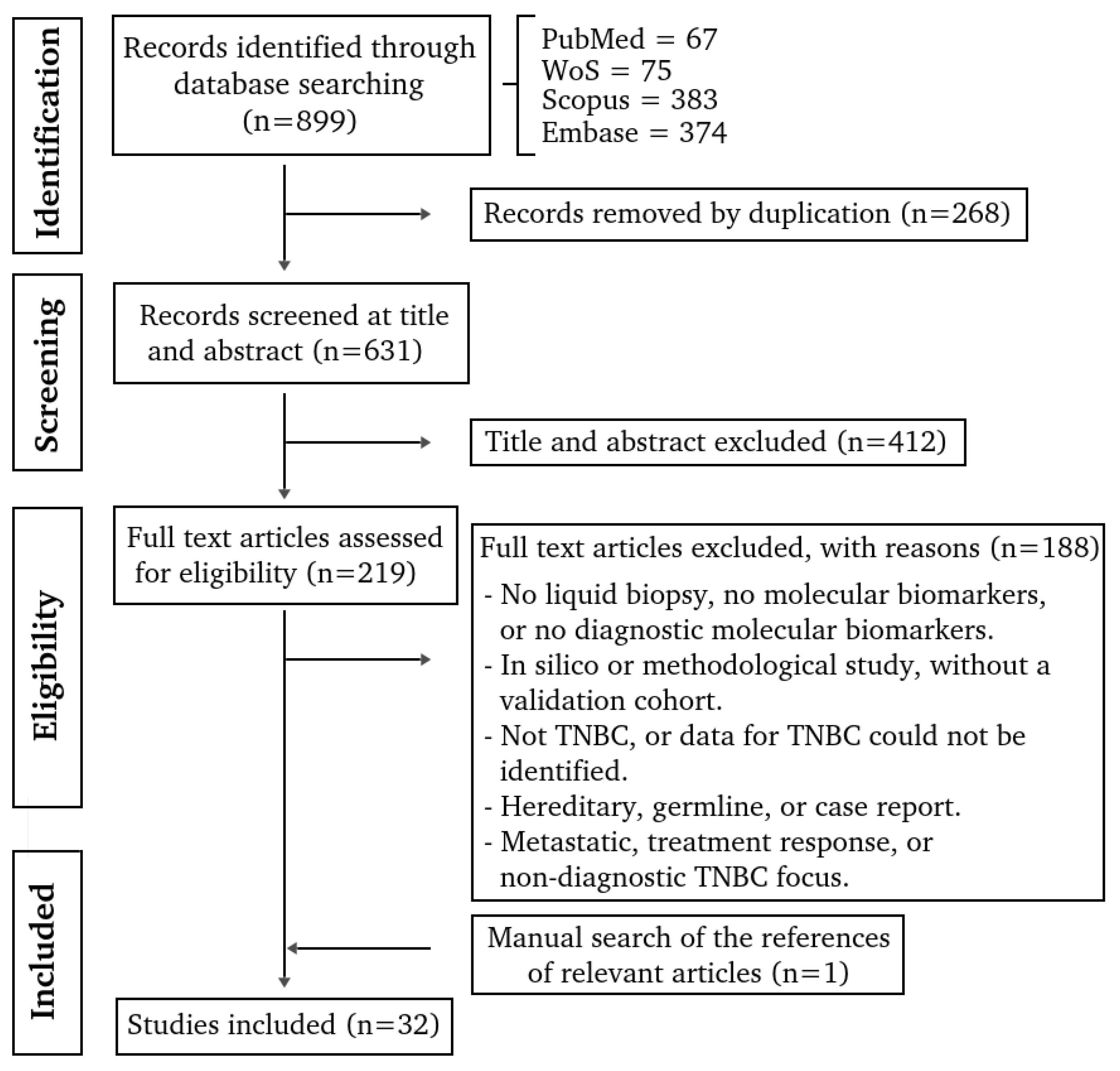
Background/Objectives: Triple-negative breast cancer (TNBC) is an aggressive subtype, with limited diagnostic options and no targeted early detection tools. Liquid biopsy represents a minimally invasive approach for detecting tumor-derived molecular alterations in body fluids. This scoping review aimed to comprehensively synthesize all liquid biopsy–derived molecular biomarkers evaluated for the diagnosis of TNBC in adults. Methods: This review followed the Arksey and O’Malley framework and PRISMA-ScR guidelines. Systematic searches of PubMed, Scopus, Embase, and Web of Science identified primary human studies evaluating circulating molecular biomarkers for TNBC diagnosis. Non-TNBC, non-human, hereditary, treatment-response, and non-molecular studies were excluded. Data on study design, patient characteristics, biospecimen type, analytical platforms, biomarker class, and diagnostic performance were extracted and synthesized descriptively by biomolecule class. Results: Thirty-two studies met inclusion criteria, comprising 15 protein-based, 11 RNA-based, and 6 DNA-based studies (one reporting both protein and RNA). In total, 1532 TNBC cases and 3137 participants in the comparator group were analyzed. Protein biomarkers were the most frequently studied, although only APOA4 appeared in more than one study, with conflicting results. RNA-based biomarkers identified promising candidates, particularly miR-21, but validation cohorts were scarce. DNA methylation markers showed promising diagnostic accuracy yet lacked replication. Most studies were small retrospective case–control designs with heterogeneous comparators and inconsistent diagnostic reporting. Conclusions: Evidence for liquid biopsy–derived biomarkers in TNBC remains limited, heterogeneous, and insufficiently validated. No biomarker currently shows reproducibility suitable for clinical implementation. Robust, prospective, and standardized studies are needed to advance liquid biopsy–based diagnostics in TNBC.

Keywords: 
;  ;  ;  ;  ;  ;  ;  ;  ;  

1. Introduction

Breast cancer was the second most frequently diagnosed malignancy worldwide in 2022, second only to lung cancer, with an estimated 2.3 million new cases, representing 11.6% of all new cancer cases. It is the leading cause of cancer-related death in women, accounting for 15.4% of deaths (666,000 deaths) [1]. It is a heterogeneous disease with variable morphological and biological characteristics, resulting in different clinical behaviors and responses to therapy [2]. Immunophenotyping using immunohistochemistry allows for molecular classification based on the expression of nuclear estrogen receptors (ER), progesterone receptors (PR), human epidermal growth factor 2 (HER2), and Ki-67, which act as prognostic factors and have predictive value for therapy response. These markers allow breast cancer to be classified into the following subtypes: luminal A (ER and/or PR+, HER2−, Ki-67 low), luminal B (ER and/or PR+, HER2−, Ki-67 high) or (ER and/or PR+, HER2+), HER2-enriched (ER and PR−, HER2+), and triple-negative (ER−, PR−, HER2−). The triple-negative breast cancer (TNBC) subtype does not express any of the aforementioned markers and accounts for 15–20% of breast cancers. These tumors present a wide spectrum of morphologies, with most being high-grade and proliferation indices exceeding 80% [2,3]. They have a more aggressive clinical course, with earlier age of presentation, a higher potential risk of metastasis, a worse clinical outcome with more frequent relapses, and a lower survival rate. This subtype includes various entities with different genetic, transcriptional, histological, and clinical profiles [4]. Therefore, early diagnosis is fundamental to enable effective and timely treatment with curative aims. Currently, diagnosis is based on imaging studies, primarily mammography and ultrasound, the latter being indicated mainly in women under 40 years of age. In cases of suspicious findings, a biopsy is performed to obtain neoplastic tissue that allows for diagnosis and classification. In this regard, diagnosis can be limited by patient access, limitations in sample collection, and access to the tissue necessary for diagnosis. Therefore, the development of diagnostic techniques that facilitate access and allow for early diagnosis is necessary. Liquid biopsy is a potential diagnostic technique, involving the collection of a sample of bodily fluid (blood, urine, or saliva, for example) and has the potential to allow the evaluation of the presence of tumor derivatives such as DNA, RNA, circulating tumor cells (CTCs), proteins, or extracellular vesicles (EVs) [5].
Although liquid biopsy has been widely studied in breast cancer [6,7], TNBC-specific evidence is limited and largely restricted to reviews focused on selected biomarker types [8,9,10]. In this scoping review, our objective is to synthesize the available evidence on all types of liquid biopsy–derived biomarkers reported for the diagnosis of TNBC up to the end of 2024.

2. Materials and Methods

2.1. Protocol and Reporting

This scoping review was conducted in accordance with the methodological framework of Arksey and O’Malley and reported following the PRISMA-ScR checklist [11]. The protocol and eligibility criteria were pre-specified prior to study initiation.

2.2. Information Sources and Search

Five reviewers (O.N-F., J.R., E.M., V.H., and A.L.R.-C.) conducted systematic searches in Medline (via PubMed), Scopus, Embase, and Web of Science (WoS) up to October 9, 2024. The search strategies combined controlled vocabulary (MeSH, Emtree, and DeCS) with free-text terms related to “triple-negative breast cancer,” “liquid biopsy,” body fluids (“plasma,” “serum,” “blood,” “saliva,” “extracellular vesicles”), and molecular analytes (DNA, cfDNA, ctDNA, methylation, miRNA, lncRNA, mRNA, proteins/proteomics, lipids/lipidomics, and glycosylation), connected using Boolean operators (AND/OR/NOT). Reference lists of all included studies were also manually screened to identify additional relevant articles. Full search strategies are provided in Supplementary Table S1.

2.3. Eligibility Criteria

We included primary human studies in adults (≥18 years) that enrolled patients with triple-negative breast cancer (TNBC) and evaluated molecular alterations detectable in liquid biopsy (circulating tumor-derived biological material in body fluids) exclusively in relation to diagnosis, without language restrictions. Excluded: in vivo/in vitro/in silico-only work or methodological study, without a validation cohort; case reports, tissue-only analyses, no liquid biopsy (not reflecting tumor-derived liquid biopsy signals), not molecular biomarkers, or not diagnostic molecular biomarkers; reviews, editorials, and letters; studies without a TNBC subgroup, or data for TNBC could not be identified; analyses restricted to Circulating Tumor Cells (CTC) counts without molecular characterization; hereditary or constitutional/germline studies; and studies focused on metastatic disease, treatment response, or non-diagnostic TNBC focus.

2.4. Study Selection and Data Extraction

Two reviewers (O.N-F. and E.M.) independently screened titles and abstracts, with disagreements resolved by a third reviewer (A.L.R.-C.). Full-text assessment and data extraction were conducted by three reviewers (O.N-F., E.M., J.R.). Extracted data items included: DOI, authorship, year, country, study design, sample size and age, biospecimen type, analytical technique, biomarker type, biomarker name/gene symbol, alteration type (e.g., hypermethylation, up-/down-regulation), and diagnostic performance metrics (p-values, AUC, sensitivity, specificity when reported). Extracted datasets are presented in Supplementary Table S3 (Sheets A–E).

2.5. Synthesis of Results

Given heterogeneity across biomarkers, analytical platforms, comparators, and endpoints, results were synthesized descriptively and grouped by analyte or biomolecule class: DNA, RNA and Protein-based. Emphasis was placed on studies reporting diagnostic performance (AUC, sensitivity, specificity). Where available, pre-analytical and analytical factors were also noted.

3. Results

3.1. Study Selection and Characteristics

A total of 899 publications up to October 9, 2024 were identified (Figure 1). Duplicated articles (n=268) and those that did not meet the inclusion criteria during the initial screening of titles and abstracts (n=412), and full-text analysis (n=188) were excluded (Supplementary file, Sheet A-C). Additionally, 1 article was incorporated by hand searching by checking the reference lists of relevant studies (Supplementary file, Sheet D). Finally, 32 studies were examined in this scoping review (Supplementary file, Sheet E).
The study design was defined in 28 studies, with 19 case–control (including variants such as nested or retrospective case–control), and the remaining were described as pilot (n = 3, including variants), experimental (n = 2), cohorts (n = 2), cross-sectional, and multi-phase. The studies were conducted primarily in Asia (n = 17), followed by Europe (n = 7), North America (n = 4), Africa (n = 2), and South America (n = 2). Among the 32 eligible studies, serum was the most commonly analyzed biological specimen (n = 17), followed by plasma (n = 11), whole or peripheral blood fractions (n = 3), buffy coat (n = 1), and a few mixed sample types. Finally, 15, 11, and 6 studies reported novel protein-based, RNA-based, and DNA-based molecular biomarkers, respectively. In one study, both protein-based and RNA-based biomarkers were reported. Of the 32 included studies, 17 reported the area under the receiver operating characteristic curve (AUC), while 14 and 13 studies provided data on sensitivity and specificity, respectively.

3.2. Patients Characteristics

Across the 32 studies, a total of 1532 TNBC cases and 3137 participants in the comparator group were analyzed. Comparator groups included healthy controls, benign breast disease, non-TNBC breast cancer, non–breast cancer participants, and, in one study, disease-free individuals. The approximate modal age was 52 years, with the majority of studies reporting mean or median ages within the 50–55-year range, with ages ranging from 21 to 93 years. Across the 32 studies, the median sample sizes were 29 TNBC cases and 48.5 controls per study. Regarding comparators, the most common study setups contrasted TNBC vs healthy controls and TNBC vs other breast cancer subtypes (e.g., luminal, HER2-positive); fewer studies included benign breast lesions or mixed comparator groups.

3.3. Novel Protein-Based Molecular Biomarkers Associated with TNBC Diagnosis

The fifteen studies reporting novel protein-based molecular biomarkers associated with TNBC diagnosis (including one mixed Protein+RNA study, Table 1) were published between 2012 and 2024. The studies included 621 patients with TNBC and 1,664 compared group, for an overall cohort of 2,285 participants. Across the studies with available data, the reported age range spanned from 26 to 86 years, the mean and modal ages were approximately 52 years. Only one study provided separate age distributions for TNBC and control groups, reporting a mean age of 43.7 ± 7.8 years for TNBC patients and 46.1 ± 10.4 years for controls (Table 1, Study [15]).
Most studies analyzed serum samples (n = 12), followed by plasma (n = 2), and whole blood fractions (n = 1). The predominant analytical techniques included ELISA-based assays, antibody microarrays, and proteomic mass spectrometry approaches (e.g., 2D-DIGE, MALDI-TOF-MS, LC–MS/MS, SWATH). These methodologies primarily aimed to identify circulating proteins differentially expressed in TNBC patients relative to healthy individuals or non-TNBC subtypes.
Among the 15 protein-based studies, al least 69 individual protein biomarkers were identify (Table 1), that could be grouped into 13 functional families, including apolipoproteins, complement components and regulators, immunoglobulins/autoantibodies, coagulation and protease inhibitors, acute-phase/transport proteins, extracellular matrix and adhesion molecules, growth factors/cytokines, and various signaling, enzymatic, and transcriptional regulators. Only APOA4 was reported in three independent studies, while TTR, FN1, APOC1, C3, and C9, were reported in two studies each. All other proteins were described in single studies.

3.4. Novel RNA-Based Molecular Biomarkers Associated with TNBC Diagnosis

Twelve studies investigated RNA-based molecular biomarkers (including one mixed Protein+RNA study). Across these investigations, the cumulative sample comprised 550 TNBC cases and 1,009 participants in the comparator group, with mean per-study sample sizes of ~48 TNBC cases and ~84 comparator participants. Publication years ranged from 2015 to 2023 (mean 2018). Diagnostic performance metrics were variably reported: AUC was provided in nine studies, while sensitivity and specificity were each reported in six studies. Serum and plasma were the most frequently analyzed biological specimens, whereas urine was used in only one study, and RT-qPCR/qRT-PCR approaches predominated, often combined with discovery arrays. Among the 12 RNA-based studies, five reported multiple RNAs or RNA panels, while seven focused on individual RNA biomarkers, resulting in a total of 40 unique RNAs identified as differentially expressed biomarkers in TNBC (Table 2). Among candidate RNAs, miR-21 was the most frequently investigated across studies (n=4), followed by miR-155 (n=2), whereas other microRNAs (e.g., miR-126-5p, miR-205, miR-199a-5p) and lncRNAs (e.g., ANRIL, SOX2OT, ANRASSF1, UCA1, HIF1A-AS2, NRIL) were each reported by single studies.

3.5. Novel DNA-Based Molecular Biomarkers Associated with TNBC Diagnosis

A total of six studies investigated DNA-based molecular biomarkers associated with TNBC diagnosis, encompassing 389 patients with TNBC and 432 participants in the comparator group. Publication years ranged from 2015 to 2023 (mean 2020). Most studies were based on plasma-derived DNA samples, with volumes ranging from 5 to 20 mL, although buffy coat and serum extracellular vesicle (EV) DNA were also analyzed. The most frequently used analytical approaches included Illumina 450K/EPIC methylation arrays, methylation-specific PCR (MSP), digital droplet PCR (ddPCR), and next-generation sequencing (NGS) platforms.
Collectively, 26 unique genes were reported across studies (Table 3). Methylation-based biomarkers, reported in four studies, included cfDNA differentially methylated regions in SPAG6, IFFO1, SPHK2, TBCD/ZNF750, LINC10606 and CPXM1, as well as promoter methylation of APC, RARB2, LINC00299, and ADAM12. Mitochondrial DNA variants were identified in MT-ND1, MT-ND2, MT-ND3, MT-ND4, MT-ND4L, MT-ND5, MT-ND6, MT-CYTB, MT-CO1, MT-CO2, MT-CO3, RNR2, MT-ATP6, and MT-ATP8 (1 study); and somatic mutations were examined in PIK3CA (1 study). Only one study reported AUC (0.74) along with sensitivity (86%) and specificity (90%).

4. Discussion

This scoping review synthesizes, for the first time to our knowledge, all molecular classes of liquid biopsy–derived biomarkers evaluated specifically for the diagnosis of TNBC. Across 32 primary studies published between 2012 and 2024, we identified proteins, RNAs, and DNA alterations as the three major biomarker categories investigated. Collectively, these findings underscore both the growing interest in minimally invasive diagnostics for TNBC and the considerable methodological and biological gaps that continue to limit their clinical translation.
A consistent observation across studies was the substantial heterogeneity in biomarker biology, study design, and analytical methods. This variability reflects not only the intrinsic molecular complexity of TNBC [44], but also differences in sample processing, controls groups, platforms, and reporting practices [45]. Protein-based biomarkers constituted the most frequently explored category (n = 15 studies), followed by RNA-based biomarkers (n = 11), while DNA-based biomarkers were less commonly evaluated (n = 6). Among RNA-derived candidates, miRNAs demonstrated comparatively stronger replication across independent studies. In contrast, somatic mutations were the least represented biomarker class (n = 1).
Despite being the predominant biomarker category, protein-based studies showed limited reproducibility. Only APOA4 was replicated in more than one independent study [15,16,24], however, the direction of change was inconsistent. In two studies, APOA4 was significantly up-regulated (p < 0.05) [15,16], whereas Santana et al. (2024) reported significant down-regulation (p < 0.05) [24]. These discrepancies highlight the sensitivity of protein measurements to pre-analytical workflows, comparator group selection, and population-level variability, reinforcing the need for standardized approaches to quantify spatial, temporal, and inter-individual heterogeneity [45,46].
RNA-based biomarkers included several promising candidates, most notably miR-21, which was significantly up-regulated in three of the four studies analyzed [28,30,33,37]. However, it has been validated in a limited number of samples, raising concerns about its reproducibility, generalizability, and suitability as a clinically reliable diagnostic biomarker [46]. Additional RNAs, including miR-199a-5p, miR-155, and miR-205 [28,30], as well as lncRNAs such as NRIL, HIF1A-AS2, and UCA1 [31], also demonstrated excellent diagnostic performance. Notably, miR-199a-5p achieved an individual AUC of 0.8838 [28], whereas the remaining markers, evaluated as part of multi-RNA panels, yielded AUC values exceeding 0.96 [30]. However, these values were reported in single studies only, underscoring the lack of external validation and the early-stage nature of this research field.
Among DNA-based biomarkers, only one study reported an AUC for a methylation panel [38] including SPAG6, LINC10606, and TBCD/ZNF750, which showed limited diagnostic performance (AUC: 0.74 in the validation set). The other three studies that analyzed methylation did not report AUC values. To date, none were replicated across independent populations, batches, or analytical platforms. The predominance of single-study biomarkers across all molecular classes reflects a fragmented evidence base where most discoveries remain unvalidated and therefore unsuitable for immediate clinical translation.
Beyond biomarker biology, study design limitations were pervasive. Most investigations employed retrospective case–control designs, which are vulnerable to spectrum bias and frequently overestimate diagnostic performance [47,48]. Diagnostic performance was inconsistently reported, with only 17 studies providing AUC values and even fewer reporting sensitivity or specificity. Validation cohorts were uncommon, and independent testing sets were rarely implemented. Clinical comparators were heterogeneous, often combining healthy individuals with patients with non-TNBC breast cancer, and demographic matching was generally inadequate, with only one study reporting age distributions separately for cases and controls. Sample sizes were typically small (median 29 TNBC cases and 35 controls), restricting subgroup analyses and limiting statistical robustness. Collectively, these issues hinder reproducibility, generalizability, and the translational potential of the biomarkers identified.
Strengths and limitations. Strengths of this scoping review include a comprehensive multi-database search strategy, adherence to PRISMA-ScR guidelines, independent screening and data extraction, and structured reporting by biomarker class. However, limitations inherent to scoping reviews also apply: no meta-analysis was conducted, and the high heterogeneity across studies prevented quantitative synthesis of diagnostic accuracy. Because only published studies were included, publication bias cannot be ruled out.
From a clinical perspective, the primary limitation is the lack of validation in independent or prospective cohorts. Most biomarkers were evaluated exclusively in discovery populations, making their real-world diagnostic utility uncertain. Until robust external replication is achieved, none of the identified circulating biomarkers (protein-, RNA-, or DNA-based), can be considered ready for clinical implementation in TNBC diagnosis.

5. Conclusions

This scoping review synthesizes the existing evidence on liquid biopsy–derived molecular biomarkers for the diagnosis of triple-negative breast cancer up to 2024. Although numerous protein-, RNA-, and DNA-based candidates have been proposed, the current evidence remains constrained by methodological heterogeneity, small cohorts, inconsistent reporting, and a lack of external and prospective validation. At present, no biomarker demonstrates sufficient reproducibility or robustness for clinical diagnostic implementation in TNBC. Advancing the field will require well-designed, adequately powered, and standardized multi-phase studies that incorporate independent validation cohorts and harmonized pre-analytical and analytical protocols.

Supplementary Materials

The following supporting information can be downloaded at the website of this paper posted on Preprints.org, Table S1: Search strategy October 10, 2024. File S1: Supplementary file detailing the scoping review process, including (A) Identification, (B) Screening, (C) Eligibility, (D) Manual Search, and (E) Data Extraction.

Author Contributions

Conceptualization; visualization; writing-original draft preparation, O.N.-F., and Á.L.R.-C.; methodology, O.N.-F., J.R., E.M., V.H., and Á.L.R.-C.; formal analysis, O.N.-F.; data curation, O.N.-F. A; writing-review and editing, O.N.-F., J.R., E.M., V.H., and Á.L.R.-C.; supervision; project administration; funding acquisition, Á.L.R.-C. All authors have read and agreed to the published version of the manuscript.

Funding

This work was supported by ANID FONDAP 152220002 (CECAN).

Institutional Review Board Statement

Not applicable.

Informed Consent Statement

Not applicable.

Conflicts of Interest

The authors declare no conflicts of interest.

Abbreviations

The following abbreviations are used in this manuscript:
AUC Area under the receiver operating characteristic curve
BC Breast cancer
cfDNA Cell-free DNA
CTC Circulating tumor cell
ctDNA Circulating tumor DNA
ddPCR Digital droplet polymerase chain reaction
DOAJ Directory of open access journals
ELISA Enzyme-linked immunosorbent assay
EVs Extracellular vesicles
HILIC Hydrophilic interaction liquid chromatography
LD Linear dichroism
lncRNA Long non-coding RNA
MALDI-TOF-MS Matrix-assisted laser desorption/ionization time-of-flight mass spectrometry
MDPI Multidisciplinary Digital Publishing Institute
miRNA MicroRNA
MSP Methylation-specific polymerase chain reaction
NGS Next-generation sequencing
RT-qPCR Reverse transcription quantitative polymerase chain reaction
SDS-PAGE Sodium dodecyl sulfate polyacrylamide gel electrophoresis
SRM Selected reaction monitoring
SWATH Sequential window acquisition of all theoretical fragment ion spectra
TLA Three letter acronym
TNBC Triple negative breast cancer
WB Western blot

References

  1. Bray F.; Laversanne M.; Sung H.; Arnold M.; et al. Global Cancer Statistics 2022: GLOBOCAN Estimates of Incidence and Mortality Worldwide for 36 Cancers in 185 Countries. CA Cancer J. Clin. 2024, 74, 229–263. [CrossRef]
  2. Tsang J.Y.S.; Tse G.M. Molecular Classification of Breast Cancer. Adv. Anat. Pathol. 2020, 27, 27–35. [CrossRef]
  3. Tan PH; Ellis I; Allison K; Brogi E; Fox SB; Lakhani S; Lazar AJ; Morris EA; Sahin A; Salgado R; Sapino A; Sasano H; Schnitt S; Sotiriou C; van Diest P; White VA; Lokuhetty D; Cree IA; WHO Classification of Tumours Editorial Board. The 2019 World Health Organization Classification of Tumours of the Breast. Histopathology 2020, 77(2), 181–185. [CrossRef]
  4. Derakhshan F; Reis-Filho J.S. Pathogenesis of Triple-Negative Breast Cancer. Annu. Rev. Pathol. Mech. Dis. 2021, 17, 181–204. [CrossRef]
  5. Tay T.K.Y.; Tan P.H. Liquid Biopsy in Breast Cancer: A Focused Review. Arch. Pathol. Lab. Med. 2021, 145(6), 678–686. [CrossRef]
  6. Qiu P.; Yu X.; Zheng F.; Gu X.; Huang Q.; Qin K.; Hu Y.; Liu B.; Xu T.; Zhang T.; Chen G.; Liu Y. Advancements in Liquid Biopsy for Breast Cancer: Molecular Biomarkers and Clinical Applications. Cancer Treat. Rev. 2025, 139, 102979. [CrossRef]
  7. Malik S.; Zaheer S. The Impact of Liquid Biopsy in Breast Cancer: Redefining the Landscape of Non-Invasive Precision Oncology. J. Liq. Biopsy 2025, 8, 100299. [CrossRef]
  8. Mazzeo R.; Sears J.; Palmero L.; Bolzonello S.; Davis A.A.; Gerratana L.; Puglisi F. Liquid Biopsy in Triple-Negative Breast Cancer: Unlocking the Potential of Precision Oncology. ESMO Open 2024, 9(10), 103700. [CrossRef]
  9. Zaikova E.; Cheng B.Y.C.; Cerda V.; et al. Circulating Tumour Mutation Detection in Triple-Negative Breast Cancer as an Adjunct to Tissue Response Assessment. npj Breast Cancer 2024, 10, 3. [CrossRef]
  10. Sheng J.; Zong X. Liquid Biopsy in TNBC: Significance in Diagnostics, Prediction, and Treatment Monitoring. Front. Oncol. 2025, 15, 1607960. [CrossRef]
  11. Levac D.; Colquhoun H.; O’Brien K.K. Scoping Studies: Advancing the Methodology. Implement. Sci. 2010, 5, 69. [CrossRef]
  12. Li C.I.; Mirus J.E.; Zhang Y.; Ramirez A.B.; Ladd J.J.; Prentice R.L.; McIntosh M.W.; Hanash S.M.; Lampe P.D. Discovery and Preliminary Confirmation of Novel Early Detection Biomarkers for Triple-Negative Breast Cancer Using Preclinical Plasma Samples from the Women’s Health Initiative Observational Study. Breast Cancer Res. Treat. 2012, 135(2), 611–618. [CrossRef]
  13. Liu A.N.; Sun P.; Liu J.N.; Yu C.Y.; Qu H.J.; Jiao A.H.; Zhang L.M. Analysis of the Differences of Serum Protein Mass Spectrometry in Patients with Triple-Negative Breast Cancer and Non-Triple-Negative Breast Cancer. Tumour Biol. 2014, 35(10), 9751–9757. [CrossRef]
  14. Suman S.; Basak T.; Gupta P.; Mishra S.; Kumar V.; Sengupta S.; Shukla Y. Quantitative Proteomics Revealed Novel Proteins Associated with Molecular Subtypes of Breast Cancer. J. Proteomics 2016, 148, 183–193. [CrossRef]
  15. Gajbhiye A.; Dabhi R.; Taunk K.; Jagadeeshaprasad M.G.; RoyChoudhury S.; Mane A.; Bayatigeri S.; Chaudhury K.; Santra M.K.; Rapole S. Multipronged Quantitative Proteomics Reveals Serum Proteome Alterations in Breast Cancer Intrinsic Subtypes. J. Proteomics 2017, 163, 1–13. [CrossRef]
  16. Del Pilar Chantada-Vázquez M.; López A.C.; Vence M.G.; Vázquez-Estévez S.; Acea-Nebril B.; Calatayud D.G.; Jardiel T.; Bravo S.B.; Núñez C. Proteomic Investigation on the Bio-Corona of Au, Ag and Fe Nanoparticles for the Discovery of Triple-Negative Breast Cancer Serum Protein Biomarkers. J. Proteomics 2020, 212, 103581. [CrossRef]
  17. Fang J.; Tao T.; Zhang Y.; Lu H. A Barcode Mode Based on Glycosylation Sites of Membrane-Type Mannose Receptor as a New Potential Diagnostic Marker for Breast Cancer. Talanta 2019, 191, 21–26. [CrossRef]
  18. Chanana P.; Pandey A.K.; Yadav B.S.; et al. Significance of Serum Vascular Endothelial Growth Factor and Cancer Antigen 15.3 in Patients with Triple-Negative Breast Cancer. J. Radiother. Pract. 2014, 13(1), 60–67. [CrossRef]
  19. Cui R.; Zou J.; Zhao Y.; Zhao T.; Ren L.; Li Y. The Dual-Crosslinked Prospective Values of RAI14 for the Diagnosis and Chemosurveillance in Triple-Negative Breast Cancer. Ann. Med. 2023, 55(1), 820–836. [CrossRef]
  20. Song D.; Yue L.; Zhang J.; Ma S.; Zhao W.; Guo F.; Fan Y.; Yang H.; Liu Q.; Zhang D.; Xia Z.; Qin P.; Jia J.; Yue M.; Yu J.; Zheng S.; Yang F.; Wang J. Diagnostic and Prognostic Significance of Serum Apolipoprotein C-I in Triple-Negative Breast Cancer Based on Mass Spectrometry. Cancer Biol. Ther. 2016, 17(6), 635–647. [CrossRef]
  21. Luo R.; Zheng C.; Song W.; Tan Q.; Shi Y.; Han X. High-Throughput and Multi-Phase Identification of Autoantibodies in Diagnosing Early-Stage Breast Cancer and Subtypes. Cancer Sci. 2022, 113(2), 770–783. [CrossRef]
  22. Chaudhary P.; Gibbs L.D.; Maji S.; Lewis C.M.; Suzuki S.; Vishwanatha J.K. Serum Exosomal Annexin A2 Is Associated with African-American Triple-Negative Breast Cancer and Promotes Angiogenesis. Breast Cancer Res. 2020, 22(1), 11. [CrossRef]
  23. Schummer M.; Thorpe J.; Giraldez M.D.; Bergan L.; Tewari M.; Urban N. Evaluating Serum Markers for Hormone Receptor-Negative Breast Cancer. PLoS ONE 2015, 10(11), e0142911. [CrossRef]
  24. Santana M.F.M.; Sawada M.I.B.A.C.; Junior D.R.S.; Giacaglia M.B.; Reis M.; Xavier J.; Côrrea-Giannella M.L.; Soriano F.G.; Gebrim L.H.; Ronsein G.E.; Passarelli M. Proteomic Profiling of HDL in Newly Diagnosed Breast Cancer Based on Tumor Molecular Classification and Clinical Stage of Disease. Cells 2024, 13(16), 1327. [CrossRef]
  25. Bilir C.; Engin H.; Can M.; Likhan S.; Demirtas D.; Kuzu F.; Bayraktaroglu T. Increased Serum Tumor Necrosis Factor Receptor-Associated Factor-6 Expression in Patients with Non-Metastatic Triple-Negative Breast Cancer. Oncol. Lett. 2015, 9(6), 2819–2824. [CrossRef]
  26. Chung J.M.; Jung Y.; Kim Y.P.; Song J.; Kim S.; Kim J.Y.; Kwon M.; Yoon J.H.; Kim M.D.; Lee J.K.; Chung D.Y.; Lee S.Y.; Kang J.; Kang H.C. Identification of the Thioredoxin-Like 2 Autoantibody as a Specific Biomarker for Triple-Negative Breast Cancer. J. Breast Cancer 2018, 21(1), 87–90. [CrossRef]
  27. Sharma U.; Barwal T.S.; Khandelwal A.; Malhotra A.; Rana M.K.; Singh Rana A.P.; Imyanitov E.N.; Vasquez K.M.; Jain A. LncRNA ZFAS1 Inhibits Triple-Negative Breast Cancer by Targeting STAT3. Biochimie 2021, 182, 99–107. [CrossRef]
  28. Shin V.Y.; Siu J.M.; Cheuk I.; Ng E.K.; Kwong A. Circulating Cell-Free miRNAs as Biomarkers for Triple-Negative Breast Cancer. Br. J. Cancer 2015, 112(11), 1751–1759. [CrossRef]
  29. Kahraman M.; Röske A.; Laufer T.; Fehlmann T.; Backes C.; Kern F.; Kohlhaas J.; Schrörs H.; Saiz A.; Zabler C.; Ludwig N.; Fasching P.A.; Strick R.; Rübner M.; Beckmann M.W.; Meese E.; Keller A.; Schrauder M.G. MicroRNA in Diagnosis and Therapy Monitoring of Early-Stage Triple-Negative Breast Cancer. Sci. Rep. 2018, 8, 11584. [CrossRef]
  30. Kumar V.; Gautam M.; Chaudhary A.; Chaurasia B. Impact of Three miRNA Signature as Potential Diagnostic Marker for Triple-Negative Breast Cancer Patients. Sci. Rep. 2023, 13, 21643. [CrossRef]
  31. Liu M.; Xing L.Q.; Liu Y.J. A Three-Long Noncoding RNA Signature as a Diagnostic Biomarker for Differentiating Between Triple-Negative and Non-Triple-Negative Breast Cancers. Medicine 2017, 96(9), e6222. [CrossRef]
  32. Souza K.C.B.; Evangelista A.F.; Leal L.F.; Souza C.P.; Vieira R.A.; Causin R.L.; Neuber A.C.; Pessoa D.P.; Passos G.A.S.; Reis R.M.V.; Marques M.M.C. Identification of Cell-Free Circulating MicroRNAs for the Detection of Early Breast Cancer and Molecular Subtyping. J. Oncol. 2019, 2019, 8393769. [CrossRef]
  33. Ibrahim A.M.; Said M.M.; Hilal A.M.; Medhat A.M.; Abd Elsalam I.M. Candidate Circulating MicroRNAs as Potential Diagnostic and Predictive Biomarkers for the Monitoring of Locally Advanced Breast Cancer Patients. Tumour Biol. 2020, 42(10), 1010428320963811. [CrossRef]
  34. Qattan A.; Intabli H.; Alkhayal W.; Eltabache C.; Tweigieri T.; Amer S.B. Robust Expression of Tumor Suppressor miRNAs let-7 and miR-195 Detected in Plasma of Saudi Female Breast Cancer Patients. BMC Cancer 2017, 17, 799. [CrossRef]
  35. Shaheen J.; Shahid S.; Shahzadi S.; Akhtar M.W.; Sadaf S. Identification of Circulating miRNAs as Non-Invasive Biomarkers of Triple-Negative Breast Cancer in the Pakistani Population. Pak. J. Zool. 2019, 51, 1113–1121. [CrossRef]
  36. Du Q.; Yang Y.; Kong X.; Lan F.; Sun J.; Zhu H.; Ni Y.; Pan A. Circulating lncRNAs Acting as Diagnostic Fingerprints for Predicting Triple-Negative Breast Cancer. Int. J. Clin. Exp. Med. 2018, 11(8), 8139–8145. https://e-century.us/files/ijcem/11/8/ijcem0073914.pdf.
  37. Ritter A.; Hirschfeld M.; Berner K.; Rücker G.; Jäger M.; Weiss D.; Medl M.; Nöthling C.; Gassner S.; Asberger J.; Erbes T. Circulating Non-Coding RNA Biomarker Potential in Neoadjuvant Chemotherapy of Triple-Negative Breast Cancer? Int. J. Oncol. 2020, 56(1), 47–68. [CrossRef]
  38. Manoochehri M.; Borhani N.; Gerhäuser C.; Assenov Y.; Schönung M.; Hielscher T.; Christensen B.C.; Lee M.K.; Gröne H.J.; Lipka D.B.; Brüning T.; Brauch H.; Ko Y.D.; Hamann U. DNA Methylation Biomarkers for Noninvasive Detection of Triple-Negative Breast Cancer Using Liquid Biopsy. Int. J. Cancer 2023, 152(5), 1025–1035. [CrossRef]
  39. Swellam M.; Abdelmaksoud M.D.; Mahmoud M.S.; Ramadan A.; Abdel-Moneem W.; Hefny M.M. Aberrant Methylation of APC and RARβ2 Genes in Breast Cancer Patients. IUBMB Life 2015, 67(1), 61–68. [CrossRef]
  40. Lee S.; Kim H.Y.; Jung Y.J.; Jung C.S.; Im D.; Kim J.Y.; Lee S.M.; Oh S.H. Comparison of Mutational Profiles Between Triple-Negative and Hormone Receptor-Positive/Human Epidermal Growth Factor Receptor 2-Negative Breast Cancers in T2N0-1M0 Stage: Implications of TP53 and PIK3CA Mutations in Korean Early-Stage Breast Cancers. Curr. Probl. Cancer 2022, 46(2), 100843. [CrossRef]
  41. Manoochehri M.; Jones M.; Tomczyk K.; Fletcher O.; Schoemaker M.J.; Swerdlow A.J.; Borhani N.; Hamann U. DNA Methylation of the Long Intergenic Noncoding RNA 299 Gene in Triple-Negative Breast Cancer: Results from a Prospective Study. Sci. Rep. 2020, 10, 11762. [CrossRef]
  42. Vikramdeo K.S.; Anand S.; Sudan S.K.; Pramanik P.; Singh S.; Godwin A.K.; Singh A.P.; Dasgupta S. Profiling Mitochondrial DNA Mutations in Tumors and Circulating Extracellular Vesicles of Triple-Negative Breast Cancer Patients for Potential Biomarker Development. FASEB Bioadv. 2023, 5(10), 412–426. [CrossRef]
  43. Mendaza S.; Ulazia-Garmendia A.; Monreal-Santesteban I.; Córdoba A.; Azúa Y.R.; Aguiar B.; Beloqui R.; Armendáriz P.; Arriola M.; Martín-Sánchez E.; Guerrero-Setas D. ADAM12 Is a Potential Therapeutic Target Regulated by Hypomethylation in Triple-Negative Breast Cancer. Int. J. Mol. Sci. 2020, 21(3), 903. [CrossRef]
  44. Asleh K.; Riaz N.; Nielsen T.O. Heterogeneity of Triple-Negative Breast Cancer: Current Advances in Subtyping and Treatment Implications. J. Exp. Clin. Cancer Res. 2022, 41, 265. [CrossRef]
  45. Gough A.; Stern A.M.; Maier J.; Lezon T.; Shun T.Y.; Chennubhotla C.; Schurdak M.E.; Haney S.A.; Taylor D.L. Biologically Relevant Heterogeneity: Metrics and Practical Insights. SLAS Discov. 2017, 22(3), 213–237. [CrossRef]
  46. Hayes D.F. Biomarker Validation and Testing. Mol. Oncol. 2015, 9(5), 960–966. [CrossRef]
  47. Hall J.A.; Brown R.; Paul J. An Exploration into Study Design for Biomarker Identification: Issues and Recommendations. Cancer Genom. Proteom. 2007, 4(3), 111–119.
  48. Ou F.S.; Michiels S.; Shyr Y.; Adjei A.A.; Oberg A.L. Biomarker Discovery and Validation: Statistical Considerations. J. Thorac. Oncol. 2021, 16(4), 537–545. [CrossRef]
Figure 1. PRISMA flow diagram of the scoping review process.
Preprints 189123 g001
Table 1. Proteins-based molecular biomarkers associated with diagnostic of TNBC.
AUC Biofluid Detection method P-value Alteration type Biomarker Comparator group Case Sample size Study Ref.
Various Plasma Antibody microarray p<0.05 Up-regulated Panel: KIT, ITGB1, EFNA5, SRP54, FAS, BRCA1, XBP1, and others 28 not BC participants 28 TNBC patients 56 [12]
Not reported Serum 2D-DIGE; MALDI-TOF-MS p<0.05 Up-regulated
Down-regulated
TTR, SERPINA1, HP 30 not BC participants 30 TNBC patients 60 [13]
0.853 Plasma iTRAQ; WB; ELISA A2M, C4BPA p<0.0001
FN1 p<0.018
Overexpression FN1, A2M, C4BPA 20 not BC participants 8 TNBC patients 28 [14]
Not reported Serum 2D-DIGE/MALDI, iTRAQ-LC-MS/MS, SWATH, WB, SRM p<0.05 Up-regulated
Down-regulated
Panel: CPN2, CO2, MYL6, HV101, APOA4, PI16, CXCL7, VTDB, IGJ, KNG1 20 not BC participants 19 TNBC patients 39 [15]
Not reported Serum NP exposure, SDS-PAGE, LC–MS/MS, SWATH/SRM p<0.05 Up-regulated
Down-regulated
Panel: APOL1, CFH, VTNC, C3, C4A, C9, LGALS3BP, FCN3, RBP4, FN1, APOA4, ORM1, ZPI, TTR, APOC1, APOC3, IGHM, IG chains 8 not BC participants 8 TNBC patients 16 [16]
Not reported Serum IP, SDS-PAGE, HILIC, MALDI-TOF MS p < 0.01 Glycosylation pattern change MR 110 participants* 35 TNBC patients 55 [17]
Not reported Serum ELISA p = 0.01 (size)
p = 0.03 (stage)
Increased serum concentration VEGF 35 not BC participants 30 TNBC patients 65 [18]
0.934 Serum ELISA p<1e-4 Increased serum concentration RAI14 60 not BC participant** 46 TNBC patients 106 [19]
0.908 Serum SELDI-TOF-MS, qRT-PCR, ELISA, WB p<1e-4 Increased serum concentration ApoC-I 215 participants*** 165TNBC patients 380 [20]
0.875 Serum Serology p<0.05 Lower concentration panel KJ901215, FAM49B, HYI, GARS, CRLF3 776 participants*** 123 TNBC patients 389 [21]
1 Serum Western blot, ELISA p<1e-4 Higher circulating concentrations ANXA2 179 participants* 58 TNBC patients 126 [22]
TP53 = <0.63 Blood ELISA Non
significant
Minimal diagnostic performance GDF15, PKM, SPARC, CA125, WFDC2, COL1A1, FN1, CTGF, S100A7, SPP1, CCL5, hsa-miR-135b, Anti-TP53, HOXA5, SFRP1 87 not BC participant 28 TNBC patients 115 [23]
Various Plasma Ultracentrifugation; LC-MS/MS Various Panel changes ApoA1, ApoA2, ApoC2, ApoC4
C3, CFB, IGLC2/3, GC, PLG, SERPINA3, IGHC1, C9, LRG1, C4B
204 participants* 20 TNBC patients 123 [24]
Not reported Serum ELISA p = 0.010 Higher serum concentration TRAF6 61 participants* 13 TNBC patients 39 [25]
Not reported Serum HuProt microarray Not reported Higher concentration Anti-TXNL2 10 not BC participants 10 TNBC patients 20 [26]
* Includes non-TNBC and not BC. ** Includes benign breast disease and not BC. *** Includes non-TNBC, benign disease and not BC. TNBC, Triple negative breast cancer; BC, Breast cancer; KIT, Mast/stem cell growth factor receptor; ITGB1, Integrin beta-1; EFNA5, Ephrin-A5; SRP54, Signal recognition particle 54 kDa; FAS, TNFR superfamily member 6 (Ab1); BRCA1, Breast and ovarian cancer susceptibility protein 1; XBP1, X box-binding protein 1; TTR, Transthyretin; SERPINA1, Alpha-1-antitrypsin; HP, Haptoglobin; FN1, Fibronectin; A2M, Alpha-2-macroglobulin; C4BPA, Complement component-4-binding protein-alpha; CFB, Complement factor-B; CPN2, Carboxypeptidase N subunit 2; CO2, Carbon dioxide; MYL6, Myosin light chain 6; HV101, Voltage-gated hydrogen channel 1; APOA4, Apolipoprotein A4; PI16, Peptidase inhibitor 16; CXCL7, Chemokine C-X-C motif Ligand 7; VTDB, Vitamin D-binding protein; IGJ, Immunoglobulin J chain; KNG1, Kininogen-1; APOL1, Apolipoprotein 1; CFH, Complement factor H-related; VTNC, Vitronectin; C3, Complement C3; C4A, Complement C4-A; C9, Complement C9; LGALS3BP, Galectin-3-binding protein, FCN3, Ficolin-3; RBP4, Retinol binding protein 4; ORM1, Orosomucoid; ZPI, protein Z-dependent protease inhibitor; APOC1, apolipoprotein C-I; APOC3, apolipoprotein C-III; IGHM, Immunoglobulin heavy constant mu; IG chains, Immunoglobulin chains; MR, Mannose receptor; MRC1, Mannose receptor C-type 1; VEGF, Vascular endothelial growth factor; CA15-3, Cancer antigen 15.3; RAI14, Retinoic acid induced 14; FAM49B, Family with sequence similarity 49, member B; HYI, Hydroxypyruvate isomerase; GARS, Glycyl-tRNA synthetase 1; CRLF3, Cytokine receptor-like factor 3; ANXA2, Annexin A2; GDF15, Growth differentiation factor 15; PKM, Pyruvate Kinase muscle; SPARC, Osteonectin; CA125, Cancer antigen 125; WFDC2, Human epididymis protein 4; COL1A1, Collagen type 1 alpha 1; CTGF, Connective tissue growth factor; S100A7, Psoriasin; SPP1, Osteoponin; CCL5, RANTES; HOXA5, Homeobox 5; SFRP1, Secreted frizzled-related protein 1; IGLC2/3, Immunoglobulin lambda constant 2/3; GC, Vitamin D-binding protein; PLG, Plasminogen; SERPINA3, Serpin family A member 3; IGHC1, Immunoglobulin Heavy Constant Gamma 1; C9, Complement Component 9; LRG1, Leucine-rich alpha-2-glycoprotein 1; C4B, Complement C4B; TWEAK, (TNF)-like weak inducer of apoptosis; TRAF6, TNF receptor-associated factor 6; Anti-TXNL2, Thioredoxin-like 2 autoantibody.
Table 2. RNA-based molecular biomarkers associated with diagnostic of TNBC.
AUC Biofluid Detection method P-value Alteration type Biomarker RNA type Comparator group Case Sample size Study Ref.
Not reported Peripheral blood RT2 lncRNA PCR Array, qRT-PCR p<1e-4 Up and down regulated ZFAS1 lncRNA 40 not BC participants 40 TNBC patients 80 [27]
0.88 Plasma miRNA arrays, RT-qPCR p<0.0001 Down-regulated miR-199a-5p, miR-16, miR-21 miRNA 255 participants* 72 TNBC patients 327 [28]
0.814 Plasma Microarray, RT-qPCR p = 1.4e-5 Concentration change miR-126-5p miRNA 21 not BC participants 21 TNBC patients 42 [29]
0.961 Serum RT-qPCR p<1e-4 Up and down regulated miR-21, miR-155, miR-205 miRNA 51 not BC participants 139 TNBC patients 190 [30]
0.934 Serum Microarray, RT-qPCR p<0.01 Overexpressed NRIL, HIF1A-AS2, UCA1 lncRNA 75 participants** 25 TNBC patients 100 [31]
0.74 Serum NanoString nCounter p≤0.05 Up-regulated miR-25-3p miRNA 69 participants** 12 TNBC patients 81 [32]
Not reported Plasma qRT-PCR Not reported Up-regulated miR-21 miRNA 46 participants** 4 TNBC patients 50 [33]
Diagnostic panel = 0.929 Plasma RT-qPCR p =0.0008–0.02 Serum differential concentration Initial and diagnostic panel. miRNA 91 participants*** 36 TNBC patients 127 [34]
Not reported Blood ELISA; RT-qPCR Non
significant
Minimal diagnostic performance miR-135b miRNA 87 not BC participants 28 TNBC patients 115 [23]
0.785 Blood RT-qPCR p<0.0001 Up- regulated miR-376c, miR-155, miR-17a, miR-10b miRNA 34 not BC participants 37 TNBC patients 71 [35]
0.959 Plasma qRT-PCR ANRIL = p<0.01
Others = p<0.05
Overexpressed ANRIL, SOX2OT, ANRASSF1 lncRNA 220 participants** 120 TNBC patients 340 [36]
Not reported Serum
Urine
RT-qPCR p<0.05 Increase and decrease in concentration Serum: let-7a, let-7e, miR-21, miR-15a, miR-17, miR-18a, miR-19b miR-30b, GlyCCC2
Urine: miR-18a, miR-19b, miR-30b, miR-222, miR-320, GlyCCC2
miRNA 20 not BC participants 16 TNBC patients 36 [37]
* Includes non-TNBC, benign disease and breast cancer. ** Includes non-TNBC and healthy controls. *** Includes free disease and luminal breast cancer. TNBC, Triple negative breast cancer; BC, Breast cancer; lncRNA, long non-coding RNA; miRNA, micro RNA.
Table 3. DNA-based molecular biomarkers associated with diagnostic of TNBC.
AUC Biofluid Detection method P-value Alteration type Biomarker Variation Comparator group Case Sample size Study Ref
Test set = 0.78
Validation set = 0.74
Plasma Illumina 450K/EPIC,
XGBoost,
ddPCR
P<0.0001 Hypermethylation and hypomethylation SPAG6, IFFO1, SPHK2; TBCD/ZNF750, LINC10606, CPXM1 cfDNA methylation 84 not BC participants 139 TNBC patients 223 [38]
Not reported Serum MSP p = 0.007
(RARB2)
Promoter methylation (RARB2 methylated in TNBC) Promoter methylation: APC, RARB2 DNA methylation 145 participants* 71 TNBC patients 216 [39]
Not reported Plasma RT-PCR --- No plasma mutations PIK3CA hotspot mutations Mutations 22 not BC participants 10 TNBC patients 32 [40]
Not reported Buffy coat ddPCR P = 0.0025
p = 0.001 (tertile 1)
Hypermethylation (leukocyte DNA) LINC00299 (cg06588802) Methylation 159 not BC participants 154 TNBC patients 313 [41]
Not reported Serum NGS
(Illumina NovaSeq PE150)
p<0.0001 (EV concentration) EV concentration
Tumor-specific/shared EV mutations
mtDNA variants (ND1, ND2, ND3, ND4, ND4L, ND5, ND6, CYTB, CO1, CO2, CO3, RNR2, ATP6, ATP8) mtDNA 9 not BC participants 9 TNBC patients 18 [42]
Not reported Plasma Illumina 450K,
Pyrosequencing
Not reported Hypomethylation Promoter methylation ADAM12 Methylation
cfDNA
13 not BC participants 6 TNBC patients 19 [43]
*Includes breast disease and healthy controls. TNBC, Triple negative breast cancer; BC, Breast cancer; cfDNA, Circulating cell-free DNA; SPAG6: Sperm Associated Antigen 6; IFFO1, Intermediate Filament Family Orphan 1; SPHK2, Sphingosine Kinase 2; TBCD, Tubulin-specific chaperone D; ZNF750, Zinc Finger Protein 750; LINC10606, Long Intergenic Non-Protein Coding RNA 606; CPXM1, Carboxypeptidase X, M14 Family Member 1; APC, Adenomatous polyposis coli; RARB2, Retinoic Acid Receptor Beta; PIK3CA, Phosphatidylinositol-4,5-Bisphosphate 3-Kinase Catalytic Subunit Alpha; LINC00299, Long Intergenic Non-Protein Coding RNA 299; ND1, Ubiquinone Oxidoreductase Core Subunit 1; ND2, Ubiquinone Oxidoreductase Subunit 2; ND3, Ubiquinone Oxidoreductase Subunit 3; ND4, Ubiquinone Oxidoreductase Subunit 4; ND4L, Ubiquinone Oxidoreductase Subunit 4L; ND5, Ubiquinone Oxidoreductase Subunit 5; ND6, Ubiquinone Oxidoreductase Subunit 6; CYTB, Cytochrome b (complejo III); CO1, Cytochrome c Oxidase Subunit 1; CO2, Cytochrome c Oxidase Subunit 2; CO3, Cytochrome c Oxidase Subunit 3; RNR2, 16S Ribosomal RNA; ATP6, ATP Synthase F0 Subunit 6; ATP8, ATP Synthase F0 Subunit 8; ADAM12, ADAM Metallopeptidase Domain 12.
Disclaimer/Publisher’s Note: The statements, opinions and data contained in all publications are solely those of the individual author(s) and contributor(s) and not of MDPI and/or the editor(s). MDPI and/or the editor(s) disclaim responsibility for any injury to people or property resulting from any ideas, methods, instructions or products referred to in the content.
Copyright: This open access article is published under a Creative Commons CC BY 4.0 license, which permit the free download, distribution, and reuse, provided that the author and preprint are cited in any reuse.
Prerpints.org logo

Preprints.org is a free preprint server supported by MDPI in Basel, Switzerland.

Subscribe

Disclaimer

Terms of Use

Privacy Policy

Privacy Settings

© 2025 MDPI (Basel, Switzerland) unless otherwise stated