Preprint
Article

This version is not peer-reviewed.

Genetic Polymorphisms of IL6-174G/C TNFα-308G/A and TNFα-238G/A and Risk to Pleural Tuberculosis in Venezuelan Patients

A peer-reviewed article of this preprint also exists.

Submitted:

25 September 2025

Posted:

26 September 2025

You are already at the latest version

Abstract

Tuberculosis (TB) has various clinical presentations; pulmonary TB (PTB) affects only the lungs, while extra-pulmonary TB is defined as the disease involving any other or-gans, including pleural TB (PLTB). Immunological studies of patients with ex-tra-pulmonary TB mainly focus on the cellular Th1 response, which produce key cyto-kines like IFN-γ, TNF-α, IL-12, and IL-6. The TNF-α, and IL-6, which play functional roles in host resistance to Mycobacterium tuberculosis (Mtb) infection. Findings suggest that TNF-α facilitates macrophage containment of Mtb; while that IL-6 increases the apoptosis of macrophages induced by Mtb. Studies involving the human genome have helped identify single nucleotide polymorphisms (SNPs) in genes encoding cytokines associated with TB susceptibility. This study aimed to determine the potential of the IL6-174G/C (rs1800795), TNFα-308G/A (rs1800629) and TNFα -238G/A (rs361525) SNPs as genetic biomarkers of susceptibility to PLTB in the Venezuelan mestizo popu-lation. A total of 269 individuals were included: 69 patients with PLTB and 200 healthy individuals. The IL6-174G/C, TNFα-308G/A and TNFα-238G/A polymorphisms were determined by sequence-specific primer polymerase chain reaction (SSP-PCR). Results showed significantly increased frequencies of the G/C, G/A and G/A genotypes in pa-tients with PLTB (94.0%, 94.2% and 83.3%) compared to controls (40.0%, 19.0% and 13.4%) for the IL6-174G/C, TNFα-308G/A and TNFα-238G/A polymorphisms, respec-tively. Logistic regression analysis showed significant associations of G/C, G/A and G/A genotypes and susceptibility to PLTB. The IL6-174G/C TNFα-308G/A and TNFα-238G/A gene polymorphisms could represent potential genetic biomarkers of susceptibility to PLTB in the Venezuelan mestizo population.

Keywords: 
;  ;  ;  ;  

1. Introduction

Tuberculosis (TB) is a contagious infectious disease caused by the bacillus Mycobacterium tuberculosis (Mtb) bacillus. The World Health Organization (WHO) 2024 Global TB Report highlights the resurgence of TB as the leading infectious killer, posing significant threats to public health [1,2]. Globally, TB has been declared one of the world’s leading infectious killers. For the first time in a decade, cases increased as a direct result of the COVID-19 pandemic. In 2022, 10.6 million people worldwide fell ill with TB, and 1.3 million died. TB is now the second deadliest infection globally, after COVID-19 and is followed by acquired immunodeficiency syndrome (AIDS), caused by the human immunodeficiency virus (HIV) [3].
The complexity of TB stems from the interaction of multiple factors that sustain this endemic disease, most notably poverty, social inequities, the impact of the HIV pandemic, and a limited understanding of key risk factors such as genetic and immunological factors. Of the people infected with Mtb, only 5% to 10% develop an active infection or active TB. The remaining 90% to 95% have a latent infection or latent TB (LTB) and are typically asymptomatic, as their immune system successfully contains the bacteria [4]. The TB has different clinical presentations. Pulmonary TB (PTB) affects only the lungs. In contrast, miliary TB (MTB) is a disseminated form of the disease, while pleural TB (PLTB) is a localized disease that affects the pleura [5,6].
The incidence of PLTB varies between countries. It is estimated to account for up to 30% of TB clinical presentations and is classified as an extrapulmonary form. Most cases are accompanied by a tuberculous pleural effusion, with an incidence of 4% to 23%. A high percentage of these patients (65%) will develop other extrapulmonary manifestations within five years if the condition is not treated appropriately [7]. In the pathogenesis of TB, the host’s cellular immune response determines whether the infection is arrested as a latent or persistent infection or progresses to active TB disease [7]. Efficient cell-mediated immunity hinders a TB infection by permanently arresting it at a latent or persistent stage. This process prevents bacterial replication within cells, increases the phagocytic activity of macrophages, and allows them to successfully eliminate (or contain) the bacteria. However, if the initial infection in the lung is not controlled or the immune system becomes weakened, the disease can progress. Therefore, genetic variants of molecules involved in innate host-defense mechanisms are expected to be associated with host susceptibility to TB [7]. Pleural TB is uncommon in young children and is more frequently reported in adolescents and adults. In young patients under 35 years of age from high-TB prevalence areas, an elevated ADA value (a marker of lymphocytic activity) has a high positive predictive value for diagnosing PLTB. Another useful inflammatory biomarker is IFN-γ, as both ADA and IFN-γ are released in the pleural space during the immune response to Mtb antigens [8].
The regulation and balance of immunity are significantly affected by various mechanisms that Mtb has acquired throughout its evolution to escape the immune response and survive within host cells. To control TB, a Th1-type cellular immune response is required, which involves the participation of CD4+ and CD8+ T lymphocytes, particularly γδ T lymphocytes, and the production of key cytokines [9,10]. The immune response to TB is regulated by interactions between lymphocytes, antigen-presenting cells, and the cytokines they secrete. Upon infection, phagocytes are activated to produce pro-inflammatory cytokines such as Tumor necrosis factor-α (TNF-α), Interleukin-6 (IL-6), and Interleukin-12 (IL-12). IL-12, in particular, drives T cells and natural killer (NK) cells to produce T helper 1 (Th1) cytokines like Interferon-γ (IFN-γ), which then activates infected macrophages to eliminate Mtb [9,10].
Antigenic stimulation of T cells in the presence of proinflammatory cytokines such as IL-6 and costimulatory molecules induces the secretion of IL-2. The binding of IL-2 to its receptor triggers the clonal expansion of antigen-specific T cells, an enhanced secretion of cytokines, and the expression of major histocompatibility complex (MHC) class II molecules. Additionally, IL-6 secreted by macrophages is known to stimulate the early production of IFN-γ [11]. In this context, cytokines play a pivotal role in the anti-mycobacterial response and can determine the type of TB. Genetic polymorphism studies on cytokines have shown that polymorphisms in the promoter or coding regions of many cytokine genes can influence gene transcription levels. These genetic variants have been associated with both the susceptibility to and severity of various diseases, including latent and active Mtb infections [12,13,14,15,16,17]. As proinflammatory cytokines, IL-6 and TNF are associated with the pathogenesis of many chronic inflammatory diseases, including TB [15]. IL-6 and TNF likely play several roles in the context of TB. It is known to block interferon (IFN)-γ-mediated signaling and downregulate IL-6 receptor (IL-6R) expression in CD4 T-cells, which is associated with T-helper cell depletion [15,18].
Studies of the human genome have helped identify single nucleotide polymorphisms (SNPs) in many genes that encode immune system proteins, which are associated with susceptibility to TB [14,15,16,19]. Therefore, this study aimed to investigate the potential of the IL6-174G/C (rs1800795)
TNFα-308G/A (rs1800629) and TNFα -238G/A (rs361525) polymorphisms as biomarkers for susceptibility to pleural tuberculosis in the Venezuelan mestizo population.

2. Materials and Methods

2.1. Study Groups

A descriptive, cross-sectional, retrospective, association (case-control) study included a total of 269 classified in two groups: Group 1: Patients with active M. tuberculosis infection and symptoms compatible with PLTB (n=69) based on the aforementioned criteria [20]. Group 2: Healthy individuals (n=200). The average age was 41.6±20,0 and 47.6±12,2 for PLTB and controls, respectively. The sex ratio (male/female) was 62.3/37.7 in the PLTB group and 37.5/62.5 in the control group. The study group consisted of diagnosed cases of PLTB or with TB pleural effusion whereas the non-TB effusion formed the control group. All the patients were HIV negative. An informed consent form was voluntarily signed by all patients and individual controls before blood and pleural effusion samples were selected. The study of patients with TB pleural effusion or PLTB was including in Project FONACIT No. 2023PGP319. All procedures were performed in compliance with relevant laws and institutional guidelines and had been approved by the Ethical Committee of the Biomedicine Institute “Dr. Jacinto Convit”-Central University of Venezuela.

2.2. Diagnosis of Tuberculous Pleural Effusions and Therapeutic Conduct

The PLTB diagnostic methods included imaging (chest X-rays), samples of pleural effusions, which were subjected to cytological, microbiological (including mycobacteriological) culture (Lowenstein-Jensen culture) and the determination of enzymes, like adenosine deaminase (ADA) assays. For molecular diagnosis, end-point polymerase chain reaction (PCR). All patients included in the study underwent thoracentesis for diagnostic purpose by the Department of Pneumonology at the Vargas Hospital of Caracas. The pleural fluid aspirate was subjected to cytological, microbiological culture and cytokine assays. Pleural biopsy was performed only when results of staining for acid-fast bacillus (smear) or culture of pleural fluid for Mtb. were negative. The PLTB was diagnosed using one of the following criteria as suggested in the national program of TB [21]. 1) M. tuberculosis isolated from pleural effusion by culture. 2) Pleural biopsy showing granulomatous inflammation together with stainable acid-fast bacilli. 3) Pleural biopsy showing granulomatous inflammation, but no stainable acid-fast bacilli, together with a sputum culture positive for Mtb or a good radio graphic response to anti-tuberculous treatment. 4) No histological or bacteriological confirmation, but with other likely alternative diagnoses excluded, together with a good clinical and radiographic response to anti-tuberculous treatment. The latter was initiated in all identified cases of PLTB, where microbiological evidence suggestive of TB and bacteriological confirmation by bacilloscopic or culture were found. Clinical and nutritional follow-up occurred for six months after anti-TB drugs treatment in order to evaluate the improvement of these aspects as therapeutic confirmation, permitting corroboration of the diagnosis. All PLTB patients were assessed for residual pleural scarring in thorax X-rays after completing anti-TB treatment.

2.3. Sample Collection and DNA Extraction and IL-6 and TNF-α Genotyping

For the genotype frequencies of the IL6-174G/C gene polymorphism between the PLTB patients and healthy controls, genomic DNA (gDNA) was extracted from peripheral blood collected before the commencement of anti-tuberculosis therapy. Briefly, 3 mL of whole blood obtained by venepuncture using EDTA as anticoagulant were spun at 3,000 rpm for 30 min; the resulting buffy coats containing peripheral blood mononuclear cells (PBMCs) were stored at 4 °C for further use. A Wizard Genomic DNA Purification Kit (Promega Corporation, WI, US) was used for isolating DNA, following the manufacturer’s instructions, which was then stored at -20oC until use; a UV spectrophotometer was used for evaluating gDNA concentration and quality. For the IL-6, TNFα genotyping, the IL-6-174G/C (rs1800795), TNFα-308G/A (rs1800629) and TNFα -238G/A (rs361525) single nucleotide polymorphisms (SNPs) were determined by sequence-specific primer polymerase chain reaction (SSP-PCR), using specific primers [22,23,24] (Table 1). The amplification products were revealed in 1.5% agarose gels stained with SYBR Green and visualized under a transilluminator.

2.4. Statistical Analysis

Genetic analyses to determine allele frequencies and the estimation of Hardy-Weinberg (H-W) equilibrium was performed using the SNPstats program [25]. Age was reported using means and standard deviations (X±SD), given its normal distribution in the study population. Chi squared or Fisher’s exact tests were used for examining age, male and female sex differences among groups. Genetic analyses to determine genotypic frequencies and binomial logistic regression for evaluating the associations between polymorphisms and TB risk, using adjusted odds ratios (OR), taking gender and age as possible confusion variables were performed using the MedCalc Software Ltd. Odds ratio calculator, IBM SPSS STATISTICS 2025 (the risk analysis Version 23.3.7).

3. Results

3.1. Study Population Characteristics

The study involved 269 individuals, 69 of whom were diagnosed as being infected by Mtb with pleural TB or PLTB based on the aforementioned criteria and 200 were healthy individuals. The Table 2 shows a comparison of the demographic data of the study groups. There were statistically significant differences regarding percentage or distribution by gender between patient and control groups; significance was found for the comparison between male (62.3%) and female (37.7%) in the patient group, p<0.001. For the control group, a significant proportion of male (37.5%) was found compared to female (62.5%), p<0.0007 (Table 2). Among the patients, 66.6% were positive for ADA positive >40u/l compared to control individuals (0%), p<0.0001. Chest X-rays were performed in all patients (Table 2).

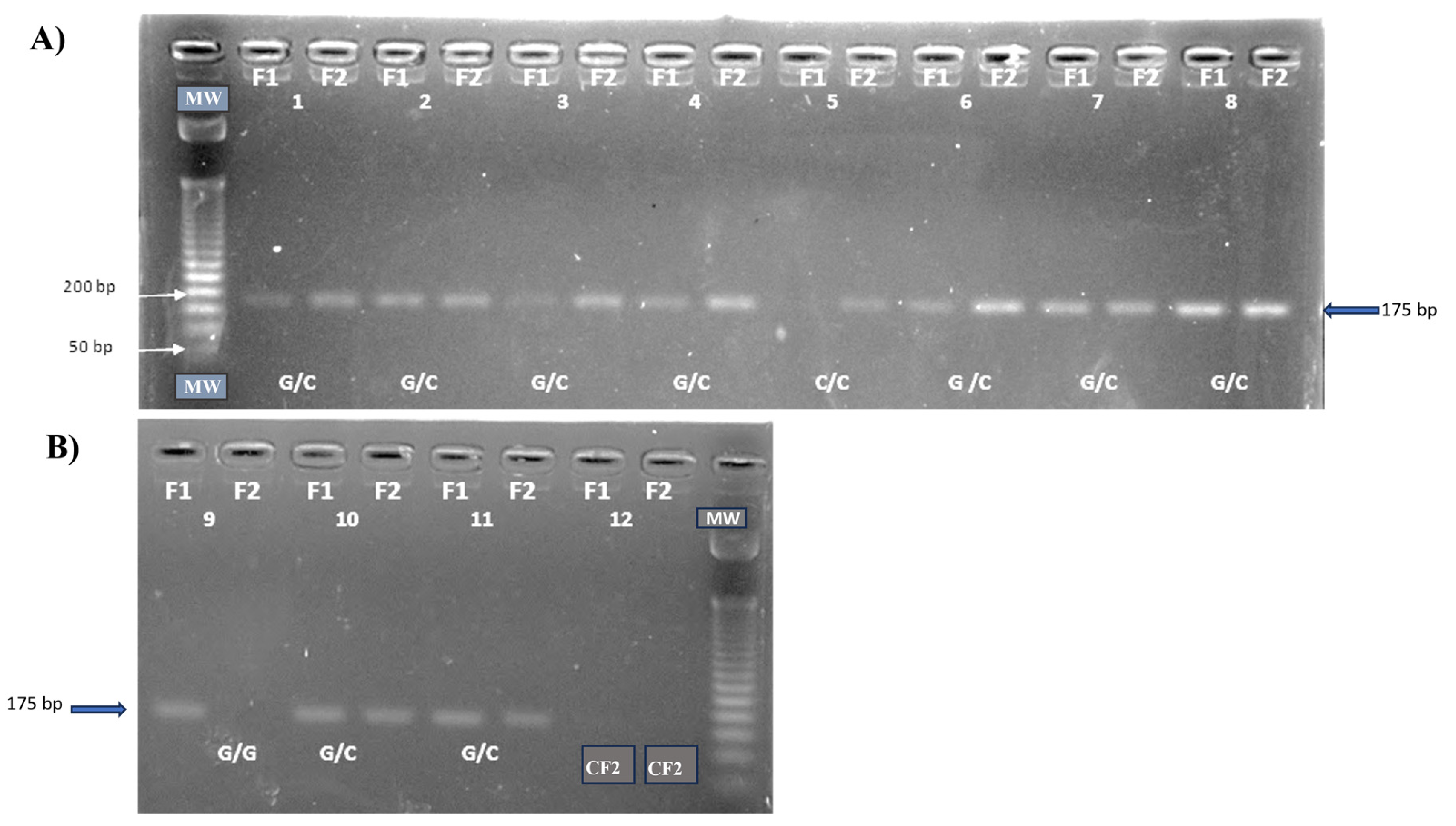
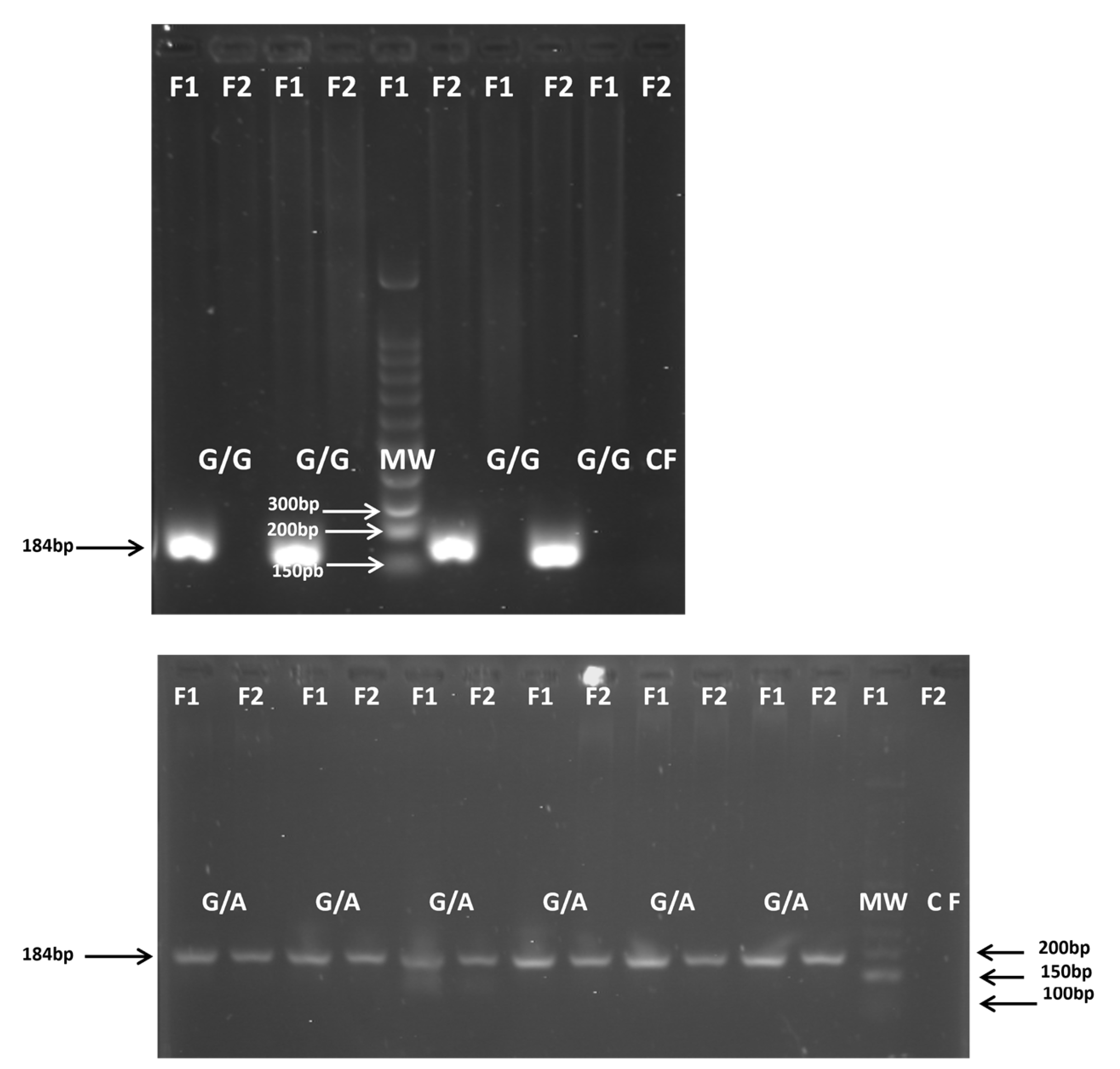
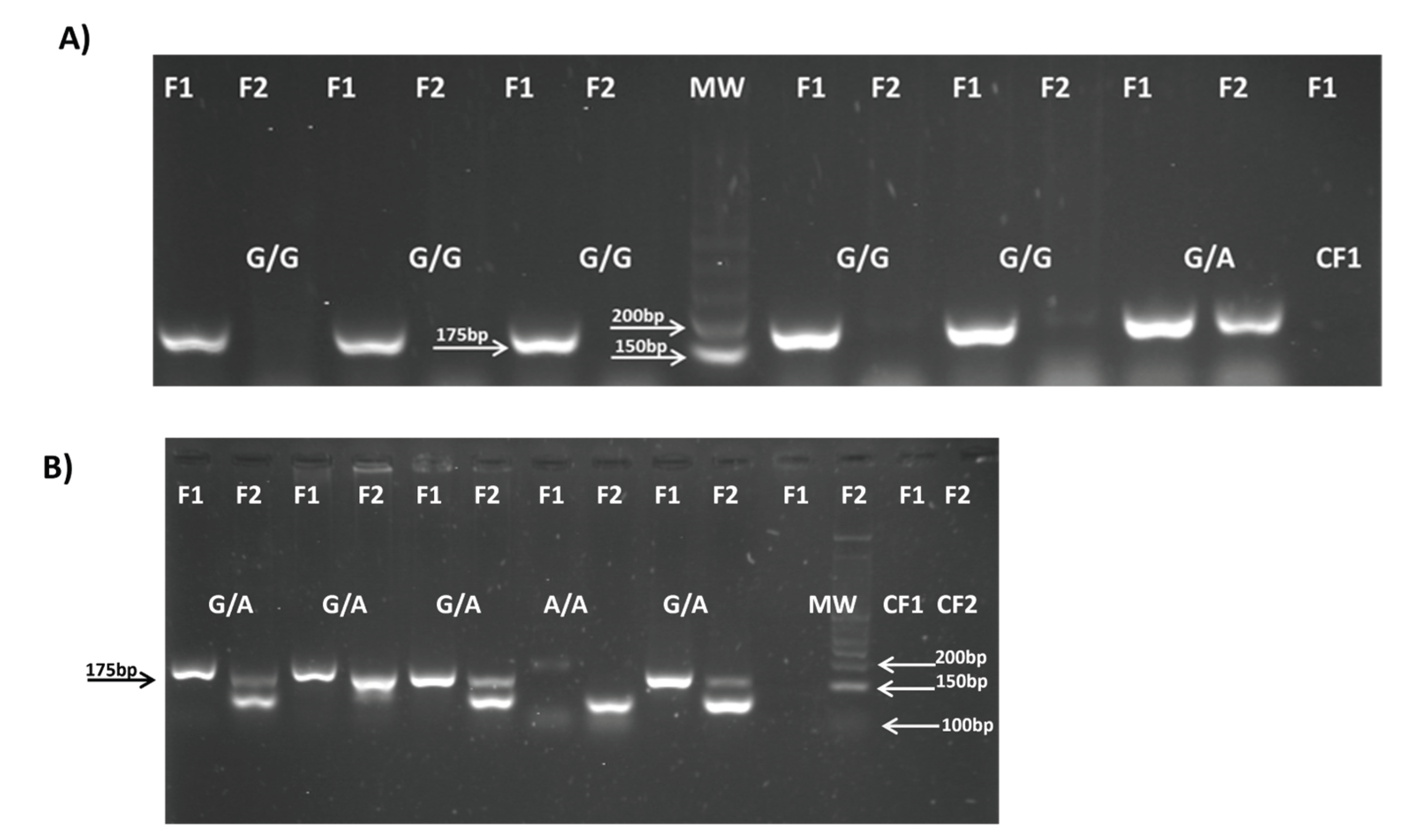
3.2. IL6 and TNFα Polymorphisms and Allele Frequencies

The results for IL6-174G/C, TNFα-308G/A and TNFα-238G/A polymorphisms showed a highly significant difference in allele frequencies between the patient and control groups (Table 3). The G for IL6-174, TNFα-308 and TNFα-238 alleles were markedly overrepresented in the control group (80.0%, 88.4% and 90.0%, respectively) compared to the patient group (50.0%, 53.0% and 58.3%, respectively), with a p-values of <0.0001 (Table 3). Conversely, the C allele, A alleles predominated significantly in the patient group (50.0%, 47.1% and 41.7%, respectively) compared to the controls (20.0%, 11.6% and 10.1%, respectively), with a p-values of <0.0001 (Table 3).

3.2. IL6 and TNFα Polymorphism Genotypes and Association with Pleural Tuberculosis

All gDNA was of high quality and purity. The amplified products for the IL6-174G/C (Figure 1), TNFα-308G/A (Figure 2) and TNFα-238 (Figure 3) gene polymorphisms were obtained at the expected molecular size.
For IL6-174G/C, TNFα-308G/A and TNFα-238G/A polymorphisms, the genotype frequencies results showed a highly significant prevalences of the heterozygous G/C, G/A and G/A genotypes (94.0%, 94.2% and 83.3%, respectively) in the patient group compared to the control group (40.0%, 19.0% and 13.4%, respectively), p<0.0001. The latter genotypes are associated with an intermediate IL-6 and TNFα production phenotypes. Conversely, the homozygous G/G, G/G and G/G genotypes (associated with high IL-6 and TNFα production phenotypes) were significantly more prevalent in the control group (59.5% for IL6-174, 78.8% for TNFα-308 and 81.1% for TNFα-238) than in the patient group (2.9%, 5.8% and 16.6%, respectively), p<0.0001. A similar low frequency of the homozygous C/C, A/A and A/A genotypes (associated with low IL-6 and TNFα productions) were observed in both the patient group (2.9% for IL6-174, 0% for TNFα-308 and 0% for TNFα-238) and the control group (0.5%, 2.1% and 1.4%, respectively) (Table 4).
Using the SNPstats program, the genotypic distribution of the studied polymorphism was confirmed to be in Hardy-Weinberg equilibrium. After adjusting for sex and age using logistic regression (IBM SPSS STATISTICS 2025,Version 23.3.7); the analysis showed a significant association between the G/C genotype and susceptibility to PLTB for IL6-174G/C (OR: 48.343; 95% CI: 11.507-203.095; p <0.0001) (Table 4) , for TNFα-308G/A (OR: 67.256; 95% CI: 22.993-196.729; p <0.0001) (Table 4) and for TNFα-238G/A (OR: 23.333; 95% CI: 9.282-58.655; p <0.0001) (Table 4). Thus, the results of the association study of IL6-174G/C, TNFα-308G/A and TNFα-238G/A heterozygous genotypes and susceptibility to PLTB through logistic regression analysis showed a significant association of the G/C, G/A and G/A genotypes, respectively and susceptibility to PLTB.

4. Discussion

There are several reports indicating that the IL6-174G/C, TNFα-308G/A and TNFα-238G/A polymorphisms may be associated with many infectious and inflammatory diseases. A functional cytokine network is a central element in maintaining the homeostasis of the immune system, and its alteration can lead to an abnormal immune response. Extensive immunological studies have been conducted to explain the various aspects of cytokine dynamics in patients with extra-pulmonary TB. These studies primarily focus on the Th1-type cellular response, which produces cytokines such as IFN-γ, TNF-α, IL-12, and IL-6. These cytokines are essential for the control of Mtb infection, due to their role in the activation and enhancement of macrophages, which will begin to synthesize reactive nitrogen species such as nitric oxide (NO) to reinforce phagocytic and microbicidal activity [26,27,28,29].
The IL6 gene is located on chromosome 7p21 and has different genetic variants associated with high, intermediate, and low levels of IL-6 production [26]. The high- and intermediate-production phenotypes are found in individuals with the −174G/G and −174G/C genotypes, respectively, resulting in elevated and intermediate levels of circulating IL-6. Conversely, the low-production phenotype is associated with the homozygous −174C/C genotype, which leads to reduced IL-6 levels. Consequently, genetic variants in the IL-6 gene have been linked to the susceptibility and severity of a wide range of diseases, including chronic hepatitis C and respiratory tract infections like tuberculosis [26]. Furthermore, IL-6 is associated with more severe TB, which includes greater radiological severity and impaired lung function. It also promotes the growth of Mtb and the expansion of monocytes following human hematopoietic stem-cell infection [27]. Additionally, IL-6 responses can drive tissue damage by facilitating Th17 cell differentiation, IL-17 activity, neutrophil chemotaxis, and fibroblast matrix metalloproteinase production [27]. Regarding tuberculous pleuritis, Antas et al. evaluated cytokine levels in pleural effusion specimens from patients with TB. Their results showed that IL-6 and IL-27 levels were higher in TB patients, and the authors identified these as significantly associated biomarkers in patients with confirmed tuberculous pleuritis or PLTB [11].
TNF-α is a potent pleiotropic proinflammatory and immunoregulatory cytokine, which contributes to the initiation, up-regulation, and perpetuation of the inflammatory response. TNF-α is produced by many cell types including neutrophils, fibroblasts, NK cells, T and B cells, and macrophages infiltrating tissue as the part of host defense against infections. Increased levels of serum TNF-α have been observed in several infectious diseases including advanced TB [30]. TNF-α, gene that encodes the cytokines TNF-α is located within the class III region of the MHC. TNF-α is expressed as a transmembrane protein that can be processed into a soluble form (sTNF) that exerts its functions via two receptors TNF receptor 1 (TNFR1) and TNFR2 [30]. The former exists in almost all cells aims to trigger apoptosis signaling pathways while the latter restricts expression and activation of cell survival. Its production is dependent on TNF-α gene expression. The TNF-α gene polymorphism affects its expression of different genotypes which can produce different proteins affecting the susceptibility and severity of diseases [30].
We found genetic variants in the TNFα and IL-6 gene polymorphisms that were associated with TB susceptibility for the SNPs rs1800795, rs1800629 and rs361525 in the analysis of alleles and genotypes between the PLTB patients and controls. The present results suggest strong associations between the IL6-174G/C, TNFα-308G/A and TNFα-238G/A polymorphisms and susceptibility to PLTB in the Venezuelan mestizo population. Our findings indicate that the heterozygous G/C genotype, which is associated with an intermediate IL-6 production phenotype, was significantly more prevalent in patients than in healthy controls. Conversely, the homozygous G/G genotype was markedly overrepresented in the control group. This pattern of genotypic distribution, along with the allelic frequencies, suggests that the C allele is a risk factor for PLTB, while the G allele may confer a protective effect in the Venezuelan mestizo population. Furthermore, an analysis using the dominant inheritance model confirmed a significant association, demonstrating a higher risk of developing PLTB in individuals carrying the C allele. This finding underscores the potential of the IL6-174G/C polymorphism as a valuable genetic biomarker for susceptibility to this specific form of TB.
A meta-analysis by Liu et al. included 25 studies to investigate the relationship between IL6 and IL18 polymorphisms and tuberculosis susceptibility. To evaluate the role of IL6 polymorphisms, 14 papers were included. The rs1800795 polymorphism was found to be significantly associated with TB in China (in a recessive model) and in Iran, Pakistan, and India (in dominant, recessive, and allele comparisons). The authors suggested that genetic polymorphisms of IL6 rs1800795 and IL18 rs187238 may increase susceptibility to TB, particularly in Asian populations [31]. Makhatadze et al. studied major histocompatibility complex polymorphisms among Venezuelan mestizos and reported that genes of Mongoloid, Negroid, and Caucasoid origin have created a unique human leukocyte antigen (HLA) genetic profile in this hybrid population. This research highlights that the Venezuelan mestizo population is genetically distinct from Asian populations [32]. The Venezuelan mestizo population is a highly admixed group with Mongoloid, Negroid, and Caucasoid ancestry, which explains the presence of heterozygous GC individuals. Thus, genetic admixture may contribute to differences in susceptibility patterns compared to monomorphic populations.
Graça et al. reported on their findings regarding cytokine genetic polymorphisms in 245 patients with clinical TB from Belém, Pará, Brazil. Of these patients, 82% had the pulmonary form and 18% had the extrapulmonary form of the disease. The study found that the wild-type GG genotype and the G allele for the IL6-174G/C polymorphism were associated with a four-fold increased risk for TB infection and development when compared to healthy controls. The GG and CC genotypes were associated with higher and lower levels of cytokines, respectively. The presence of the CC genotype was, therefore, linked to a lower risk of TB. However, the same authors noted a contrasting study from India where the CC genotype was associated with a higher risk of the disease [33].
Most studies on the IL6 polymorphism have reported a relationship primarily with susceptibility to pulmonary tuberculosis. However, a recent study by Silva et al. demonstrated a significant association between IL6-174G/C and TB risk, including cases of extra-pulmonary TB, in a Brazilian population. Silva et al. reported their findings based on the IL6-174 G > C SNP in patients from northeastern Brazil. They observed that the heterozygous GC genotype predominated in patients with PTB, while the homozygous GG genotype was significantly more frequent in those with miliary TB, a form of extra-pulmonary TB [34]. This contrasts with our findings in the Venezuelan mestizo population. While the GC genotype was associated with PTB in the Brazilian study, our research found this same genotype to be associated with pleural tuberculosis, which is also a form of extra-pulmonary TB. This suggests that the specific genetic association of the IL6-174G/C polymorphism may vary between different populations and even between different clinical forms of extra-pulmonary tuberculosis.
Evidence suggests TNF-a gene is associated with the development of TB [30]. However, there are controversial studies regarding the association of -308 TNF-α genotypes. Correa et al. (2004 and 2005) found an association of the -308 TNF-α genotype G/G with TB and concluded that the A/A genotype was a protective factor against TB; while other authors observed an increase in A/A -308 TNF-α in PTB and chronic TB when compared to controls [35,36]. However, the strongest evidence of a protective role of TNF-α in human TB is the high incidence of TB reactivation in patients with rheumatoid arthritis treated with monoclonal antibodies against TNF-α [37].
Our findings about TNFα-308G/A polymorphism indicate that the heterozygous G/A genotype, which is associated with an intermediate TNF-α production phenotype, was significantly more prevalent in patients (94.2%) than in healthy controls (19.0%). Conversely, the homozygous G/G genotype was markedly overrepresented in the control group (78.8% vs 5.8%). This pattern of genotypic distribution, along with the allelic frequencies, suggests that the A allele is a risk factor for PLTB, while the G allele may confer a protective effect in the Venezuelan mestizo population. In relation to TNFα-238G/A polymorphism findings indicate that also the heterozygous G/A genotype, which is associated with an intermediate TNF-α production phenotype, was significantly more prevalent in patients (83.3%) than in healthy controls (13.4%). Conversely, the homozygous G/G genotype was markedly overrepresented in the control group (81.1% vs 16.6%). This pattern of genotypic distribution, along with the allelic frequencies, suggests that the A allele is a risk factor for PLTB, while the G allele may confer a protective effect in the Venezuelan mestizo population.
There are some reports on the correlation between the TNF-α-308, TNF-α-238 gene polymorphism and TB infection in different populations, but the results are contrary [30]. A comprehensive understanding of the relationship between IL6 and TNFα polymorphisms and TB susceptibility requires the collection of data from diverse populations, as differences in results may be related to ethnic variations. Our results indicate that the IL6-174G/C (rs1800795), TNFα-308G/A (rs1800629) and TNFα -238G/A (rs361525) polymorphisms are interesting candidates to explain individual differences in susceptibility to extrapulmonary TB among the Venezuelan mestizo population. These findings are supported by a broader analysis of existing literature. For instance, subgroup analyses based on ethnicity have found significant associations between the IL6-174G/C polymorphism and TB risk in Asian and Latino populations, while no significant association was observed in Caucasian populations. This strongly suggests that ancestral genetic factors have a considerable impact on TB risk [38,39]. Further supporting the role of ethnicity in TB pathogenesis, our studies on Warao indigenous and non-indigenous populations revealed a low incidence of pleural TB (PLTB) among the Warao people compared to non-indigenous populations. This is a notable finding, given the high incidence and prevalence of pulmonary TB (PTB) among Warao indigenous people. This observation suggests that genetic background may not only influence overall TB risk but also the specific clinical form the disease presents [40,41]. Ultimately, more studies are needed to investigate the precise mechanisms by which the IL6 and TNFα polymorphisms regulate IL-6 and TNFα productions and influence susceptibility to various forms of TB, specially PLTB.

5. Conclusions

The present study has highlighted genetic association of the intermediate IL-6 and TNF-α production phenotypes, which could be genetic biomarkers that could contribute to a better understanding of TB pathogenesis and identifying genetic and immunological risk factors for PLTB. An allele frequency where in dominant models GC+CC vs GG (IL6-174G/C), and GA+AA vs GG (TNFα-308G/A and TNFα-238G/A) are predominates indicates a greater probability of developing PLTB in the Venezuelan mestizo population.

Author Contributions

Z.A.: Conceptualization, writing- review & editing. J.H.d.W.: Data curation, molecular and ADA assays for the confirmatory diagnosis of pleural tuberculosis. M.F.M.: Methodology, formal analysis, investigation, supervision. D.S.: developed in polymorphism assays and molecular characterization. C.J.S.: Formal analysis. L.A.D.J.G.: Conceptualization, writing- Review & editing. J.E.L.R.: Data curation. B.R.S.: Investigation, writing- review & editing.

Funding

This study was supported by the budget provided by the FONACIT of the Ministry of Science and Technology (Project FONACIT No. 2023PGP319).

Institutional Review Board Statement

Not applicable.

Informed Consent Statement

It was mentioned in the section of Material and Methods.

Data Availability Statement

The original contributions presented in this study are included in the article. Further inquiries can be directed to the corresponding author.

Acknowledgments

Our gratitude to the patients, the Tuberculosis Laboratory of the “Dr. Jacinto Convit” Institute of Biomedicine (UCV).

Conflicts of Interest

The authors declare no competing interests.

References

  1. World Health Organization (WHO). Global Tuberculosis Report 2024. Geneva 2024.
  2. Castro, V.; Celiz, J. Tuberculosis: Una vieja enfermedad conocida que no deja de sorprendernos. 2017. Available online: https://www.academia.edu/35362011/Tuberculosis_Una_Vieja_enfermedad_conocida_que_no_deja_de_sorprendernos.
  3. World Health Organization. Global Tuberculosis Programme. Global tuberculosis report 2023. Geneva 2023. ISBN 978-92-4-008385.
  4. MacNeil, A.; Glaziou, P.; Sismanidis, C.; Date, A.; Maloney, S.; Floyd, K. Global epidemiology of tuberculosis and progress toward meeting global targets — worldwide, 2018. MMWR Morb Mortal Wkly Rep. 2020 69, 281−285.
  5. Porcel, J.M. Tuberculous Pleural Effusion. Lung. 2009, 187, 263–27. [Google Scholar] [PubMed]
  6. Leung, C.C.; Feller-Kopman, D.; Niederman, M.S.; Spiro, S.G. Year in review 2010: Tuberculosis, pleural diseases, respiratory infections. Respirology, 2011. 16, 564–573.
  7. Barssa, L.; Connors, W.J.A.; Fisher, D. Chapter 7: Extra-pulmonary tuberculosis. Canadian J. Resp. Critical Care Sleep Med. 2022, 6, 87−108. [Google Scholar]
  8. Naik, M.; Prakash-Nayak, O.; Murmu, M.; Ranjan-Partra, S.; Baa, M. Role of CBNAAT in Suspected Cases of Tubercular Pleural Effusion. J. Dental Medical Sci. 2019, 18, 46–77. [Google Scholar] [CrossRef]
  9. Ferreiro L, San Jose E, Valdes L. Tuberculous pleural effusion. Arch. Bronco neumol. 2014, 50, 435e43. [Google Scholar]
  10. Yang L, Hu YJ, Li FG, Chang XJ, Zhang TH, Wang ZT. Analysis of cytokine levers in pleural effusions of tuberculous pleurisy and tuberculous empyema. Mediat. Inflamm. 2016, 2016, 3068103.
  11. Antas, P.; Borchert, J.; Ponte, C.; Lima, J.; Georg, I.; Bastos, M.; Trajman, A. Interleukin-6 and-27 as potential novel biomarkers for human pleural tuberculosis regardless of the immunological status. Microbes Infect. 2024, 26, 105238. [Google Scholar] [CrossRef]
  12. Hamilton, F. , Schurz, H. , ates, T.A., Gilchrist, J.J., Möller, M., Naranbhai, V., Ghazal, P., Timpson, N.J., Tom Parks, T., Pollara. G. Altered IL-6 signalling and risk of tuberculosis: a multi-ancestry mendelian randomisation study. Lancet Microbe 2025, 6, 100922. [Google Scholar]
  13. Mohan, V.P.; Scanga, C.A.; Yu, K.; Scott, H.M. ; Tanaka, K,E. ; Tsangm E.; Chih, T.M.; Flynn, J.L.; John, C.J. Effects of tumor necrosis factor alpha on host immune response in chronic persistent tuberculosis: possible role for limiting pathology. Infect. Immun. 2001, 69, 1847−1855. [Google Scholar]
  14. Romero-Adrian, T.B.; Leal-Montiel, J.; Fernández, G.; Valecillo, A. Role of cytokines and others factors involved in the Mycobacterium tuberculosis infection. World J. Immunol. 2015, 5, 16−50. [Google Scholar]
  15. Selvaraj, P.; Alagarasu, K.; Harishankar, M.; Vidyarani, M.; Nisha-Rajeswari, D. ; Narayanan. P.R. Cytokine gene polymorphisms and cytokine levels in pulmonary tuberculosis. Cytokine. 2008, 43, 26−33. [Google Scholar]
  16. Henao, M.I.; Montes, C.; París, S.C.; García, L.F. Cytokine gene polymorphisms in Colombian patients with different clinical presentations of tuberculosis. Tuberculosis (Edinb) 2006, 86, 11−19. [Google Scholar]
  17. Ates, Ö.; Musellim, B.; Ongen, G.; Topal-Sarıkaya, A. IL-10 and TNF Polymorphisms in tuberculosis. J. Clin. Immunol. 2008, 28, 232−236. [Google Scholar] [CrossRef]
  18. Varahram, M.; Farnia, P.; Javad-Nasir, M.; Afraei-Karahrudi, M.; Kazempour-Dizag, M.; Akbar-Velayati, A. Association of Mycobacterium Tuberculosis Lineages with IFN-γ and TNF-α Gene Polymorphisms among Pulmonary Tuberculosis Patient. Mediterr. J. Hematol. Infect. Dis. 2014, 6, e2014015. [Google Scholar] [CrossRef] [PubMed]
  19. Khalilullah, S.A.; Harapan, H.; Hasan, N.A.; Winardi, W.; Ichsan, I.; Mulyadi, M. Host genome polymorphisms and tuberculosis infection: What we have to say? . Egypt J. Chest Dis. Tuberc. 2013, 63, 173−185. [Google Scholar] [CrossRef]
  20. Fernández de Larrea, C.; Giampietro, F.; Luna, J.; Singh, M.; de Waard, J.H.; Araujo, Z. Diagnosis accuracy of immunological methods in patients with tuberculous pleural effusion from Venezuela. Inves. Clin. 2011, 52, 23−34. [Google Scholar]
  21. MPPS (Ministerio del Poder Popular para la Salud de Venezuela), 2016. Available online: http://www.minsalud.com.
  22. Ambruzova, Z. , Mrazek, F. , Raida, L., Jindra, P., Vidan-Jeras, B., Faber, E., Pretnar, J., Indrak, K., Petrek, M. Association of IL6 and CCL2 gene polymorphisms with the outcome of allogeneic haematopoietic stem cell transplantation. Bone Marrow Transplant. 2009, 44, 227−235. [Google Scholar]
  23. Verjans, G.M.; Brinkman, B.M.N.; Van Doornik, C.E.M.; Kijlstra, A.; Verweij, C.L. Polymorphism of tumour necrosis factor-alpha (TNF-a) at position -308 in relation to ankylosing spondylitis. Clin. Exp. Immunol. 1994, 97, 45−47. [Google Scholar] [CrossRef] [PubMed]
  24. Magalhaes, R.F.; Ana Cristina Biral, A.C.; Pancoto, J.A.T.; Donadi, E.A.; Texeira Mendes-Júnior, C. Human leukocyte antigen (HLA) and single nucleotide polymorphisms (SNPs) tumor necrosis factor (TNF)-alpha -238 and -308 as genetic markers of susceptibility to psoriasis and severity of the disease in a long-term follow-up Brazilian study. 2010. Available online. [CrossRef]
  25. Sole, X.; Guino, E.; Valls, J.; Iniesta, R.; Moreno, V. SNPStats: a web tool for the analysis of association studies. Bioinformatics. 2006, 22, 1928−1929. [Google Scholar] [CrossRef]
  26. Fu, H.; Sun, W.; Xu, Y.; Zhang, H. Advances in cytokine gene polymorphisms in tuberculosis. Clin. Microbiol. 2025, 10, e0094424. [Google Scholar] [CrossRef]
  27. Scriba, T.J.; Coussens, A.K.; Fletcher, H.A. Human Immunology of Tuberculosis. Microbiol. Spectr. 2017, 5, TBTB2–0016. [Google Scholar] [CrossRef]
  28. Rivas Santiago B, Vieyra Reyes P, Araujo Z. Respuesta de inmunidad celular en la tuberculosis pulmonar: Invest. Clín. 2005, 46, 391–412.
  29. Araujo Z, Acosta M, Escobar H, Baños R, Fernández de Larrea C, Rivas Santiago B. Respuesta inmunitaria en tuberculosis y el papel de los antígenos de secreción de Mycobacterium tuberculosis en la protección, patología y diagnóstico: Revisión. Invest Clin. 2008, 49, 411–441.
  30. Huang W, Zhou R, Li J, Wang J, Xiao H. Association of the TNF-α-308, TNF-α-238 gene polymorphisms with risk of bone-joint and spinal tuberculosis: a meta-analysis. Biosci. Rep. 2019; 39, BSR20182217.
  31. Liu, Q.; Chen, X.; Dai, X. The association of cytokine gene polymorphisms with tuberculosis susceptibility in several regional populations. Cytokine. 2022, 156, 155915. [Google Scholar] [CrossRef] [PubMed]
  32. Makhatadze, N.J. , Franco, M. T., Layrisse, Z. HLA class I and class II allele and haplotype distribution in the Venezuelan population, Hum. Immunol. 1997, 55, 53−58. [Google Scholar]
  33. Graça-Amoras, E.S.; Gouvea-de Morais, T.; do Nascimento-Ferreira, R.; Monteiro-Gomes, S.T.; Martins-de Sousa, F.D.; Rosário-Vallinoto, A.C.; Freitas-Queiroz, M.A. Association of Cytokine Gene Polymorphisms and Their Impact on Active and Latent Tuberculosis in Brazil’s Amazon Region. Biomolecules. 2023, 13, 1541. [Google Scholar]
  34. Silva-Luiz, R.D.S.; Alves-Campelo, T.; Soares-Silva, C.; de Lima-Nogueira, L.; de Oliveira- Sancho, S.; Alves-da Silva, A.K.; Cunha-Frota, C.; Carvalho-Costa, F.A. Single nucleotide polymorphisms in IFN-gamma, TNF-alpha, IL-6, IL-10, and TGF-beta in pulmonary and extrapulmonary tuberculosis in the State of Ceará, northeastern Brazil. Mem. Inst. Oswaldo Cruz. 2025, 120, e240147. [Google Scholar] [CrossRef]
  35. Correa P, Gómez L, Anaya J. Polimorfismo del TNF- alfa en autoinmunidad y tuberculosis. 2004, 24, 43–51.
  36. Correa P, Gómez L, Cadena J, Anaya J. Autoimmunity and tuberculosis. Opposite association with TNF polymorphism. J Rheumatol. 2005, 32, 219–224.
  37. Casas LA, Gómez Gutiérrez A. Asociación de polimorfismos genéticos de FNT-a e IL-10, citocinas reguladoras de la respuesta inmune, en enfermedades infecciosas, alérgicas y autoinmunes. Infection. 2008, 12, 38–53.
  38. Ramirez, M.C.D. Bases genéticas de la susceptibilidad a enfermedades infecciosas humanas. Rev. Inst. Nac. Hyg. Rafael Rangel. INHRR. 2007, 38, 1−16. [Google Scholar]
  39. Araujo, Z.; Camargo, M.; Moreno-Pérez, D.A.; Wide, A.; Pacheco, D.; Díaz-Arévalo, D.; Celis- Giraldo, C.T.; Salas, S.; de Waard, J.H.; Patarroyo, M.A. Differential NRAMP1gene’s D543N genotype frequency: Increased risk of contracting tuberculosis among Venezuelan populations. Hum. Immunol. 2023, 84, 484−491. [Google Scholar] [CrossRef] [PubMed]
  40. Giampietro, F.; de Waard, J.H.; Rivas-Santiago, B.; Enciso-Moreno, J.A; Salgado, A.; Araujo, Z. In vitro levels of cytokines in response to purified protein derivative (PPD) antigen in a population with high prevalence of pulmonary tuberculosis. Hum. Immunol. 2010, 71, 1099−1104. [Google Scholar] [CrossRef]
  41. Araujo, Z.; Palacios, A. ; Enciso-Moreno, L,; Lopez-Ramos, J. E.; Wide, A.; de Waard, J.H., Rivas-Santiago, B.; Serrano, C.J; Bastian-Hernandez, Y.; Castañeda-Delgado, J.E,; et al. Evaluation of the transcriptional immune biomarkers in peripheral blood from Warao indigenous associate with the infection by Mycobacterium tuberculosis. Rev. Soc. Brasileira Med. Trop. 2019, 52, e20180516. [Google Scholar]
Figure 1. Amplification of the IL6 -174G/C (rs1800795) polymorphism; A) G/C and C/C genotypes, B) G/G and G/C genotypes, CF1: Negative Control F1, CF2: Negative Control F2, MW: Molecular size marker, Amplicon size 175 bp.
Preprints 178240 g001
Figure 2. Amplification of the TNFα(-308G/A) (rs1800629) polymorphism; A) G/G genotype. B) G/A genotype, CF: Negative control forward primer, MW: Molecular size marker. Amplicon size 184bp.
Preprints 178240 g002
Figure 3. Amplification of the TNFα(-238G/A) (rs361525) polymorphism; A) G/G and G/A genotypes. B) G/A and.A/A genotypes. CF1: Negative Control F1, CF2: Negative Control F2, MW: Molecular size marker, Amplicon Size 175bp.
Preprints 178240 g003
Table 1. Primer sequences used for IL6-174G/C, TNFα-308G/A and TNFα-238G/A polymorphisms, F1 and F2: Forward primers, R: Forward reverse, bp: Base pair.
Polymorphism Primer 5′—3′ Amplicon
Size
(bp)
References
IL6-174G/C
(rs1800795)
F1:5′AAT GTG ACG TCC TTT AGC ATC-3′
F2:5′-AAT GTG ACG TCC TTT AGC ATG-3′
R:5′-TCG TGC ATG ACT TCA GCT TTA-3′

175 (G)
175 (C)
(Ambruzova et al., 2008) [22]
TNFα-308G/A
(rs1800629)
F1:5′-ATAGGTTTTGAGGGGCATGG-3′
F2:5′-ATAGGTTTTGAGGGGCATGA-3′
R:5′-TCTCGGTTTCTTCTCCATCG-3′

184 (G)
184 (A)
(Verjans GM et al., 1994) [23]
TNFα-238G/A
(rs361525)
F1:5′- CCCCATCCTCCCTGCTCC -3′
F2: 5′- TCCCCATCCTCCCTGCTCT -3′
R:5′-AGGCAATAGGTTTTGAGGGCCAT -3′

175 (G)
175 (A)
(Magalhaes R et al., 2010) [24]
Table 2. Demographic and clinical information of the individuals enrolled in the study.

Marker
Patients
(n=69)
Controls
(n=200)

p-value
Age 41.6±20,0 47.6±12,2 NS
Male (%) 62.3(a) 37.5(c) (a) vs (b)0.001
Female (%) 37,7(b) 62.5(d) (c) vs (d)0.0007
ADA+ ˃ 40u/l (%) 66.6 0 0.0001
PPD+ (%)
Chest X-rays (CXRs) (%)
ND
100.0
35.0
ND
-
-
NS: No Significan, ND: No Determinate.
Table 3. Allele Frequencies of IL6-174G/C, TNFα-308G/A and TNFα-238G/A.

Allele
Frequency

Patients Controls
(%) (%)
Odds Ratio
OR
(95% IC)

X2 Test
(p value)
IL-6(-174G/C)
(rs1800795)
G
C

69(50.0)(a) 318(80.0)(b)
69(50.0)(c) 82(20.0)(d)

0.250(0.133-0.468)
4.000(2.135-7.493)

(a) vs (b) 0.0001
(c) vs (d) 0.0001
TNFα(-308G/A)
(rs1800629)
G
A

73(53.0)(a) 334(88.4)(b)
65(47.1)(c) 44(11.6)(d)

0.153(0.074-0.315)
6.503(3.166-13.359)

(a) vs (b) 0.0001
(c) vs (d) 0.0001
TNFα(-238G/A)
(rs361525)
G
A

70(58.3)(a) 124(90.0)(b)
50(41.7)(c) 14(10.1)(d)

0.153(0.071-0.329)
6.517(3.034-14.002)

(a) vs (b) 0.0001
(c) vs (d) 0.0001
Table 4. Genotype frequency and logistic regression for modelling the relationship between the genotype and the risk of Tuberculosis.
Patients Controls
Polymorphism/
Genotype
n % n % OR† 95%CI p-value
IL6(-174 G/C)
(rs1800795)
G/G (W) 2 2.90 119 59.5 Reference
G/C (He) 65 94.02 80 40.0 48.343 11.507–203.095 0.0001
C/C (Ho) 2 2.90 1 0.5 119.000 7.399 -1913.710 0.0007
TNFα(-308G/A)
(rs1800629)


TNFα(-308G/A)
(rs1800629)
TNFα(-308G/A)
(rs1800629)
G/G (W) 4 5.80 149 78.84 Reference
G/A (He) 65 94.20 36 19.05 67.256 22.993–196.729 0.0001
A/A (Ho) 0 0 4 2.11 3.691 0.171–79.407 0.4042
TNFα(238-G/A)
(rs361525)


TNFα(238-G/A)
(rs361525)
TNFα(238-G/A)
(rs361525)
G/G (W) 10 16.67 56 81.16 Reference
G/A (He) 50 83.33 12 13.40 23.333 9.282–58.655 0.0001
A/A (Ho) 0 0 1 1.44 1.793 0.068- 47.086 0.7260
OR: odds ratio, CI: confidence interval. Univariate analysis odds ratios were adjusted for age. and gender. Values in bold had p<0.0001 when it was adjusted.
Disclaimer/Publisher’s Note: The statements, opinions and data contained in all publications are solely those of the individual author(s) and contributor(s) and not of MDPI and/or the editor(s). MDPI and/or the editor(s) disclaim responsibility for any injury to people or property resulting from any ideas, methods, instructions or products referred to in the content.
Copyright: This open access article is published under a Creative Commons CC BY 4.0 license, which permit the free download, distribution, and reuse, provided that the author and preprint are cited in any reuse.
Prerpints.org logo

Preprints.org is a free preprint server supported by MDPI in Basel, Switzerland.

Subscribe

Disclaimer

Terms of Use

Privacy Policy

Privacy Settings

© 2026 MDPI (Basel, Switzerland) unless otherwise stated