Preprint
Article

This version is not peer-reviewed.

Phthalates, Bisphenol A, and Microbiological Investigations in Deep-Sea Shrimp Aristaeomorpha foliacea from Mediterranean Sea: Signs of the Marine Anthropological Pollution

A peer-reviewed article of this preprint also exists.

Submitted:

28 August 2025

Posted:

05 October 2025

You are already at the latest version

Abstract

Sea pollution caused by anthropological activities represents a risk both for the organisms that inhabit it and for humans themselves. Great attention is paid to plastic waste because it takes decades to decompose and fragments into microscopic pieces that can be easily dispersed and ingested by marine fauna. Polymeric materials, in general, are rich in plasticizers (phthalates, PAEs; and bisphenol A, BPA), substances recognized as toxic both for aquatic organisms and for humans who could ingest them once contaminated marine organisms were to enter their diet. In this work, effective analytical protocols based on the use of solid phase microextraction (SPME) coupled with chromatography techniques were employed to evaluate the presence of PAEs and BPA in the extracted pulp of shrimps of the commercial species Aristaemorpha foliacea from 4 different fishing stations in the Mediterranean Sea. In addition to chemical analysis, a comprehensive microbiological characterization was carried out to assess microbiological risk due to shrimps’ consumption. This dual approach provides a more complete evaluation of the impact of human pollution on these crustaceans, revealing both chemical contamination and potential biological disruptions that could pose a danger to food safety.

Keywords: 
;  ;  ;  ;  ;  ;  ;  ;  

1. Introduction

Worldwide, coastal and offshore ecosystems are facing diverse environmental pressures from overexploitation, soil erosion, urban and industrial waste discharges. Therefore, the consequences of pollution in the marine environment pose a risk to human health through contaminated seafood and affect marine ecosystems which provide valuable services.
Although quali-quantitative pollution by different contaminants may vary across regions, it is not questionable how plastic material represents a critical threat especially because polymeric waste is voluntarily or involuntarily abandoned by humans in the different ecosystems [1,2,3,4] and then widely dispersed by atmospheric agents (wind, rain, etc.), sewage or drainage systems, erosion, and rivers in coastal and offshore ecosystems where they tend to accumulate on the seafloor [5]. Every year, approximately 275 million tons of plastic waste is produced worldwide; between 4.8 and 12.7 million tons are deliberately dragged or dumped into the sea. It is estimated that the planet's municipal solid waste will double within 15 years, much of it in the form of single-use plastic items (e.g. bottles, bags, balloons, packaging, etc.) and it is forecasted to reach 53 million tons per year in 2030, for plastic alone [6]. As this waste takes decades to decompose, European Union (EU) has classified plastic pollution as a global problem that is being addressed through regular monitoring programs and policy measures [7,8].
Sunlight and sea waves are the main agents that degrade plastic debris that reaches the marine environment, until they are reduced to micro (size < 5 mm) or nanoscopic (size < 1 mm) fragments [11,12]. Microplastics (MPs) are particularly insidious in the marine environment as they can be easily carried by water currents and wind even at great distances from their source, so much so that they have been found both in surface or deep waters as well as in ocean sediments and biota worldwide [11,12]. Microplastics have become ubiquitous in the marine environment, although benthic ecosystems are considered the largest sink for plastic and MP contamination, leading to multiple interactions with the biota present. Various potential effects have been detected in species with different trophic levels [13], also in those with high commercial value. Recent studies highlighted how some decapod crustaceans living on the seabed and valuable resources are particularly exposed to MPs [14]. This triggers concern about potential economic impacts and the risks of dietary exposure, especially for coastal human communities.
Plastic fragments are ingested by all marine animals, including fish, dolphins, seals, turtles, and crustaceans (e.g. shrimps), organisms that live at variable depths and feed on plants and small animals [7,15,16,17,18]. Although it is not yet clear how much of this plastic is ingested by marine organisms, it is true that in the last decades plastic fragments, from 1 to 20, have been found in numerous fish species that proliferate in different habitats (e.g. deep sea, estuarine waters, etc.) [16,17,19,20], with a frequency between 2% and 100% of the fish analyzed [16,21].
The effects of microplastics on the organisms that ingest them are not yet fully understood, but the possible consequences include reduced sense of smell [22], gastrointestinal damage, and false feeling of satiety that could even cause death from malnutrition [23]. Furthermore, once they enter the food chain, microplastics could accumulate and be ingested by top predators, including humans, with potential harmful effects on their health [24]. This risk exposure is principally connected to recognized toxic molecules widely used by plastic producers as “plasticizers” such as phthalic acid esters, better known as phthalates (PAEs), and bisphenol A (BPA).
PAEs are a synthetic group of molecules produced from ortho-phthalic acid and aliphatic/aryl alcohols, generally employed to improve the plasticity, strength and flexibility of plastic materials (e.g. polyethylene, PE, polystyrene, PS; polyvinyl chloride, PVC; etc.), simultaneously reducing their fragility [25]. The use of BPA, on the other hand, is essential in the production of polycarbonate (PC), PVC, and epoxy resins that coat metal cans for food and beverages [26].
PAEs and BPA are toxic compounds, and their toxicity is mainly due to their ability to damage the endocrine system of humans, to cause carcinomas, dysplasia, and adversely affect the reproductive system [27,28].
The dangers of PAEs and BPA have long attracted global attention, so much so that as early as 1977 the United States Environmental Protection Agency (USA EPA) listed six PAEs as priority pollutants, including dimethyl phthalate (DMP), diethyl phthalate (DEP), dibutyl phthalate (DBP), benzyl butyl phthalate (BBP), diethyl hexyl phthalate DEHP and di-n-octyl phthalate (DnOP). In 2017, the Committee for Risk Assessment and the Committee for Socioeconomic Analysis proposed to ban the use of DEHP, DnBP, diisobutyl phthalate (DiBP) and BBP in all plastic materials that could cause exposure to PAEs by contact or inhalation such as: textiles, mattresses, footwear, flooring, office supplies and equipment, etc. Commission regulation (EU) 2024/3190 of 19 December 2024 prohibited the use of BPA and other bisphenol derivatives with harmonized classification for specific hazardous properties in materials and articles intended to meet food starting from January 2025, granting a transitional period of 18 months [29].
Crustaceans, furthermore, may serve as reservoirs for pathogenic bacteria, acquired both from the marine environment and during post-harvest handling. Specifically, the health and hygiene risks associated with the consumption of raw shrimp are influenced by multiple factors, including the quality of source water, environmental pollution, harvesting and transportation methods, storage conditions, and hygiene standards maintained throughout processing and distribution [30,31]. The increasing consumption of raw or minimally processed seafood has raised considerable public health concerns. In particular, the rising popularity of raw seafood products - such as shellfishes, shrimps, and raw fish preparations like sushi, tartare, and carpaccio or shrimp-derived pulp that often is consumed without prior cooking may contribute to foodborne illnesses due to potential contamination with pathogenic microorganisms [32].
Therefore, the assessment of microbial contamination in seafood products such as shrimps is of particular interest to improve food safety, maintain product quality, and ensure consumer’s health.
A crustacean of high commercial value, caught through trawl fishing in the Mediterranean, at depths between 400 and 800 m, is the giant red shrimp (Aristaemorpha foliacea) [33]. This species is also an important fishery resource in the central Mediterranean, between the southern Adriatic and the north-western Ionian Sea, where its biology, population dynamics and the effects of the environmental factors on its distribution and abundance have long been studied [32,34,35]. Recently, a significant increase in the abundance of the giant red shrimp correlated with the reduction of fishing effort, the increase of the sea-bottom temperature and presence of refuge areas scarcely accessible to fishing, has been observed in the north-western Ionian Sea [36].
Unfortunately, despite the importance of this species as a seafood, apart from a study on heavy metals concentration in muscle and cephalothorax [37], and recent evidence on the plastic ingestion by the giant red shrimp [38,39], there are no data on both its chemical and microbiological contamination.
The aim of this study was to investigate the potential ingestion of plastic residues by Aristaemorpha foliacea shrimps from Mediterranean Sea by determining the concentration of plasticizers (PAEs and BPA) in their pulp (edible part even raw) by solid phase microextraction (SPME) coupled with chromatography techniques.
A comprehensive microbiological analysis was also conducted to put in evidence the overall chemical and biological risk on human health, following ingestion of these crustaceans.

2. Materials and Methods

2.1. Sampling

A total of 80 individuals of the giant red shrimp Aristaeomorpha foliacea were collected during the Mediterranean Trawl Survey (MEDITS) program, included in the EU Data Collection Framework, carried out in the north-western Ionian Sea during the summer of 2024.
During this survey a standardized protocol, that includes gear characteristics, haul duration and sampling procedures, following a depth-stratified random design, from 10 to 800 m in depth, was adopted [40].
The specimens of Aristomorpha foliacea were caught at different depths from four stations in two different areas of Mediterranean the north-western Ionian Sea, along the southern coast of Italy (Table 1; Figure 1). After collection, specimens were immediately frozen on board and stored at − 20 ◦C. until further processing and analysis.

2.2. Analytical Protocols for PAEs and BPA Determination

PAEs and BPA were quantified by applying effective analytical protocols based on the use of SPME coupled to gas chromatography/mass spectrometry (GC/MS) or high-performance liquid chromatography (HPLC), respectively, on shrimp extracted pulp (edible part of the crustacean even without prior cooking).

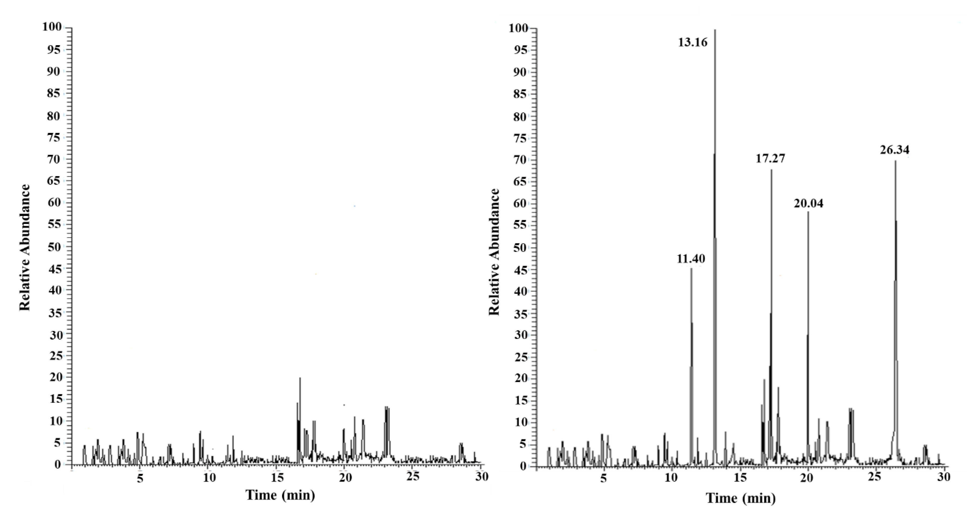
2.2.1. PAEs Determination

The selected target molecules were BBP, DBP, DEP, DMP, and DnOP. They are widely used in the production of plastic materials for food and non-food packaging, toys, etc. and are, as previously underlined, among the substances recognized as a priority by the USA EPA.
Standard compounds (purity > 99%) were purchased from Sigma Aldrich (St. Louis, Missouri, USA). Stock solutions, at a concentration level of 1 mg/mL, were prepared in sterile filtered ultrapure water (SFUW; Sigma Aldrich) with 20% (w/v) NaCl (Sigma Aldrich) and stored in glass vials at 8 °C. Working solutions, daily obtained by diluting stock solutions with SFUW, were stored at 8 °C until use.
Following an experimental protocol previously successfully optimized for meat [41,42,43], 2 g of shrimp extracted pulp sample were cut into small pieces, transferred into a centrifuge tube, and added with 12 mL of an n-pentane/methanol (Sigma Aldrich) mixture in a 5:9 (v/v) ratio. The tube was shaken at room temperature on a vortex mixer for 5 min, followed by centrifugation for 10 min at 1150 RCF (SBS-LZ-4000/20, Steinberg Systems, Hamburg, Germany). The extraction process was repeated twice, and the supernatants were combined and dried by evaporation. The resulting residue was dissolved in 1.5 mL of a 20% NaCl aqueous solution and transferred into a 1.7 mL vial, before being subjected to SPME-GC/MS analysis.
A polydimethylsiloxane/divinylbenzene fiber (PDMS/DVB, diameter 65 µm, Sigma Aldrich) was used for the direct immersion SPME procedure (DI-SPME). PAE extraction was carried out under constant stirring for 20 min at 40 °C. To prevent a possible carryover effect, the fiber was kept at 200 °C for 30 min in the GC injector after the desorption step and before each subsequent extraction.
To preserve the robustness of the SPME fibers to more than 100 cycles, they were rinsed after each cycle and stored in fresh water overnight.
The GC/MS system used was a Finnigan TRACE GC ultra gas chromatograph (Thermo Fisher Scientific, Waltham, MA, USA) equipped with a split/splitless injector interfaced with an ion trap MS (Finnigan Polaris Q, Thermo Fisher Scientific). The capillary column used was a Sigma Aldrich SPB-5 fused silica (30 m length, 0.25 µm i.d., 0.25 µm film thickness) with helium (purity 99.9999%, S.I.A.D., Bergamo, Italy) as the carrier gas at a flow rate of 1 mL/min. The transfer line temperature was set at 220 °C, while the injector (in splitless mode for 1 min) was maintained at 270 °C. The oven temperature program was the same as Aresta et Al. [43] modified as follows to reduce the eluition times: initial temperature of 60 °C; ramp: 10 °C/min from 60 °C to 260 °C; hold at 260 °C for 3 min. The mass spectrometer was operated in electron impact positive-ion mode (EI+), with the ion source temperature set at 250 °C. The electron energy was 70 eV, and the filament current was 150 µA.
Detection of individual PAEs was carried out by comparing the mass spectrum of each single analyte, obtained from a chromatogram purchased in TIC (total ion current; m/z range 50-450) mode of standard solutions (1 ng/mL) of each one of the five considered analytes, with the data contained in the NIST (National Institute of Standards & Technology) library implemented in the GC/MS system management software. For all 5 PAEs the relative retention times (TR) and the characteristic ions, used for the subsequent acquisitions in SIM (selected ion monitoring) mode, were therefore identified and summarized in Table 2.
For the quantitative evaluation of PAEs in shrimp using the standard addiction method (SAM), 100 µL of mixed standard solutions at known PAEs concentration levels were added to 2.0 g of the uncontaminated shrimp extracted pulp sample, purchased from a local supplier and coming from Atlantic Ocean, which was then extracted and analyzed as described above. Each measurement was repeated in triplicate.
Throughout all experimental procedures, the use of plastic objects of any kind (tips, containers, etc.) was always avoided.

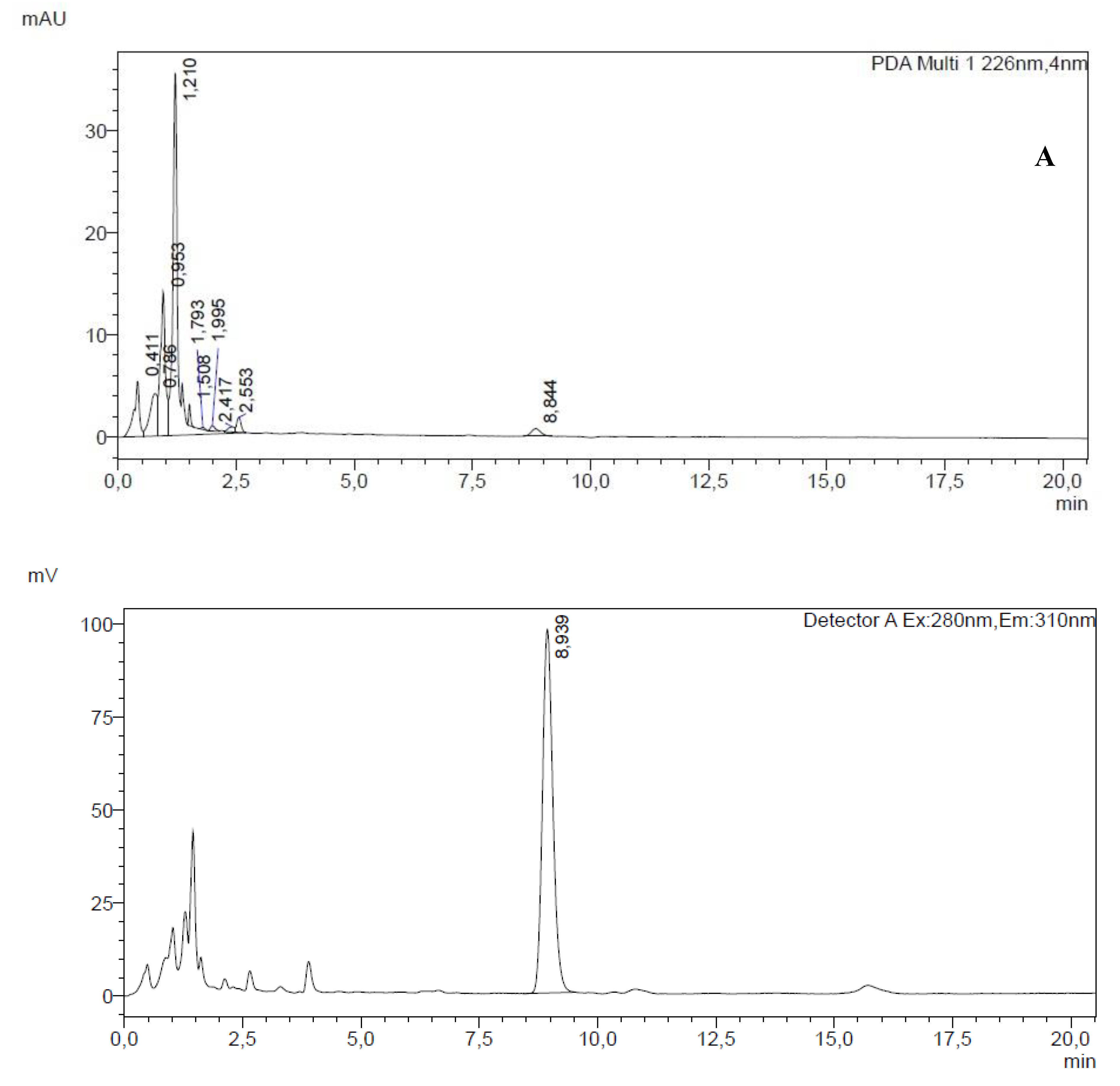
2.2.2. BPA Determination

BPA (purity ≥ 99%) was purchased from Sigma-Aldrich. HPLC-grade acetonitrile (purity ≥ 99.9%), hexane (purity ≥ 98%), and SFUW were also obtained from Sigma-Aldrich. Stock solutions of BPA were prepared in acetonitrile and stored in amber glass vials at 8 °C.
The analytical protocol used for shrimps’ analysis was developed by combining effective methodologies from previous studies [46,47,48].
Approximately 1 g of shrimp extracted pulp was homogenized in 2 mL of hexane, used as a defatting phase to remove lipids and reduce potential matrix interferences. Then, 2.5 mL of acetonitrile was added due to its intermediate polarity, which helps denature proteins and selectively solubilize BPA. The sample was vortexed for 5 minutes at room temperature, and the resulting suspension was centrifuged at 1150 RCF for 10 minutes (SBS-LZ-4000/20, Steinberg Systems). This led to the formation of two layers: an upper hexane-rich phase and a lower acetonitrile phase containing BPA.
The acetonitrile fraction was carefully collected and evaporated to dryness; the residue was reconstituted in 1.5 mL of saline solution (7.5% w/v NaCl) and transferred into a 1.7 mL screw-cap vial with septum [46].
The aqueous solution was analysed by SPME, using a polyacrylate (PA) fiber (85 µm thickness; Sigma Aldrich) selected for its high efficiency in extracting BPA from food matrices and simulants [46,47].
Before use, the fiber was conditioned for one hour in a vial containing the mobile phase; it was then immersed in the aqueous solution for 20 minutes at room temperature under constant magnetic stirring. After extraction, the fiber was desorbed for 5 minutes in a 200 µL microvial containing 150 µL of mobile phase (static desorption mode). A 50 µL aliquot of the desorbed solution was injected into the HPLC system for BPA quantification.
To prevent potential carry-over phenomena, the fiber was immersed for 30 minutes in a vial containing the mobile phase, following desorption and prior to each subsequent extraction.
Chromatographic analyses were performed on a Shimadzu (Kyoto, Japan) Nexera system equipped with dual LC-30AD pumps, an RF-20AXS fluorescence detector (FLD), a SPD-M20A diode array detector (DAD), and an Accucore™ C18 analytical column (100 × 4.6 mm, 4 µm; Thermo Scientific). A matching guard column was employed. Data acquisition was managed using LabService software (v5.03).
HPLC analyses were carried out under isocratic conditions at room temperature [48], with a mobile phase consisting of acetonitrile and water (30:70, v/v) at a flow rate of 1 mL/min. [49].
The identification of BPA was confirmed by comparing the signals obtained through fluorescence and UV detection. The FLD was set at an excitation wavelength of 280 nm and an emission wavelength of 310 nm, while UV spectra were acquired in the 220–500 nm range using a DAD. The peak observed at a retention time of 8.9 minutes in the fluorescence detector was attributed to BPA, based on the matching TR in both detectors and the overlap of the UV absorption spectrum with that of the analytical standard, thereby confirming the identity of the analyte.
To ensure accuracy in BPA quantification, the SAM was employed. A calibration curve was built using FLD detection by adding BPA standard solutions, with concentrations ranging from 0.0008 to 0.08 mg/kg to 1 g of uncontaminated oceanic shrimp extracted pulp sourced from a local supplier. These spiked samples were then processed and analysed as previously described. Each measurement was performed in triplicate.
Also in this case, the use of plastic objects during all the experimental procedure was avoided.

2.3. Microbiological Characterization

2.3.1. Bacteria Investigation

The crustaceans, collected from the designed areas (Table 1), were transported under refrigeration (+4 °C) and processed within 24 hours. The pulp extracted from shrimps were examined for the following mesophilic microorganisms: Total Bacteria Count (TBC), β-glucuronidase positive Escherichia coli, Enterobacteriaceae, Enterococci, positive-coagulase staphylococci, Listeria monocytogenes, Salmonella spp., Shiga Toxing-Producing E. coli (STEC), and Vibrio spp. according to the standardized methods detailed below.
For the quantitative analysis, 10 grams of each sample was added to 90 mL of Buffered Peptone Water (Biolife Italiana srl, Italy) and homogeneized in a stomacher device (Biosigma, Cona, Italy), followed by serial diluition up to 10^-3 prior to testing.
For the total bacteria count (TBC) 1 mL of each dilution was inoculated on Plate Count Agar (Biolife Italiana srl) by pour plate method and incubated at 30°C for 72 hours (UNI EN ISO 4833-1:2013) [50].
For the quantitative analysis of b-glucuronidase positive Escherichia coli, each dilution was tested by pour plate method on Tryptone Bile X-Glucuronide Agar (Biolife Italiana srl). The plates were incubated at 44 °C for 18-24 hours. b-glucuronidase E. coli coli positive colonies appear blue to blue green after incubation (UNI ISO 16649-2:2010) [51].
The enumeration of coagulase-positive staphylococci was carried out by plating the samples on Baird Parker Agar (Biolife Italiana srl) and incubating at 37°C for 48 hours (ISO 6888-1:2018) [52]. Typical colonies, which appear black and grey, shiny and convex, surrounded by a zone of clearing in the medium, were sub-cultured on Brain Heart Infusion Broth (Biolife Italiana srl) and incubated at 37 °C for 24 hours and subsequent coagulase testing with EDTA Plasma Coagulase (Biolife Italiana srl).
For the evaluation of Enterococci, samples were plated on Slanetz Bartley Agar (Biolife Italiana srl) at 36 + 2 °C for 48 hours. Presumptive colonies, with red, maroon or pink colonies were subjected with biochemical and MALDI-TOF identification.
For the enumeration of Enterobacteriaceae, each dilution was tested by pour plate method on Red bile Glucose (VRBG) agar (Microbiol s.r.l.) and incubated at 37 °C for 24 hours (ISO 21528-2: 2017) [53]. Presumptive pink or red-violet colonies with or without halos were subculture dog Nutrient agar (Biolife Italiana srl) for 24 hours at 37°C and subjected to confirmatory biochemical test.
For the evaluation of Listeria monocytogenes, 25 g of each sample was added to 225 mL of pre-enrichment Half-Fraser broth and incubated at 30°C for 24 hours. Subsequently, 100 mL was inoculated in 10 mL of Half-Fraser broth and incubated at 37 °C for 24 hours. Each enrichment medium was plated on Ottaviani and Agosti Listeria agar (ALOA; Microbiol s.r.l.) and Palcam agar (Biolife Italiana srl) and incubated at 37 °C for 24 hours, respectively. Presumptive colonies were subcultured on Columbia Blood Agar prior to confirmation tests (ISO 11290-1:2017) [54].
For the detection of Salmonella spp. 25 g of each sample was transferred to 225 mL of Buffered Peptone Water pre-enrichment (Biolife Italiana srl) and incubated at 37 °C for 24 hours (ISO 6579-1: 2017) [55]. Subsequently, 100 mL of each sample was plated in three equidistant drops on Modified Semisolid Rappaport Vassiliadis (MSRV) agar selective enrichment medium (Microbiol s.r.l.) and incubated at 41.5 °C for 24 h, and 1 mL was inoculated to the Muller–Kauffmann Tetrathionate–Novobiocin Broth (MKTTn, Biolife Italiana srl) at 37 °C for 24 h. Then, the enrichment was plated on Xylose Lysine Desoxycholate agar specific selective culture medium (Biolife Italiana srl) and Salmonella–Shigella agar (Biomèrieux, Marcy l’Etoile, France) and incubated at 37 °C for 24 h and 24–48 h, respectively. Presumptive colonies, with typical, black-centered colonies underwent further biochemical testing for identification.
The detection of Shiga Toxing-Producing E. coli (STEC) was performed by molecular methods: 25 g of shrimp pulp was transferred to 225 mL of Buffered Peptone Water pre-enrichment prewarmed (Biolife Italiana srl) and incubated at 41,5 + 1 for 16-24 hours. The DNA was extracted from the enrichment and tested by Real Time polymerase chain reaction (PCR) PCR-iQ-Check STEC PCR Detection Kits (BioRad, Hercules, California, USA) for the Shiga-toxin genes (stx1 and stx2) and for the eae gene (intimin). At the same time, cultural investigation was performed by directly striking pre-enrichment on CHROMAgarTM STEC (CHROMagarTM, Saint-Denise, France) and incubating at 35-37 °C for 18-24 hours. No STEC was detected by either molecular or cultured methods.
For the evaluation of Vibrio spp. 25 g of each sample was homogenized in 225 mL alkaline saline peptone water (ASPW) (Microbiol s.r.l.) first enrichment and incubated at 41,5 °C for 6 + 1 hours (ISO 21872-1:2023) [56]. Subsequently 1 mL of the enrichment was inoculated into 10 mL of ASPW and incubated at 41,5 °C for 18 + 1 hour. Both enrichments were stroked on Thiosulfate Citrate Bile and Sucrose (TCBS) (Biolife Italiana srl) agar plate and Soya peptone triphenyl tetrazolium chloride (TSTA) agar (HiMedia GmbH, Einhausen, Germany) at 37 °C for 24 + 3 hours. Presumptive colonies were subjected to biochemical identification.

2.3.2. Fungi Investigation

The enumeration of fungi, 0,1 mL of each dilution was inoculated on Dichloran Rose Bengal Cloramphenicol (DRBC) agar (Liofilchem Srl, Roseto degli Abruzzi, Termoli, Italy) and incubated at 25 °C for 5 days (ISO 21527:2018) [57].

3. Experimental Results and Discussion

3.1. PAEs and BPA Determination

Figure 1. shows a chromatogram acquired in SIM (acquisition parameters in paragraph 2.2.2) of the extracted pulp of shrimp (2 g) from the Atlantic Ocean, as purchased by a local supermarket (A) and after the addition of 200 µL of a standard solution of the five selected PAEs at a concentration level of 10 ng/mL each one (B).
No traces of considered PAEs were found in the reference extracted pulp shrimp.
The analytical method was validated in terms of linearity, limits of detection (LOD) and quantification (LOQ), and precision. The results showed good linearity for all the analytes in the range 0.005–1 mg/Kg with correlation coefficients always better than 0.9924 (DEP). The LOD and LOQ, determined based on the signal-to-noise (S/N) ratio, ranged from 0.023 (DBP) to 0.037 (DEP) mg/Kg and from 0.076 (DBP)–0.124 (DEP) mg/Kg, respectively. The intermediate precision of the method (expressed in % relative standard deviation, RSD%) for the same replicate sample ranged from 10.8 (DEP) to 24.5 (DnOP) and from 22.3 (DMP) to 34.3 (DBP) for within-day and between-day reliability, respectively. Table 3 summarizes all the analytical method validation parameters.
Table 4 shows the concentrations of the PAEs under examination found in the extracted pulp of shrimps from 4 caught stations of 2 different areas of the Mediterranean Sea.
All the crustaceans analyzed were contaminated by DEP and DMP at concentration levels between 0.145-0.267 mg/Kg and 0.127-0.171 mg/Kg, respectively. The shrimps from St_21 (South Calabria), moreover, contained 0.096 mg/Kg of DBP. The presence of such quantities of plasticizers in the extracted pulp of the crustaceans analyzed can be traced back to the ingestion, by the crustaceans themselves, of fragments of plastic materials dispersed in marine waters or deposited on the seabed.
DEP and DMP, recognized by recent studies as able to induce male and female reproductive toxicity, developmental damage and liver effects [58,59], together with DnOP, are among the most detected persistent organic pollutants in the environment. These substances are not authorized to produce plastic materials and objects intended for contact with food [60] and so there is no specific migration limit (SML) for these compounds, therefore the generic threshold limit of 60 mg/kg of food applies. According to Ministerial Decree 123 of 28 March 2003 [61], DEP may be present in quantities not exceeding 5% as the sum of all phthalates in plastic materials intended for contact with food.
As regards DBP, included by the EU list of substances that interfere with the endocrine system [45], as well as BBP, it is important to note that, according to EU Regulation 2023/1442 [62], the use of this substance is permitted to produce reusable plastic objects in contact with foods not containing fatty substances. Its SML is set at 0.3 mg/kg of food [60].
Analyzing our experimental results, it is possible to conclude that the concentration levels found for DBP, DEP and DMP exceed the respective LOQ values and are always below the LMS permitted by law.
The use of SPME coupled with HPLC-FLD detection is crucial for the analysis of non-volatile contaminants in shrimp extracted pulp, as it offers pre-concentration and highly sensitive separation of low-concentration compounds. The fluorescence detector enhances the ability to detect specific contaminants even at extremely low levels due to its high selectivity and sensitivity. In Figure 2 is showed a HPLC chromatograms with diode array (DAD) (A) and fluorescence (FLD) (B) detection of shrimp extracted pulp after the addition of BPA standard at a concentration level of 0.1 µg/g.
Appling the selected optimized experimental conditions (see paragraph 2.2.2), the instrument response (peak area) was proportional to the concentration, with a R² value greater than 0.998. Method validation included assessment of linearity, LOD and LOQ, and %RSD, too. LOD and LOQ, determined based on the S/N ratio as for PAEs, showed values of 0.0008 mg/kg for LOD and 0.0042 mg/kg for LOQ. Intra-day and inter-day precision values were 6.8 and 8.3 %RSD, respectively.
Table 5 resumes the BPA concentration levels in shrimp extracted pulp samples from the selected four fishing stations of the Mediterranean Sea.
Observing Table 4 is evident that the BPA concentration level in the considered analysed crustaceans extracted pulp ranged from 0.0010 to 0.0075 mg/kg. All values, exceeding the LOD and, in most cases, the LOQ, were significantly below the specific SML set by the EU for BPA [62], which is 0.05 mg/kg.
The detection of the considered residues of plasticizers in the extracted pulp of the shrimps analyzed confirms that plastic pollution of the seas undoubtedly has a negative effect also on the organisms that populate it since, evidently, small polymer fragments end up in the food they feed on and are not completely metabolized. Such contamination can therefore represent a source of danger also for human beings now in which the crustaceans considered enter our food chain.

3.2. Microbiological Investigation

The total bacterial count showed significant variability between the samples, with values ranging from 920 to 84,000 CFU/g. The highest levels were observed at station 67, while the lowest count was recorded at station 21.
The fungal count was below the limit of quantification (<1 CFU/g) in all samples.
Similar results were observed for E. coli and Enterococci enumeration that resulted <1 CFU/g for all samples, except for station 57 and 67 which showed 180 UFC/g and 50 UFC/g, respectively. Moreover, MALDI-TOF identification revealed the presence of E. faecalis for station 57 and 67.
The presence of Enterobacteriaceae was also generally very limited, with values below 1 CFU/g, except at station 9 (160 CFU/g), station 21 (80 CFU/g), and station 57 (220 CFU/g).
Coagulase-positive staphylococci were detected in all samples: station 9 (3000 CFU/g), station 21 (790 CFU/g), station 57 (1000 CFU/g) and station 67 (7300 CFU/g) (experimental results in Table 6).
None of the considered pathogens were detected in the sample analyzed (Table 7). However, Proteus mirabilis which is a Gram-negative, rod-shaped, facultative anaerobic bacterium, was identified in the sample collected from station 57 using MALDI-TOF.
None of the pathogens were detected in the samples analysed (see Table 7).

4. Conclusions

Marine pollution due to human activities, and especially plastic waste that voluntarily or involuntary ends up in the sea, poses a serious risk both to the marine ecosystem and, indirectly, to human health. Marine organisms (e.g., fish, crustaceans, etc.) can ingest microscopic plastic residues, which then end up in our diet, as top predators at the end of the food chain.
Plastic contains plasticizers (e.g., PAEs and BPA), substances that, if ingested, can be particularly harmful to human health being recognized, for example, as endocrine disruptors, potentially responsible of carcinomas, dysplasia, and adversely effects on the reproductive system [27,28].
The plastics ingestion, with the occurrence of plastic fibers and MPS in the stomachs of A. foliacea, was recently documented in the Western and Eastern Mediterranean [39,63] but no data on the concentration of the toxic plasticizers in the edible muscle of this deep-sea shrimp was reported so far.
In this study, effective analytical protocols based on the use of SPME coupled with chromatographic techniques were applied to determine PAEs and BPA residues in the giant red shrimp Aristaeomorpha foliacea , in two different areas of the Mediterranean Sea. The results confirm and further extend the awareness of the high exposure of the deep-sea crustaceans to plastic pollution. All samples were found to be contaminated with DEP, DMP (in one case also by DBP), and BPA at concentration levels always below the maximum limits permitted by current regulations, emerging the possible risk for human health related to their consumption. Furthermore, a comprehensive microbiological analysis completed the work and permit to evaluate the risks for human health associated with the ingestion of pathogens and fungi, if the examined crustaceans were consumed without prior cooking.
In conclusion, this dual approach provides a complete evaluation of the impact of human pollution on these crustaceans, revealing both chemical contamination and potential biological disruptions that could pose a danger to food safety.

Founding

This research was supported by: project funded under the National Recovery and Resilience Plan (NRRP), Mission 4 Component 2 Investment 1.3—Call for tender No. 341 of 15 March 2022 of the Italian Ministry of University and Research funded by the European Union— NextGenerationEU; Project code PE00000003, Concession Decree No. 1550 of 11 October 2022 adopted by the Italian Ministry of University and Research, CUP D93C22000890001, Project title “ON Foods— Research and innovation network on food and nutrition Sustainability, Safety and Security—Working ON Foods”; the Italian Ministry of Agriculture, Food Sovereignty and Forestry (MASAF) and the European Commission (EU Regulations 1004/2017) under the Data Collection Framework (DCF).

References

  1. Geyer, R.; Jambeck, J.R.; Law, K.L. Production, use, and fate of all plastics ever made. Sci Adv. 2017, 3, e1700782. [Google Scholar] [CrossRef] [PubMed]
  2. Karbalaei, S.; Hanachi, P.; Walker, T.R.; Cole, M. Occurrence, sources, human health impacts and mitigation of microplastic pollution. Environ. Sci. Pollut. Res. Int. 2018, 25, 36046–36063. [Google Scholar] [PubMed]
  3. Galgani, L.; Tsapakis, M.; Pitta, P.; Tsiola, A.; Tzempelikou, E.; Kalantzi, I.; Esposito, C.; Loiselle, A.; Tsotskou, A.; Zivanovic, S.; Dafnomili, E.; Diliberto, S.; Mylona, K.; Magiopoulos, I.; Zeri, C.; Pitta, E.; Loiselle, S.A. Microplastics increase the marine production of particulate forms of organic matter. Environ. Res. Letters 2019, 14, 124085. [Google Scholar]
  4. Ozturk, R.C.; Altinok, I. Interaction of plastics with marine species. Turkish J. of Fisheries and Aquatic Sci. 2020, 20, 647–658. [Google Scholar] [CrossRef]
  5. Stanev, E.V.; Ricker. M. The fate of marine litter in semi-enclosed seas: a case study of the Black Sea. Front. Mar. Sci. 2019, 660.
  6. Borrelle, S.B., Ringma, J., Lavender Law, K., Monnahan, C.C., Lebreton, L., McGivern, A., Murphy, E., Jambeck, J., Leonard, G.H., Hilleary, M.A., Eriksen, M., Possingham, H.P., De Frond, H., Gerber, L.R., Polidoro, B., Tahir, A., Bernard, M., Mallos, N., Barnes, M., Rochman, C.M. Predicted growth in plastic waste exceeds efforts to mitigate plastic pollution. Science 2020,1515–1518.
  7. https://www.futureagenda.org/foresights/plastic-oceans.
  8. Galgani, F.; Lusher, A.L.; Strand, J.; Haarr, M.L.; Vinci, M.; Jack, E.M.; Kagi, R.; Aliani, S.; Herzke, D.; Nikiforov, V.; Primpke, S.; Schmidt, N.; Fabres, F.; De Witte, B.; Solbakken, V.S.; Van Bavel, B. Revisiting the strategy for marine litter monitoring within the european marine strategy framework directive (MSFD). Ocean & Coastal Manag. 2024, 255, 107254. [Google Scholar]
  9. da Costa, J.P. Micro- and nanoplastics in the environment: research and policymaking. Current Opinion in Environ. Sci. & Health 2018, 1, 12–16. [Google Scholar]
  10. Aransiola, S.A.; Ekwebelem, M.O.V.; Daza, B.X.; Oladoye, P.O.; Alli, Y.A.; Bamisaye, A.; Aransiola, A.B.; Oni, S.O.; Maddela, N.R. Micro- and nano-plastics pollution in the marine environment: Progresses, drawbacks and future guidelines, Chemosphere 2025, 374, 14421114.
  11. Hantoro, I. , Löhr, A. J., Van Belleghem, F. G. A. J., Widianarko, B., & Ragas, A. M. J. Microplastics in coastal areas and seafood: implications for food safety. Food Additives & Contaminants: Part A 2019, 36, 674–711. [Google Scholar]
  12. Ferreira, H.C.; Lôbo-Hajdu, G. Microplastics in coastal and oceanic surface waters and their role as carriers of pollutants of emerging concern in marine organisms, Marine Environ. Res. 2023, 188, 106021. [Google Scholar]
  13. Pitacco, V.; Orlando-Bonaca, M.; Avio, C.G. 4 - Plastic impact on marine benthic organisms and food webs, Editor(s): Giuseppe Bonanno, Martina Orlando-Bonaca, Plastic Pollution and Marine Conservation, Academic Press 2022, 95-151.
  14. Cau, A.; Gorule, P.A.; Bellodi, A.; Carreras-Colom, E.; Moccia, D.; Pittura, L.; Regoli, F.; Follesa, M.C. Comparative microplastic load in two decapod crustaceans Palinurus elephas (Fabricius, 1787) and Nephrops norvegicus (Linnaeus, 1758). Mar. Pollut. Bull. 2023, 191, 114912. [Google Scholar] [CrossRef]
  15. Burkhardt-Holm, P.; N'Guyen, A. Ingestion of microplastics by fish and other prey organisms of cetaceans, exemplified for two large baleen whale species. Mar. Pollut. Bull. 2019, 44, 224–234. [Google Scholar] [CrossRef]
  16. Jabeen, K.; Su, L.; Li, J.; Yang, D.; Tong, C.; Mu, J.; Shi, H. Microplastics and mesoplastics in fish from coastal and fresh waters of China. Environ. Pollut. 2017, 221, 141–149. [Google Scholar] [CrossRef]
  17. Pozo, K.; Gomez, V.; Torres, M.; Vera, L.; Nuñez, D.; Oyarzún, P.; Mendoza, G.; Clarke, B.; Fossi, M.C.; Baini, M.; Přibylová, P.; Klánová, J. Presence and characterization of microplastics in fish of commercial importance from the Biobío region in central Chile. Mar. Pollut. Bull. 2019, 140, 315–319. [Google Scholar] [CrossRef]
  18. Rocha-Santos, T.; Duarte, A.C. A critical overview of the analytical approaches to the occurrence, the fate and the behavior of microplastics in the environment. TrAC Trends in Anal. Chem. 2015, 65, 47–53. [Google Scholar] [CrossRef]
  19. Wootton, N.; Reis-Santos, P.; Gillanders, B.M. Microplastic in fish – A global synthesis. Rev Fish Biol Fisheries 2021, 31, 753–771. [Google Scholar] [CrossRef]
  20. Hossain, M.S.; Sobhan, F.; Uddin, M.N.; Sharifuzzaman, S.M.; Chowdhury, S.R.; Sarker, S.; Chowdhury, M.S.N. Microplastics in fishes from the Northern Bay of Bengal. Sci. of The Total Environ. 2019, 690, 821–830. [Google Scholar] [CrossRef] [PubMed]
  21. Arias, A.H.; Ronda, A.C.; Oliva, A.L.; Marcovecchio, J.E. Evidence of microplastic ingestion by fish from the Bahía Blanca estuary in Argentina, South America. Bull. Environ. Contam. Toxicol. 2019, 102, 750–756. [Google Scholar] [CrossRef] [PubMed]
  22. Jovanović, B. Ingestion of microplastics by fish and its potential consequences from a physical perspective. Integr. Environ. Assess. Manag. 2017, 13, 510–515. [Google Scholar] [CrossRef]
  23. Cole, M.; Lindeque, P.; Fileman, E.; Halsband, C.; Goodhead, R.; Moger, J.; Galloway, T.S. Microplastic ingestion by zooplankton. Environ. Sci. Technol. 2013, 47, 6646–55. [Google Scholar] [CrossRef]
  24. Cole, M.; Lindeque, P.; Halsband, C.; Galloway, T.S. Microplastics as contaminants in the marine environment: A review. Mar. Pollut. Bull. 2011, 62, 2588–2597. [Google Scholar] [CrossRef]
  25. Huang, L.; Zhu, X.; Zhou, S.; Cheng, Z.; Shi, K.; Zhang, C.; Shao, H. Phthalic acid esters: natural sources and biological activities. Toxins (Basel). 2021, 13, 495. [Google Scholar] [CrossRef]
  26. Braunrath, R.; Cichna, M. Sample preparation including sol–gel immunoaffinity chromatography for determination of bisphenol A in canned beverages, fruits and vegetables. J. of Chrom. A 2005, 1062, 189–198. [Google Scholar] [CrossRef] [PubMed]
  27. Wang, Y.; Qian, H. Phthalates and their impacts on human health. Healthcare (Basel) 2021, 9, 603. [Google Scholar] [CrossRef] [PubMed]
  28. Rochester, J.R. Bisphenol A and human health: A review of the literature. Reproduct. Toxicol. 2013, 42, 132–155. [Google Scholar] [CrossRef] [PubMed]
  29. https://eur-lex.europa.eu/eli/reg/2024/3190/oj/eng.
  30. Dumen, E.; Ekici, G.; Ergin, S.; Bayrakal, G.M. Presence of Foodborne Pathogens in Seafood and Risk Ranking for Pathogens. Foodborne Pathog. Dis. 2020, 17, 541–546. [Google Scholar] [CrossRef]
  31. Lehel, J.; Yaucat-Guendi, R.; Darnay, L.; Palotás, P.; Laczay, P. Possible food safety hazards of ready-to-eat raw fish containing product (sushi, sashimi). Critical Reviews. Food Sci. and Nutrition 2020, 61, 867–888. [Google Scholar]
  32. Chintagari, S.; Hazard, N.; Edwards, G.; Jadeja, R.; Janes, M. Risks Associated with Fish and Seafood. Microbiol. Spectr. 2017, 5, 10. [Google Scholar] [CrossRef]
  33. Guijarro, B.; Bitetto, I.; D’Onghia, G.; Follesa, M.C.; Kapiris, K.; Mannini, A.; Markovic, O.; Micallef, R.; Ragonese, S.; Skarvelis, K.; Cau, A. Spatial and temporal patterns in the Mediterranean populations of Aristaeomorpha foliacea and Aristeus antennatus (Crustacea: Decapoda: Aristeidae) based on the MEDITS surveys. Scientia Marina 2019, 83S1, 57–70. [Google Scholar] [CrossRef]
  34. D’Onghia, G.; Maiorano, P.; Matarrese, A.; Tursi, A. Distribution, biology and population dynamics of Aristaeomorpha foliacea (Risso, 1827) (Crustacea, Decapoda) from the north-western Ionian Sea (Mediterranean Sea). Crustaceana 1998, 71(5), 518–544. [Google Scholar] [CrossRef]
  35. D’Onghia, G.; Giove, A.; Maiorano, P.; Carlucci, R.; Minerva, M.; Capezzuto, F.; Sion, L.; Tursi, A. Exploring relationships between demersal resources and environmental factors in the Ionian Sea (Central Mediterranean). J. of Marine Biology 2012, 2, 279406. [Google Scholar] [CrossRef]
  36. Maiorano, P.; Capezzuto, F.; Carluccio, A.; Calculli, C.; Cipriano, G.; Carlucci, R.; Ricci, P.; Sion, L.; Tursi, A.; D’Onghia, G. Food from the Depths of the Mediterranean: The Role of Habitats, Changesin the Sea-Bottom Temperature and Fishing Pressure. Foods 2022, 2022. 11, 1420. [Google Scholar] [CrossRef]
  37. Soultani, G.; Sele, V.; Rasmussen, R.R.; Pasias, I.; Stathopoulou, E.; Thomaidis, N.S.; Sinanoglou, V.J.; Sloth, J.J. Elements of toxicological concern and the Arsenolipids’ profile in the giant-red Mediterranean shrimp, Aristaeomorpha foliacea. J. of Food Composition and Analysis 2021, 97, 103786. [Google Scholar] [CrossRef]
  38. D’Iglio, C. , Di Fresco, D. , Spanò, N., Albano, M., Panarello, G., Laface, F., Faggio, C., Capillo, G., Savoca, S., 2022. Occurrence of anthropogenic debris in three commercial shrimp species from south-western ionian sea. Biology 2022, 11, 1616. [Google Scholar]
  39. Chiacchio, L.; Cau, A.; Soler-Membrives, A.; Follesa, M.C.; Bellodi, A.; Carreras-Colom, E. Comparative assessment of microplastic ingestion among deep sea decapods: Distribution analysis in Sardinian and Catalan waters. Environ Res. 2025, 270, 120962. [Google Scholar] [CrossRef] [PubMed]
  40. Spedicato, M.T.; Massutí, E.; Mérigot, B.; Tserpes, G.; Jadaud, A.; Relini, G. 2019.The MEDITS TrawlSurvey Specifications in an Ecosystem Approach to Fishery Management. Sci. Mar. 2019, 83, 9–20. [Google Scholar] [CrossRef]
  41. De Vietro, N.; Aresta, A.M.; Gubitosa, J.; Rizzi, V.; Zambonin, C. Assessing the conformity of plasticizer-free polymers for foodstuff packaging using solid phase microextraction coupled to gas chromatography/mass spectrometry. Separations 2024, 11, 25. [Google Scholar] [CrossRef]
  42. Liu, W.; Lun, Y. Determination of sub-ppb level of phthalates in water by auto-SPME and GC–MS. Application 5989-7726EN; Agilent Technologies: Santa Clara, CA, USA, 2008. [Google Scholar]
  43. Aresta, A.M.; De Vietro, N.; Mancini, G.; Zambonin, C. Effect of a chitosan-based packaging material on the domestic storage of “ready-to-cook” meat products: evaluation of biogenic amines production, phthalates migration, and in vitro antimicrobic activity’s impact on Aspergillus Niger. Separations 2024, 11, 159. [Google Scholar] [CrossRef]
  44. Moreira, M.A.; Coelho, L.A.; de Lourdes Cardeal, Z. Analysis of plasticiser migration to meat roasted in plastic bags by SPME–GC/MS. Food Chem. 2015, 178, 195–200. [Google Scholar] [CrossRef]
  45. De Vietro, N.; Triggiano, F.; Cotugno, P.; Palmisani, J.; Di Gilio, A.; Zambonin, C.; de Gennaro, G.; Mancini, G.; Aresta, A.M.; Diella, G.; Marcotrigiano, V.; Trerotoli, P.; Marzocca, P.; Lorusso, L.; Spagnolo, A.M.; Sorrenti, G.T.; Lampedecchia, M.; Sorrenti, D.P.; D’Aniello, E.; Gramegna, M.; Nancha, A. Analytical investigation of phthalates and heavy metals in edible ice from vending machines connected to the Italian water supply. Foods 2024, 13, 2910. [Google Scholar] [CrossRef]
  46. Sarkis, N.; Shaghel, J.A. A new analytical method for determination bisphenol A in canned meat. Reas. Square 2022, 1, 14. [Google Scholar]
  47. Salafranca, J.; Batlle, R.; Nerı́n, C. Use of solid-phase microextraction for the analysis of bisphenol A and bisphenol A diglycidyl ether in food simulants. J. of Chrom. A 1999, 864, 137–144. [Google Scholar] [CrossRef]
  48. Aristiawan, Y.; Aryana, N.; Putri, D.; Styarini, D. Analytical method development for bisphenol A in tuna by using high performance liquid chromatography-UV. Proc. Chem. 2015, 16, 202–208. [Google Scholar] [CrossRef]
  49. Mita, L.; Bianco, M.; Viggiano, E.; Zollo, F.; Bencivenga, U.; Sica, V.; Monaco, G.; Portaccio, M.; Diano, N.; Colonna, A.; Lepore, M.; Canciglia, P.; Mita, D.G. Bisphenol A content in fish caught in two different sites of the Tyrrhenian Sea (Italy). Chemospere 2011, 82, 405–410. [Google Scholar] [CrossRef]
  50. Microbiologia Della Catena Alimentare—Metodo Orizzontale per la Conta dei Microrganismi—Parte 1: Conta Delle Colonie a 30 °C con la Tecnica Dell’inseminazione in Profondità. UNI EN ISO 4833-1:2013. 3 October 2013.
  51. Microbiologia di Alimenti e Mangimi per Animali—Metodo Orizzontale per la Conta di Escherichia coli beta Glucuronidasi-positiva—Parte 2: Tecnica Della Conta Delle Colonie a 44 °C che Utilizza 5-bromo-4-cloro-3- Indolil beta-D-glucuronide. UNI ISO 16649-2:2010. 28 April 2010.
  52. Microbiologia di Alimenti e Mangimi per Animali—Metodo Orizzontale per la Conta di Stafilococchi Coagulasi-positivi (Staphylococcus Aureus e Altre Specie)—Parte 1: Tecnica che Utilizza il Terreno Agar Baird-Parker. UNI EN ISO 6888-1:2018. 27 September 2018.
  53. Microbiology of the Food Chain—Horizontal Method for the Detection and Enumeration of Enterobacteriaceae—Part 2: Colony-count Technique. ISO 21528-2:2017. June 2017.
  54. Microbiology of the Food Chain—Horizontal Method for the Detection and Enumeration of Listeria Monocytogenes and of Listeria spp. Detection Method. ISO 11290-1:2017. May 2017.
  55. Microbiology of the Food Chain—Horizontal Method for the Detection, Enumeration and Serotyping of Salmonella Detection of Salmonella spp. ISO 6579-1:2017. February 2017.
  56. Microbiologia della catena alimentare - Metodo orizzontale per la determinazione di Vibrio spp - Parte 1: Ricerca delle specie di Vibrio parahaemolyticus, Vibrio cholerae e Vibrio vulnificus potenzialmente enteropatogene UNI EN ISO 21872-1:2023. 04 May 2023.
  57. Microbiology of food and animal feeding stuffs — Horizontal method for the enumeration of yeasts and moulds Part 1: Colony count technique in products with water activity greater than 0,95. ISO 21527-1:2008.
  58. Weaver, J.A.; Beverly, B.E.J.; Keshava, N.; Mudipalli, A.; Arzuaga, X.; Cai, C.; Hotchkiss, A.K.; Makris, S.L.; Yost, E.E. Hazards of diethyl phthalate (DEP) exposure: A systematic review of animal toxicology studies. Environ. Int. 2020, 145, 105848. [Google Scholar] [CrossRef]
  59. Cao, B.; Kong, H.; Shen, C.; She, G.; Tian, S.; Liu, H.; Cui, L.; Zhang, Y.; He, Q.; Xia, Q.; Liu, K. Dimethyl phthalate induced cardiovascular developmental toxicity in zebrafish embryos by regulating MAPK and calcium signaling pathways. Sci. of The Tot. Environ. 2024, 926, 171902. [Google Scholar] [CrossRef]
  60. European Commission Regulation (EU). No 10/2011 on plastic materials and articles intended to come into contact with food. Off. J. Eur. Comm. 2011, 12, 1–1394. [Google Scholar]
  61. https://www.normattiva.it/uri-res/N2Ls?urn:nir:ministero.salute:decreto:2003-03-28;123.
  62. https://eur-lex.europa.eu/eli/reg/2023/1442/oj/eng.
  63. Bordbar, L.; Sedlacek, P.; Anastasopoulou, A. Plastic pollution in the deep-sea Giant red shrimp, Aristaeomorpha foliacea, in the Eastern Ionian Sea; an alarm point on stock and human health safety. Sci. Total Environ. 2023, 877, 162783. [Google Scholar]
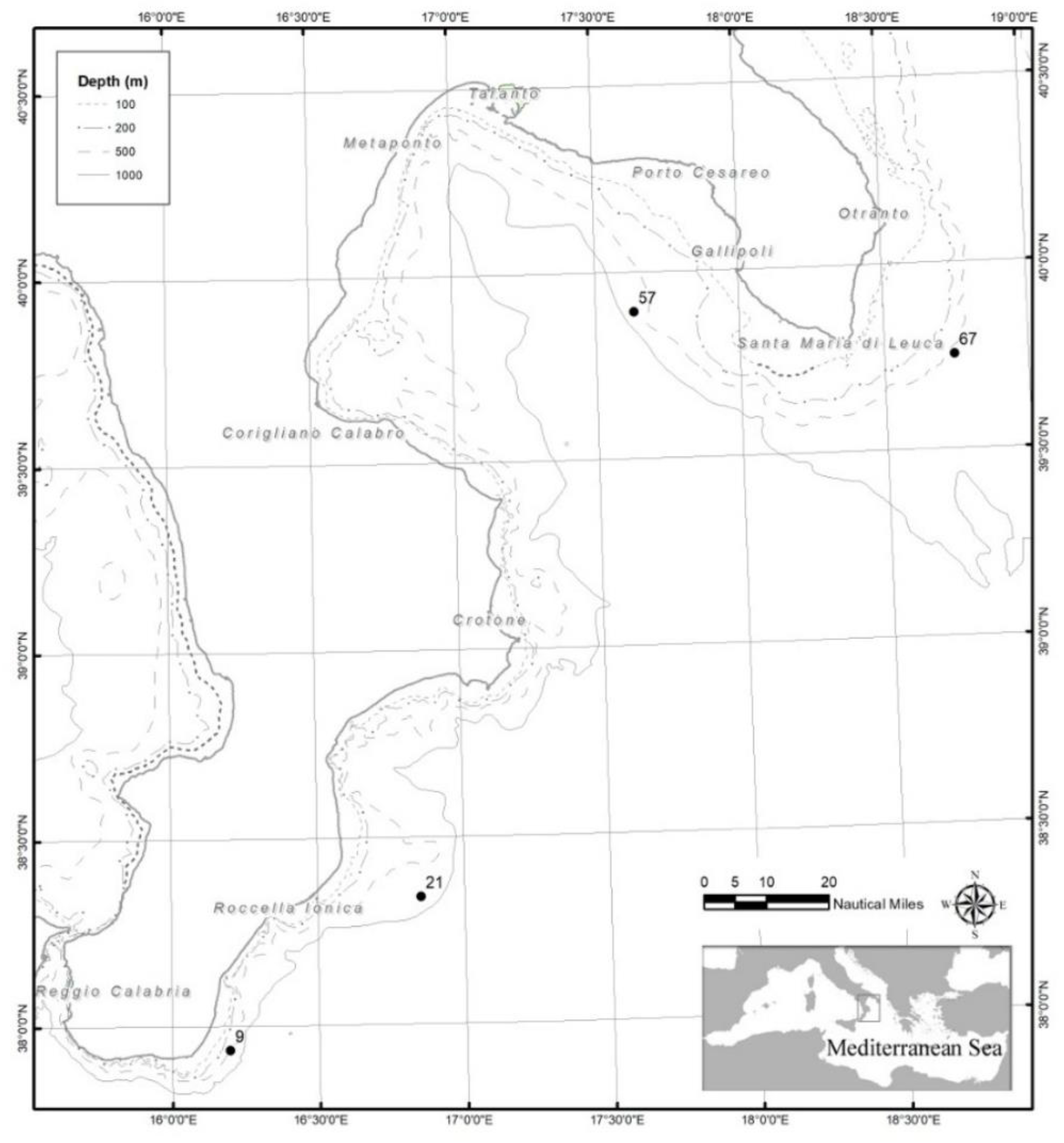
Figure 1. Map of the study areas in the notth-western Ionian Sea, with indication of the sampling stations in the Apulian and southern Calabria.
Preprints 174271 g001
Figure 1. Chromatogram acquired in SIM of the extracted pulp sample (2 g) of Atlantic shrimp as purchased (A) and after the addition of 2 ng of each standard PAE (B). .
Preprints 174271 g002
Figure 2. HPLC chromatograms with diode array (DAD) (A) and fluorescence (FLD) (B) detection of 1 g of shrimp extracted pulp after the addition of 1 µg of BPA standard.
Preprints 174271 g003
Table 1. Sampling station, with code, depth and geographic coordinates of Aristaeomorpha foliacea specimens collected in two different areas of the north-western Ionian Sea (central Mediterranean Sea).
Area Station code Depth (m) Latitude Longitude
Southern Calabria 9 543 3756163 N 01611752 E
21 656 3820496 N 01651188 E
Apulia 57 592 3953622 N 01738057 E
67 527 3945234 N 01844612 E
Table 2. TR and selected ions (m/z) for considered 5 PAEs.
PAE TR (min) m/z
BBP
DBP
DEP
DMP
DnOP
20.04±0.02
17.27±0.02
13.16±0.02
11.40±0.02
26.34±0.02
149, 206 [44]
149, 205, 223 [41,43,44,45]
105, 149, 177 [41,43,44,45]
135, 163, 194 [41,44,45]
149, 279 [44]
Table 3. Analytical method validation parameters.
PAE slope Correlation coefficient (R2) LOD (mg/Kg) LOQ (mg/Kg) Concentration level (mg/Kg)
Within-day
0.05      0.25      2.5
Between-day
0.05      0.25      2.5
BBP 707295 0.9940 0.030 0.100 22.1% 21.0% 19.7% 30.7% 28.8% 27.6%
DBP 501948 0.9965 0.023 0.076 20.3% 21.6% 22.0% 33.8% 34.3% 30.2%
DEP 6x10⁶ 0.9924 0.037 0.124 11.5% 10.8% 12.1% 22.4% 24.7% 25.3%
DMP 2x10⁶ 0.9940 0.033 0.110 13.7% 12.5% 11.9% 23.5% 25.5% 22.3%
DnOP 607682 0.9957 0.026 0.085 24.4% 23.1% 24.5% 30.2% 28.6% 29.4%
Table 4. Selected PAEs concentration in shrimps extracted pulp from 4 caught stations of 2 different areas of the Mediterranean Sea.
MEDITERRANEAN SEA BBP (MG/KG) DBP (MG/KG) DEP (MG/KG) DMP (MG/KG) DNOP (MG/KG)
ST_57 (SOUTH CALABRIA) / / 0.145±0.003 0.162±0.003 /
ST_67 (SOUTH CALABRIA) / / 0.267±0.005 0.165±0.003 /
ST_09 (APULIA) / / 0.164±0.003 0.127±0.002 /
ST_21 (APULIA) / 0.096±0.003 0.175±0.003 0.171±0.003 /
Table 5. Concentration levels of BPA in shrimps extracted pulp samples from 4 caught stations of 2 different areas of the Mediterranean Sea.
MEDITERRANEAN SEA BPA CONCENTRATION LEVEL (MG/KG)
ST_57 (SOUTH CALABRIA) 0.0058±0.0003
ST_67 (SOUTH CALABRIA) 0.001±0.00006
ST_09 (APULIA) 0.0058±0.0003
ST_21_ (APULIA) 0.0075±0.0005
Table 6. Microorganism counts obtained shrimps from the Mediterranean Sea.
Microorganism count (CFU/g)
Station Total bacteria count Total fungi count
Escherichia coli
Enterobacteriaceae
positive-coagulase staphylococci
Enterococci
9 14000 <1 <1 160 3000 <1
21 920 <1 <1 80 790 <1
57 1610 <1 <1 220 1000 180
67 84000 <1 <1 <1 7300 50
Table 7. Detection of Listeria monocytogenes, Salmonella spp. and Shiga-toxin-producing Escherichia coli (STEC), Vibrio spp. in samples collected at four monitoring stations (9, 21, 57, 67).
Detection
Station Listeria monocytogenes Salmonella spp. Shiga Toxing-Producing E.coli Vibrio spp.
9 Not detected Not detected Not detected Not detected
21 Not detected Not detected Not detected Not detected
57 Not detected Not detected Not detected Not detected
67 Not detected Not detected Not detected Not detected
Disclaimer/Publisher’s Note: The statements, opinions and data contained in all publications are solely those of the individual author(s) and contributor(s) and not of MDPI and/or the editor(s). MDPI and/or the editor(s) disclaim responsibility for any injury to people or property resulting from any ideas, methods, instructions or products referred to in the content.
Copyright: This open access article is published under a Creative Commons CC BY 4.0 license, which permit the free download, distribution, and reuse, provided that the author and preprint are cited in any reuse.
Prerpints.org logo

Preprints.org is a free preprint server supported by MDPI in Basel, Switzerland.

Subscribe

Disclaimer

Terms of Use

Privacy Policy

Privacy Settings

© 2025 MDPI (Basel, Switzerland) unless otherwise stated