Submitted:
28 February 2025
Posted:
28 February 2025
You are already at the latest version
Abstract
This study revealed the significant environmental and health risks posed by heavy metals contamination in Oreochromis niloticus (Nile tilapia) near landfill sites. It also assessed potential human health risks and investigated genotoxicity and oxidative stress in fish. Heavy metal concentrations were measured using Inductively Coupled Plasma Optical Emission Spectrometry. Results indicated that all four metals in water samples exceeded safety limits, and Cd levels in sediment were also above regulatory thresholds. In fish muscle, metal concentrations followed the order Cr > Pb > Cd > As, with Pb exceeding food quality standards. The bioaccumulation factor ranked as Cr > Pb > As > Cd. Health risk assessments, including the health risk index and carcinogenic risk analysis, suggested Pb contamination poses significant health risks through fish consumption. Genetic analysis showed greater genetic differentiation in landfill-site fish compared to reference-site fish, indicating genotoxic effects. Oxidative stress markers revealed higher levels of malondialdehyde, hydrogen peroxide, but lower activities of catalase and superoxide dismutase in landfill fish. These results emphasize the urgent need for regular environmental monitoring, stricter pollution controls, and improved waste management strategies to protect aquatic ecosystems and human health.
Keywords:
animals
; genetic differentiation
; oxidation
; public health
; toxic substances
Introduction
Rapid economic growth and rising industrial and socio-commercial activities have led to a significant increase in solid waste generation in Khon Kaen Province, Thailand [1]. The primary disposal method remains landfilling, but inadequate waste management has resulted in toxic leachate seeping into the surrounding environment [2]. At the Khon Kaen municipal landfill, high concentrations of major heavy metals (HM) such as As, Cd, Cr, and Pb have been detected in both leachate and local organisms [3,4,5,6,7]. Notably, this pollution extends beyond landfill sites and can have widespread environmental impacts [5].
The accumulation of HMs in fish can have harmful health effects, as these metals may be transferred to the human body upon consumption [8]. The severity of health impacts depends on the quantity of fish consumed [9]. Therefore, assessing human health risks is crucial when evaluating the potential effects of fish consumption in this area. This study aimed to analyze HM concentrations in water, sediment, and fish near the landfill. Additionally, it examined the bioaccumulation factor (BAF) of HMs in fish and the potential health risks associated with their consumption. The findings of this research can raise awareness among consumers and authorities about the possible hazards of fish consumption and contribute to future improvements in landfill environmental management.
Fish play a crucial role in aquatic ecosystems, occupying a high position in the food chain and being highly susceptible to heavy metal pollution [10]. Nile tilapia, a key protein source for humans, is widely farmed in tropical and subtropical regions worldwide. To assess pollution levels, monitoring HM concentrations in water and sediments is essential. Additionally, tracking fish health provides further evidence of HM contamination. HMs enter aquatic organisms through two primary pathways: direct ingestion of water and biota or non-dietary absorption via the gills and skin [11]. Research has shown that HM exposure in fish can lead to chromosomal damage and alterations in DNA structure [12,13]. Moreover, HMs induce oxidative stress by disrupting the balance between free radical production and elimination [14]. Beyond their direct impact on fish, HMs accumulate in aquatic organisms and transfer through the food chain to predators such as birds, animals, and humans, amplifying their harmful effects on the biosphere. To date, reports on the toxic effects of HMs on Tilapia fish health and molecular genetics have been limited. Therefore, this study aims to measure the concentrations of As, Cd, Cr, and Pb in water, sediments, and Tilapia fish, assess potential human health risks, and investigate genetic toxicity and oxidative stress biomarkers following exposure to HMs in a landfill site.
Materials and Methods
Study Sites
Study site was conducted in the Municipal landfill site, Kham Bon village, Khon Kaen Province, Thailand (Latitude: 16.59608238433, Longitude: 102.806853323728) (Figure 1). The water, sediment and fish samples were collected from landfill leachate water site and compared to samples from a non-polluted reference site as in commercial fish farm, Thapra subdistrict, Muang Khon Kaen, Khon Kaen Province (Latitude: 16.334442760000, Longitude: 102.79644053868).
Sample Collection
Water, sediment and tilapia were randomly selected from each of the 5 spots at the landfill site and the reference site (n=5).
The Analysis of Water Quality
An analysis of water quality was conducted by comparing samples from a Khon Kaen landfill with those from a reference site. The parameters assessed included temperature, total dissolved solids, dissolved oxygen, electrical conductivity and pH levels.

Table 1.
Analytical methods used for analysis of water quality indicators.
| Water Quality indicators | Analytical instruments |
|---|---|
| Temperature | Thermometer |
| Total dissolved solid | Total dissolved solids, Mettler Toledo, Model CH-8603 |
| Dissolved oxygen | DO meter, Mettler Toledo, Model 966 |
| Electro-conductivity | EC meter, Mettler Toledo, Model CH-8603 |
| pH | pH meter, Eutech, Model EcoScan pH 5 |
Measurement of Heavy Metal Concentration in Water
Add 1.25 ml of 30% HNO3 to the 25 ml water sample. Heat the mixture on a heating plate to a temperature of 95 ± 5 °C and maintain this temperature for a duration of 60 min. After cooling, add deionized water to bring the volume up to 25 ml. Filter the solution using filter paper and analyze it using induction coupled plasma-mass spectrometry (ICP-OES) detection [15].
Measurement of Heavy Metal Concentration in Sediment
Exact 1 g of sediment was treated with 5 mL of nitric acid, 10 mL of hydrogen peroxide and 15 mL of sulfuric acid for two hours at 95 ± 5 °C on a hot plate. After cooling, the volume was filtered through No. 42 cellulose filter paper and increased to 50 mL using deionized water. The final samples were subjected to the same protocol for ICP-OES analysis, together with a standard reference material and a blank [16].
Measurement of Heavy Metal Concentration in Tilapia Fish
Add nitric acid and hydrochloric acid to 1 g of fish muscle samples, and put on hot plate 60 ℃ for 30 min. Then add 10 ml of hydrogen peroxide, put it on a hot plate at 95 ± 5 ℃ for 1h. After cooling, add deionized water to make 25 ml. The mixture was filtered through filter paper No. 1 and examined by ICP-OES [17].
Quality Control and Quality Assurance
Every ten to twenty samples, across all tested samples, are evaluated in relation to quality control standards and method blanks (MB). During the preparation and digestion stages, the concentrations of HMs found in the method blanks are deducted from those found in the test series samples. The Laboratory Fortified Matrix (LFM) verifies the accuracy of the analysis. Standard reference materials were added at regular concentrations or one sample test per set was selected using standard reference material levels [15]. The value of metals recovery is determined based on acceptance criteria that fall within the range of 85% to 115% [18].
Bioaccumulation Factor (BAF)
BAF indicates how much metal an organism accumulates through uptake from its environment. The BAF can be calculated using the equation, where Cm represents the concentration of HMs in soil or water and fish represents the concentration of HMs in fish based on wet weight [7].
BAF=C tissue/Cm
Fish muscles concentrations of HMs are expressed as Cm, while fish tissue concentrations (C tissue) are measured based on their wet weight.
Estimated Daily Intake (EDI)
Three factors are taken into account by the EDI when evaluating exposure to HMs through fish consumption: the human body weight (Bw), the daily quantity of fish consumed (D), and the concentration of metal in the fish (C).
The EDI is calculated using the formula below [5].
EDI= (C metal x W fish)/Bw
Normal fish eaters consume 0.0215 kg of W fish per day [19].
The Health Risk Index (HRI)
The HRI was computed using the formula as the ratio of the oral reference dose (RfD) to the EDI for each metal for non-cancerous oral exposure [20].
HRI=EDI/RfD
where 0.001, 0.003, 0.004 and 0.0003 mg/kg/day were the oral reference doses for Cd, Cr, Pb and As, respectively [21].
Carcinogenic Risk (CR)
CR stands for cancer risk. The following formula was used to determine the CR [22].
CR=EDI/CSFo
where Pb and As had oral carcinogenic slope factors (CSFo) of 0.0085 mg/kg/day and 1.5 mg/kg/day, respectively [21].
DNA Extraction and PCR Analysis
The DNA molecules extracted from the gills and livers of the fish were effectively amplified using 25 inter-simple sequence repeat (ISSR) primers as detailed in Table 2 in a PCR cycler (Flex Cycler2, Analytikjena) [23]. Each DNA band was assessed and documented using the following diallelic coding system: presence was denoted as 1, while absence was indicated as 0. The results of all assessed bands were incorporated into the dendrogram to evaluate the genetic similarity and differentiation of O. niloticus from the sites under investigation [24].
Oxidative Stress Biomarkers
Statistical Analysis
The human health risk assessment (BAF, EDI, HRI, CR) was calculated according to the mentioned formula. The statistical analysis of the genotoxicity study involved transferring each assessed DNA band to the dendrogram configuration utilizing the NTSYSpc 2.1 software [24]. The concentrations of HMs in water, sediment, tilapia fish samples and oxidative stress levels were analyzed and evaluated using an independent t-test in SPSS. The statistical analyses were performed at a confidence level of 95%.
Results
Water Quality
Indicators of water quality in the reference and landfill sites are presented (Table 3).
Temperature, total dissolved solids, dissolved oxygen, electrical conductivity and pH from the landfill and reference site were within Thailand's surface water quality standards [29].
Heavy Metal Concentrations in Water and Sediment
The concentrations of HMs present in the water and sediment from both the reference and landfill sites are respectively presented (Table 4 and Table 5). The concentration of As, Cd, Cr and Pb in water and the concentration of Cd in sediment from landfill exceeded the standard of the Pollution Control Department of Thailand [30]. Statistical analysis showed a significant difference between the Cd, Cr, Pb in water and As, Cd, Cr, Pb in sediment from reference site and landfill site (p < 0.05).
Heavy Metal Concentrations in Tilapia Fish
The concentrations of HMs in O. niloticus from the reference and landfill sites are presented (Table 6). The concentration of Pb in O. niloticus exceeded the standard for contaminants in food according to the Notification of the Ministry of Public Health of Thailand [32]. Statistical analysis showed a significant difference between the As, Cd, Cr, Pb concentration in O. niloticus from reference site and landfill site (p < 0.05).
The Fish Sample's BAFs of Heavy Metals
The fish BAF of HMs is shown in relative order as Cr>Pb>Cd>As (Table 7). BAF values greater than 1 were found in the fish cases of Cr when the BAF was calculated for water.
Possible Hazards to Human Health from Consuming Fish That Contains Heavy Metals
EDI, HRI and CR were used to assess the possible health risks in the study population resulting from HM exposure from eating tilapia. Table 8 displays the findings. The order of EDI values for HMs consumed by tilapia was Pb> Cr>Cd>As. The order of HRI values for HMs consumed by tilapia was Cr> As> Pb> Cd. The HRI values of all tilapia samples did not exceed 1. The CR value of Pb was well than 1×10⁻⁴, while the CR value of As was below 1×10⁻⁴.
Genotoxicity
The application of 13 ISSR primers resulted in the generation of a total of 621 bands, which included 83 distinct characteristics, comprising 27 similar band profiles and 56 unique band profiles. The dendrogram analysis classified the tilapia fish into two distinct clusters based on their respective sampling locations. As shown in Table 9 and depicted in Figure 2 and Figure 3, the genetic similarity values between the fish samples varied, ranging from 0.87 to 0.96 for samples 1.1 to 1.5 and from 0.61 to 0.71 for samples 2.1 to 2.5.
Oxidative Stress Biomarkers
Table 10 presents the oxidative stress biomarkers data for tilapia fish samples. The oxidative biomarker levels revealed that the concentrations of MDA and H₂O₂ were greater in the landfill fish than of the reference fish, while the SOD and CAT values were lower in the landfill fish compared to the reference fish. Statistically significant differences were observed in the MDA, H₂O₂, SOD and CAT levels of O. niloticus liver samples from the reference and landfill sites (p<0.05).
Discussion
Water Quality Parameters
This study showed that the values of the reference site and the landfill site were within the range of the Thai surface water standards [34]. Monitoring water quality, including temperature, TDS, EC, DO and pH can support the presence of landfill leachate and its potential impact on the suitability of the aquatic ecosystem [34,35]. The lower dissolved oxygen value in the landfill site in this study could exert on various biological processes, including stress response, metabolism, reproduction, genetics and health condition of fish [36,37,38].
Heavy Metal Concentrations in Water, Sediment and Tilapia Fish
HM levels in the water exceeded Thailand's surface water quality standards. Cd concentrations in sediment samples surpassed Thailand's soil quality standards. HM concentrations were higher in sediment than in water, likely due to human activities and ecological factors around the landfill, which contribute to HM accumulation in sediments. Additionally, Oreochromis niloticus from the reference site exhibited higher HM concentrations than those from the landfill site. Notably, lead (Pb) levels in O. niloticus exceeded Thailand's food quality standards [32]. Consequently, these HMs are deposited in sediments and accumulate in Oreochromis niloticus. Numerous studies have documented HM accumulation in water, sediment, and fish. Intamat et al. [4] also reported that As levels in landfill-associated fish species, such as Barbonymus gonionotus, Raiamas tornieri, Anabas testudineus, and Oreochromis niloticus, exceeded Thailand's food quality standards. Additionally, Intamat et al. [4] and Promsid [39] observed As accumulation in various fish and plant species, including Monopterus albus, Clarias batrachus, and Channa striata. Noudeng et al. [40] found that while leachate metal concentrations remained within international standards, Cd, Cr and Pb levels in fish from a Laos landfill exceeded permissible limits. Sriuttha et al. [41] reported that Cr concentrations in A. testudineus and R. tornieri, along with Cd levels in B. gonionotus and O. niloticus, exceeded international standards set by organizations such as the FAO and USA. Conversely, Aytekin et al. [42] observed the lowest accumulation of Cd, Cr, and Pb in fish muscle. Additionally, Sriuttha et al. [41] found that Pb levels in R. tornieri, A. testudineus, O. niloticus, and B. gonionotus surpassed international guidelines. Aly et al. [43] noted that Fe, Zn, Cu, Cd, and Pb levels in tilapia fish remained within WHO standards; however, Pb concentrations in water samples exceeded the Egyptian chemical standards (1993) at Ismailia Canal, Egypt. Waichman et al. [44] investigated HM concentrations (Cd, Mn, Fe, Ni, Cu, Zn, As, Cr, Pb, and Hg) in 11 fish samples from the Brazilian Amazon River and found that Cr and Hg levels in fish meat exceeded the Brazilian safety limit for human consumption.
BAFs of Heavy Metals in the Fish Samples
The BAF is more ecologically relevant as it accounts for environmental exposure. The relative order of BAF values for HMs in fish absorbed from water was Cr > Pb > As > Cd. When calculating the BAF for water, fish containing Cr exhibited BAF values greater than 1. This suggests that Cr has the potential for bioaccumulation from the water. As can damage the integument, nervous system, and digestive organs [45]. Thitiyan et al. [46] reported that Barbonymus gonionotus can accumulate low concentrations of As, Cr, Cd, and Pb from both soil and water. Similarly, Sriuttha et al. [41] observed BAF values exceeding 1 for Cd, Cr, and Pb in Anabas testudineus, Rasbora tornieri, and Oreochromis niloticus in a reservoir near a municipal landfill. Vaseem et al. [47] conducted an experiment to examine the effects of Cr, Pb, and Cu contamination in water on their accumulation in the gills, skin, muscle, and liver of Labeo rohita. These findings highlight the potential health risks posed by HM accumulation in fish, particularly for consumers who regularly ingest contaminated fish. Thus, a comprehensive health risk assessment is crucial to ensure food safety by estimating HM levels in consumed fish [48,49,50,51]. Phoonaploy et al. [52] reported that Cd can damage genetic material, impair development and fertility, and negatively affect the nervous system. Cr exposure can cause acute symptoms such as vomiting, abdominal pain, diarrhea, and stomach bleeding. Chronic exposure may lead to skin irritation, ulcers, osteoporosis, or cancer. The toxicity of Cr depends on its oxidation state, with Cr⁶⁺ being more harmful than Cr3⁺ [53,54]. Additionally, prolonged exposure to Cr can result in lung cancer and death. Pb can also cause significant harm, affecting the kidneys, intestines, hemoglobin production, brain, and nerve cells [55].
Possible Harmful Effects of Heavy Metals on Health via Fish Consumption
Heavy metal (HM) pollution poses health risks through fish consumption, a key food source in Thailand. As, Cd, Cr and Pb are particularly hazardous metals [3]. To assess potential health risks from consuming HMs in fish, the EDI, HRI and CR were analyzed. HRI indicates non-carcinogenic health risks, while CR represents carcinogenic risks.
The relative EDI values followed the order: Cr > Pb > Cd > As. All EDI values were below the provisional maximum tolerable daily intake levels for As (0.42 μg/kg/day), Cd (1 μg/kg/day), Cr (100 μg/kg/day), and Pb (3.57 μg/kg/day). This indicates no significant health risk from HM exposure through the sampled fish. For tilapia consumption, all detected HM levels had HRI values below 1, based on the oral reference dose, suggesting it is generally safe for the local population. However, Pb concentrations in tilapia exceeded the recommended food safety levels, warranting caution. HM contamination in Thailand's Nam Phong River, which is located near a municipal landfill. Local residents frequently consume aquatic plants and animals from the river, leading to HM accumulation in their bloodstreams [57,58]. The United States Environmental Protection Agency (USEPA) classifies As and Pb as carcinogenic based on their CR values. According to USEPA (2015), CR values exceeding 1×10⁻⁴ are generally considered unacceptable [59]. In this study, the CR value of Pb was above the unacceptable threshold, while the CR value of As remained below it. Specifically, Pb contamination in tilapia exceeded the acceptable limit, whereas As levels did not pose a significant risk. Even low-level Pb exposure can lead to subtle neurological changes and has been associated with various cancers, including those of the skin, lungs, liver, prostate, and bladder. Additionally, Pb toxicity has been linked to diabetes, neurological disorders, cardiovascular diseases, and reproductive issues [60,61]. Therefore, in this study area, local residents consuming tilapia from the Nam Phong River are at risk of developing cancer due to Pb contamination. The CR values suggest that consuming tilapia near the landfill site may pose significant health risks to the local population.
Genetic Differentiation
A total of 621 DNA bands were produced by the ISSR patterns of 13 successful primers of which 56 were unique. In this study, the dendrogram results categorized the fish DNA samples into two distinct groups according to the sampling locations examined. The findings indicate that metal accumulations in fish from landfill may impact their genotoxicity as evidenced by alterations in the DNA bands. In environments contaminated with HMs, exposure can result in genotoxic effects in fish, characterized by various forms of DNA damage. These effects may include single- and double-strand breaks, disruptions in DNA repair mechanisms, oxidation of nucleobases, and the formation of DNA-protein cross-links [62,63,64]. Research on the interaction of HMs with cell membranes has demonstrated genotoxicity through multiple mechanisms. For instance, Jha et al. [65] reported that As induced DNA alterations in Channa punctatus. Cd has been linked to oxidative stress, ploidy changes, gene base oxidation, DNA damage, mutagenesis, deletions, and point mutations [66,67]. Cr has been shown to cause nitrogenous base alterations, DNA strand breaks, and the formation of protein-Cr-DNA adducts [68,69,70]. Pb has been associated with carcinogenic events, DNA damage, oxidative stress, changes in gene transcription, and increased mitogenesis [71]. The loss of DNA structural or functional integrity in exposed organisms can have adverse effects at both individual and population levels, particularly in terms of growth and reproduction [71,72,73]. In this study, experimental tilapia demonstrate potential as a valuable genotoxic indicator in aquatic ecosystems.
Oxidative Stress
Oxidative stress is the result of an imbalance between antioxidants and ROS products and affects DNA molecules and cellular enzymes (Fatima et al., 2015). Accumulation of HMs and metalloid ions in organisms induces free radicals such as superoxide, hydrogen peroxide and hydrogen ions, which can have potential effects on the health of organisms [25,74]. In this study, the liver of fish will be measured for MDA, H2O2, SOD and CAT as oxidative stress biomarkers to monitor the extent of oxidative damage to the fish liver by HMs. The levels of MDA, H2O2, SOD and CAT in fish collected from the landfill site were significantly different (p < 0.05) from those in fish collected from the reference area (Table 10).
MDA is a major byproduct of polyunsaturated fatty acid peroxidation in cellular membranes, and its levels rise with increased free radical activity. Measuring MDA concentration provides insight into the extent of oxidative damage caused by toxic substances [75,76]. This study found that fish from the landfill site had higher MDA concentrations compared to those from the reference site. Research has shown that toxic substances can accumulate in organisms, leading to elevated MDA levels [25,77,78]. Thitiyan et al. [46] observed a significant increase in liver MDA levels in Barbonymus gonionotus after exposure to As, Cd, Cr, Pb.
H₂O₂ is a crucial precursor to the hydroxyl radical (OH), the most harmful reactive oxygen species (ROS) within cells [26]. This study revealed that fish from the landfill site had higher H₂O₂ concentrations than those from the reference site. The accumulation of HMs in fish contributed to increased H₂O₂ levels. Thitiyan et al. [46] reported that B. gonionotus from a municipal landfill site exhibited higher H₂O₂ concentrations than fish from a reference site, with an increasing trend linked to elevated As, Cd, Cr, and Pb levels. Similarly, Cd accumulation in Channa striata led to higher H₂O₂ concentrations at the study site compared to the reference site [26]. Additionally, Cd(VI) exposure resulted in increased H₂O₂ levels in Channa punctatus [79,80].
CAT and SOD levels were lower in the liver of tilapia from the landfill site compared to those from the reference site. These enzymes play a crucial role in the antioxidant defense system of organisms, helping to counteract the harmful effects of free radicals on vital biomolecules and tissues [81]. Specifically, SOD catalyzes the dismutation of superoxide anions into H₂O₂, while CAT subsequently breaks down H₂O₂ into oxygen and water [82].
Weber et al. [83] reported that Hoplias intermedius exposed to As, Pb, and Ni from tin mining waste exhibited reduced SOD and CAT levels compared to reference fish. As oxidative stress biomarkers influenced by As, Cd, Cr, and Pb, the levels of SOD, CAT, and MDA in Barbonymus gonionotus from a reservoir near a municipal landfill could serve as indicators for monitoring metal- and metalloid-induced oxidative stress. Similarly, Fatima et al. [84] found that Channa striata and Heteropneustes fossilis accumulated Pb, Cd, Cr, and Ni from the Kali River in Northern India, showing decreased SOD and CAT levels compared to reference fish.
Additionally, variations in oxidative stress biomarkers among fish species can be influenced by habitat differences, water quality parameters, feeding habits, species-specific responses, and the concentration and toxicity of contaminants.
Conclusion
HMs (As, Cd, Cr, Pb) in the water exceeded Thailand's surface water standards. Sediment samples also showed Cd concentrations above Thailand's soil quality standards, while lead levels in fish samples surpassed the country's food safety limits. Health risk assessment using the HRI indicated that consuming local tilapia did not pose a cancer risk. However, CR values suggested that lead exposure could increase the likelihood of cancer development among locals. As a result, fish from the landfill site were deemed unsafe for human consumption. Additionally, heavy metals in the contaminated environment may have induced genetic alterations in tilapia near the landfill. Fish from this site exhibited higher concentrations of MDA and H2O2, along with reduced levels of SOD and CAT compared to those from the reference site.
Author Contributions
Conceptualization, L.N. and B.T.; methodology, B.T., L.N. and N.Y..; software, N.Y..; validation, B.T. and L.N.; formal analysis, B.T., L.N. and N.Y..; investigation, B.T., L.N., S.I., P.M. and N.Y..; resources, L.N. and B.T.; writing—original draft preparation, B.T., L.N. S.I., P.M. and N.Y..; writing—review and editing, B.T. and L.N.; supervision, B.T. and L.N.; project administration, B.T. and L.N.; supervision, B.T. and L.N.; project administration, B.T. and L.N.; funding acquisition, B.T. All authors have read and agreed to the published version of the manuscript.
Funding
This research was funded by Toxic Substances, Microbials and Feed Additives in Livestock and Aquatic Animals for Food Safety Program (TMFLAFP), Khon Kaen University, Khon Kaen, Thailand.
Institutional Review Board Statement
The study was conducted according to the guidelines and approved by the Institution Animal Care and Use Committee of Khon Kaen University (IACUCKKU-1/67).
Informed Consent Statement
Informed consent was obtained from all subjects involved in the study.
Data Availability Statement
Data presented in this study are available on request from the corresponding author.
Acknowledgments
We thank TMFLAF Program, Khon Kaen University for providing research support. We thank the Molecular Biochemistry Laboratory, Faculty of Science, Khon Kaen University for providing genetic diversity analysis.
References
- Siriratpiriya, O. Municipal Solid Waste Management in Thailand: Challenges and Strategic Solution. Municipal solid waste management in Asia and the Pacific Islands: Challenges and strategic solutions.
- Ali, H.; Khan, E.; Sajad, M. A. Phytoremediation of Heavy Metals-Concepts and Applications. Chemosphere, 2013. [Google Scholar]
- Chuangcham, U.; Wirojanagud, W.; Charusiri, P.; Milne-Home, W.; Lertsirivorakul, R. Assessment of Heavy Metals from Landfill Leachate Contaminated to Soil: A Case Study of Kham Bon Landfill, Khon Kaen Province, NE Thailand. Journal of Applied Sciences 2008, 8, 1383–1394. [Google Scholar] [CrossRef]
- Intamat, S.; Buasriyot, P.; Sriuttha, M.; Tengjaroenkul, B.; Neeratanaphan, L. Bioaccumulation of Arsenic in Aquatic Plants and Animals near a Municipal Landfill. International Journal of Environmental Studies 2017, 74, 303–314. [Google Scholar] [CrossRef]
- Ruchuwararak, P.; Intamat, S.; Tengjaroenkul, B.; Neeratanaphan, L. Bioaccumulation of Heavy Metals in Local Edible Plants near a Municipal Landfill and the Related Human Health Risk Assessment. Human and ecological risk assessment: an international journal.
- Ruchuwararak, P.; Intamat, S.; Neeratanaphan, L. Genetic Differentiation and Bioaccumulation Factor After Heavy Metal Exposure in Edible Aquatic Plants Near a Municipal Landfill. EnvironmentAsia 2020, 13. [Google Scholar]
- Thanomsangad, P.; Tengjaroenkul, B.; Sriuttha, M.; Neeratanaphan, L. Heavy Metal Accumulation in Frogs Surrounding an E-Waste Dump Site and Human Health Risk Assessment. Human and Ecological Risk Assessment: An International Journal.
- Ali, H.; Khan, E. Bioaccumulation of Non-Essential Hazardous Heavy Metals and Metalloids in Freshwater Fish. Risk to Human Health. Environ Chem Lett 2018, 16, 903–917. [Google Scholar] [CrossRef]
- Castro-González, M. I.; Méndez-Armenta, M. Heavy Metals: Implications Associated to Fish Consumption. Environ Toxicol Pharmacol 2008, 26, 263–271. [Google Scholar] [CrossRef]
- Authman, M. M. N.; Zaki, M. S.; Khallaf, E. A.; Abbas, H. H. Use of Fish as Bio-Indicator of the Effects of Heavy Metals Pollution. J Aquac Res Dev 2015, 6, 1–13. [Google Scholar] [CrossRef]
- Rahman, M. S.; Molla, A. H.; Saha, N.; Rahman, A. Study on Heavy Metals Levels and Its Risk Assessment in Some Edible Fishes from Bangshi River, Savar, Dhaka, Bangladesh. Food Chem 2012, 134, 1847–1854. [Google Scholar] [CrossRef]
- Neeratanaphan, L.; Khamlerd, C.; Chowrong, S.; Intamat, S.; Sriuttha, M.; Tengjaroenkul, B. Cytotoxic Assessment of Flying Barb Fish (Esomus Metallicus) from a Gold Mine Area with Heavy Metal Contamination. International Journal of Environmental Studies 2017, 74, 613–624. [Google Scholar] [CrossRef]
- Van der Oost, R.; Beyer, J.; Vermeulen, N. P. E. Fish Bioaccumulation and Biomarkers in Environmental Risk Assessment: A Review. Environ Toxicol Pharmacol 2003, 13, 57–149. [Google Scholar] [CrossRef]
- Onwuemesi, F. E.; Onuba, L. N.; Chiaghanam, O. I.; Anudu, G. K.; Akanwa, A. O. Heavy Metal Accumulation in Fish of Ivo River, Ishiagu Nigeria. Research Journal of Environmental and Earth Sciences 2013, 5, 189–192. [Google Scholar] [CrossRef]
- American Public Health Association (2005). APHA Standard Methods for the Examination of Water and Wastewater. American Water Works Association and Water Environment Federation.
- Sriuttha, M.; Khammanichanh, A.; Patawang, I.; Tanomtong, A.; Tengjaroenkul, B.; Neeratanaphan, L. Cytotoxic Assessment of Nile Tilapia (Oreochromis Niloticus) from a Domestic Wastewater Canal with Heavy Metal Contamination. Cytologia (Tokyo) 2017, 82, 41–50. [Google Scholar] [CrossRef]
- Yang, L. I.; Li, Y.; Xj, G.; Ma, X.; Yan, Q. Comparison of Dry Ashing, Wet Ashing and Microwave Digestion for Determination of Trace Elements in Periostracum Serpentis and Periostracum Cicadae by ICP-AES. Journal of the Chilean Chemical Society 2013, 58, 1876–1879. [Google Scholar] [CrossRef]
- Chand, V.; Prasad, S. ICP-OES Assessment of Heavy Metal Contamination in Tropical Marine Sediments: A Comparative Study of Two Digestion Techniques. Microchemical Journal 2013, 111, 53–61. [Google Scholar] [CrossRef]
- Mingkhwan, R.; Worakhunpiset, S. Heavy Metal Contamination near Industrial Estate Areas in Phra Nakhon Si Ayutthaya Province, Thailand and Human Health Risk Assessment. Int J Environ Res Public Health 2018, 15, 1890. [Google Scholar] [CrossRef]
- Vrhovnik, P.; Arrebola, J. P.; Serafimovski, T.; Dolenec, T.; Šmuc, N. R.; Dolenec, M.; Mutch, E. Potentially Toxic Contamination of Sediments, Water and Two Animal Species in Lake Kalimanci, FYR Macedonia: Relevance to Human Health. Environmental pollution 2013, 180, 92–100. [Google Scholar] [CrossRef]
- USEPA. 2017. Regional Screening Level (RSL) Resident Soil to GW Table (TR = 1E-06, HQ = 1) 17. Available at https://semspub.epa.gov/work/HQ/197049. 20 November.
- Shaheen, N.; Irfan, N. M.; Khan, I. N.; Islam, S.; Islam, M. S.; Ahmed, M. K. Presence of Heavy Metals in Fruits and Vegetables: Health Risk Implications in Bangladesh. Chemosphere 2016, 152, 431–438. [Google Scholar] [CrossRef]
- Neeratanaphan, L.; Kamollerd, C.; Suwannathada, P.; Suwannathada, P.; Tengjaroenkul, B. Genotoxicity and Oxidative Stress in Experimental Hybrid Catfish Exposed to Heavy Metals in a Municipal Landfill Reservoir. Int J Environ Res Public Health 2020, 17, 1980. [Google Scholar] [CrossRef]
- Rohlf, F. J. NTSYSpc Numerical Taxonomy and Multivariate Analysis System Version 2. 0 User Guide. 1998.
- Farombi, E. O.; Adelowo, O. A.; Ajimoko, Y. R. Biomarkers of Oxidative Stress and Heavy Metal Levels as Indicators of Environmental Pollution in African Cat Fish (Clarias Gariepinus) from Nigeria Ogun River. Int J Environ Res Public Health 2007, 4, 158–165. [Google Scholar] [CrossRef]
- Suhartono, E.; Yunanto, A.; Firdaus, R. T. Chronic Cadmium Hepatooxidative in Rats: Treatment with Haruan Fish (Channa Striata) Extract. APCBEE procedia 2013, 5, 441–445. [Google Scholar] [CrossRef]
- Majeed, S. A.; Nambi, K. S. N.; Taju, G.; Vimal, S.; Venkatesan, C.; Hameed, A. S. S. Cytotoxicity, Genotoxicity and Oxidative Stress of Malachite Green on the Kidney and Gill Cell Lines of Freshwater Air Breathing Fish Channa Striata. Environmental Science and Pollution Research 2014, 21, 13539–13550. [Google Scholar] [CrossRef] [PubMed]
- Hadwan, M. H.; kadhum Ali, S. New Spectrophotometric Assay for Assessments of Catalase Activity in Biological Samples. Anal Biochem 2018, 542, 29–33. [Google Scholar] [CrossRef] [PubMed]
- Thailand Pollution Control Department (TPCD), 2001, Water Quality Standards. Notification in Ministry of Public Health. No. 98, (Bangkok: Thailand Pollution Control Department).
- Thailand Pollution Control Department (TPCD) 1994. Surface Water Quality Standards. Notification of the National Environmental Board, No. 8. Thailand Pollution Control Department (TPCD) 1994. Surface Water Quality Standards. Notification of the National Environmental Board, No. 8. TPCD, Bangkok.
- Thailand Pollution Control Department (TPCD) 2004. Soil Quality Standards for Habitat and Agriculture. Notification of the Nation al Environmental Board, No. 25. Thailand Pollution Control Department (TPCD) 2004. Soil Quality Standards for Habitat and Agriculture. Notification of the Nation al Environmental Board, No. 25. TPCD, Bangkok.
- Ministry of Public Health. Standard of Contaminants in Food, Notification of the Ministry of Public Health No. 273/2; Ministry of Public Health: Bangkok, Thailand, 2003. [Google Scholar]
- Ullah, A. K. M. A.; Maksud, M. A.; Khan, S. R.; Lutfa, L. N.; Quraishi, S. B. Dietary Intake of Heavy Metals from Eight Highly Consumed Species of Cultured Fish and Possible Human Health Risk Implications in Bangladesh. Toxicol Rep 2017, 4, 574–579. [Google Scholar] [CrossRef]
- Simachaya, W.; Yolthantham, T. Policy and Implementation on Water Environment in Thailand. Pollution Control Department of Thailand. Bangkok, Thailand.
- Ip, Y. K.; Chew, S. F.; Randall, D. J. Ammonia Toxicity, Tolerance, and Excretion. Fish physiology 2001, 20, 109–148. [Google Scholar]
- Buet, A.; Banas, D.; Vollaire, Y.; Coulet, E.; Roche, H. Biomarker Responses in European Eel (Anguilla Anguilla) Exposed to Persistent Organic Pollutants. A Field Study in the Vaccarès Lagoon (Camargue, France). Chemosphere 2006, 65, 1846–1858. [Google Scholar] [CrossRef]
- Da Rocha, A. M.; De Freitas, D. P. S.; Burns, M.; Vieira, J. P.; De La Torre, F. R.; Monserrat, J. M. Seasonal and Organ Variations in Antioxidant Capacity, Detoxifying Competence and Oxidative Damage in Freshwater and Estuarine Fishes from Southern Brazil. Comparative Biochemistry and Physiology Part C: Toxicology & Pharmacology 2009, 150, 512–520. [Google Scholar]
- Jezierska, B.; Witeska, M. Metal Toxicity to Fish. Monografie. University of Podlasie (Poland).
- Promsid, P. Chromosomal Aberration Assessment of Fish in Reservoir Affected by Leachate in Municipal Landfill. Master of Science Thesis, Department of Environmental Science, Faculty of Science, Khon Kaen University 2014. [Google Scholar]
- Noudeng, V.; Pheakdey, D. V.; Xuan, T. D. Toxic Heavy Metals in a Landfill Environment (Vientiane, Laos): Fish Species and Associated Health Risk Assessment. Environ Toxicol Pharmacol 2024, 108, 104460. [Google Scholar] [CrossRef]
- Sriuttha, M.; Tengjaroenkul, B.; Intamat, S.; Phoonaploy, U.; Thanomsangad, P.; Neeratanaphan, L. Cadmium, Chromium, and Lead Accumulation in Aquatic Plants and Animals near a Municipal Landfill. Human and Ecological Risk Assessment: An International Journal 2017, 23, 350–363. [Google Scholar] [CrossRef]
- Aytekin, T.; Kargın, D.; Çoğun, H. Y.; Temiz, Ö.; Varkal, H. S.; Kargın, F. Accumulation and Health Risk Assessment of Heavy Metals in Tissues of the Shrimp and Fish Species from the Yumurtalik Coast of Iskenderun Gulf, Turkey. Heliyon 2019, 5. [Google Scholar] [CrossRef] [PubMed]
- Aly, M.; Dalia, M.; Ghada, S. Impact of Some Heavy Metals on Muscles, Hematological and Biochemical Parameters of the Nile Tilapia in Ismailia Canal, Egypt. Egypt J Aquat Biol Fish 2023, 27, 335–348. [Google Scholar] [CrossRef]
- Waichman, A. V; de Souza Nunes, G. S.; de Oliveira, R.; López-Heras, I.; Rico, A. Human Health Risks Associated to Trace Elements and Metals in Commercial Fish from the Brazilian Amazon. Journal of Environmental Sciences 2025, 148, 230–242. [Google Scholar] [CrossRef] [PubMed]
- Phuphisut, O.; Sangrajang, S. Electronic Waste and Hazardous Substances. Thai Journal of Toxicology 2010, 25, 67. [Google Scholar]
- Thitiyan, T.; Pongdontri, P.; Tengjaroenkul, B.; Neeratanaphan, L. Bioaccumulation and Oxidative Stress in Barbonymus Gonionotus Affected by Heavy Metals and Metalloid in Municipal Landfill Reservoir. International Journal of Environmental Studies 2022, 79, 98–113. [Google Scholar] [CrossRef]
- Vaseem, H.; Banerjee, T. K. Metal Bioaccumulation in Fish Labeo Rohita Exposed to Effluent Generated during Metals Extraction from Polymetallic Sea Nodules. International Journal of Environmental Science and Technology 2015, 12, 53–60. [Google Scholar] [CrossRef]
- El-Sadaawy, M. M.; El-Said, G. F.; Sallam, N. A. Bioavailability of Heavy Metals in Fresh Water Tilapia Nilotica (Oreachromis Niloticus Linnaeus, 1758): Potential Risk to Fishermen and Consumers. Journal of Environmental Science and Health, Part B 2013, 48, 402–409. [Google Scholar] [CrossRef]
- Rajeshkumar, S.; Li, X. Bioaccumulation of Heavy Metals in Fish Species from the Meiliang Bay, Taihu Lake, China. Toxicol Rep 2018, 5, 288–295. [Google Scholar] [CrossRef]
- Ezemonye, L. I.; Adebayo, P. O.; Enuneku, A. A.; Tongo, I.; Ogbomida, E. Potential Health Risk Consequences of Heavy Metal Concentrations in Surface Water, Shrimp (Macrobrachium Macrobrachion) and Fish (Brycinus Longipinnis) from Benin River, Nigeria. Toxicol Rep 2019, 6, 1–9. [Google Scholar] [CrossRef]
- Maurya, P. K.; Malik, D. S.; Yadav, K. K.; Kumar, A.; Kumar, S.; Kamyab, H. Bioaccumulation and Potential Sources of Heavy Metal Contamination in Fish Species in River Ganga Basin: Possible Human Health Risks Evaluation. Toxicol Rep 2019, 6, 472–481. [Google Scholar] [CrossRef]
- Phoonaploy, U.; Tengjaroenkul, B.; Neeratanaphan, L. Effects of Electronic Waste on Cytogenetic and Physiological Changes in Snakehead Fish (Channa Striata). Environ Monit Assess 2019, 191. [Google Scholar] [CrossRef]
- Tchounwou, P. B.; Yedjou, C. G.; Patlolla, A. K.; Sutton, D. J. Heavy Metal Toxicity and the Environment. Molecular, clinical and environmental toxicology: volume 3: environmental toxicology.
- Pavesi, T.; Moreira, J. C. Mechanisms and Individuality in Chromium Toxicity in Humans. Journal of Applied Toxicology 2020, 40, 1183–1197. [Google Scholar] [CrossRef]
- Flora, G.; Gupta, D.; Tiwari, A. Toxicity of Lead: A Review with Recent Updates. Interdiscip Toxicol 2012, 5, 47–58. [Google Scholar] [CrossRef] [PubMed]
- Mohamed, H.; Haris, P. I.; Brima, E. I. Estimated Dietary Intakes of Toxic Elements from Four Staple Foods in Najran City, Saudi Arabia. Int J Environ Res Public Health 2017, 14, 1575. [Google Scholar] [CrossRef] [PubMed]
- Laohasiriwong, W.; Srathonghon, W.; Pitaksanurat, S.; Nathapindhu, G.; Setheetham, D.; Intamat, S.; Phajan, T.; Neeratanaphan, L. Factors Associated with Blood Zinc, Chromium, and Lead Concentrations in Residents of the Nam Pong River in Thailand. Human and Ecological Risk Assessment: An International Journal 2016, 22, 1583–1592. [Google Scholar] [CrossRef]
- Sratlionghon, W.; Laohasiriwong, W.; Pitaksanurat, S.; Nathapindhu, G.; Setheetham, D.; Intamat, S.; Phajan, T.; Neeratanaphane, L. Factors Influencing Blood Cadmium and Mercury Concentrations in Residents of Agro-Industries along Nam Phong River, Thailand. EnvironmentAsia 2016, 9. [Google Scholar]
- USEPA. 2015. Risk Based Screening Table. Composite Table: Summary Tab 0615. Available at http://www2.epa.gov/risk/risk based screening table generic tables.
- Debnath, B.; Singh, W. S.; Manna, K. Sources and Toxicological Effects of Lead on Human Health. Indian Journal of Medical Specialities 2019, 10, 66–71. [Google Scholar]
- Hong, Y.-S.; Song, K.-H.; Chung, J.-Y. Health Effects of Chronic Arsenic Exposure. Journal of preventive medicine and public health 2014, 47, 245. [Google Scholar] [CrossRef]
- Wood, R. D.; Mitchell, M.; Sgouros, J.; Lindahl, T. Human DNA Repair Genes. Science (1979) 2001, 291, 1284–1289. [Google Scholar] [CrossRef]
- Monserrat, J. M.; Martínez, P. E.; Geracitano, L. A.; Amado, L. L.; Martins, C. M. G.; Pinho, G. L. L.; Chaves, I. S.; Ferreira-Cravo, M.; Ventura-Lima, J.; Bianchini, A. Pollution Biomarkers in Estuarine Animals: Critical Review and New Perspectives. Comparative Biochemistry and Physiology Part C: Toxicology & Pharmacology.
- Vilela, C. L. S.; Bassin, J. P.; Peixoto, R. S. Water Contamination by Endocrine Disruptors: Impacts, Microbiological Aspects and Trends for Environmental Protection. Environmental pollution 2018, 235, 546–559. [Google Scholar] [CrossRef]
- Jha, D. K.; Yashvardhini, N.; Bhattacharya, S.; Sayrav, K.; Kumar, A.; Khan, P. Study of Arsenic Genotoxicity in a Freshwater Fish (Channa Punctatus) Using RAPD as Molecular Marker. 2021.
- Castaño, A.; Becerril, C. In Vitro Assessment of DNA Damage after Short-and Long-Term Exposure to Benzo (a) Pyrene Using RAPD and the RTG-2 Fish Cell Line. Mutation Research/Fundamental and Molecular Mechanisms of Mutagenesis.
- Waalkes, M. P. Cadmium Carcinogenesis. Mutation Research/Fundamental and Molecular Mechanisms of Mutagenesis 2003, 533 (1–2), 107–120.
- Wise, S. S.; Holmes, A. L.; Wise John Pierce, Sr. Hexavalent Chromium-Induced DNA Damage and Repair Mechanisms. Rev Environ Health 2008, 23, 39–58. [Google Scholar] [CrossRef]
- Velma, V.; Tchounwou, P. B. Oxidative Stress and DNA Damage Induced by Chromium in Liver and Kidney of Goldfish, Carassius Auratus. Biomark Insights 2013, 8, BMI–S11456. [Google Scholar] [CrossRef]
- Arunachalam, K. D.; Annamalai, S. K.; Kuruva, J. K. In-Vivo Evaluation of Hexavalent Chromium Induced DNA Damage by Alkaline Comet Assay and Oxidative Stress in Catla Catla. Am J Environ Sci 2013, 9, 470. [Google Scholar] [CrossRef]
- Silbergeld, E. K. Facilitative Mechanisms of Lead as a Carcinogen. Mutation Research/Fundamental and Molecular Mechanisms of Mutagenesis.
- Bolognesi, C.; Hayashi, M. Micronucleus Assay in Aquatic Animals. Mutagenesis 2011, 26, 205–213. [Google Scholar] [CrossRef] [PubMed]
- Mustafa, A.; Widodo, M. A.; Kristianto, Y. Albumin and Zinc Content of Snakehead Fish (Channa Striata) Extract and Its Role in Health. IEESE International Journal of Science and Technology 2012, 1, 1. [Google Scholar]
- Luoma, S. N.; Rainbow, P. S. Sources and Cycles of Trace Metals. Metal contamination in aquatic environments: science and lateral management. Cambridge University Press, Cambridge 2008, 66.
- Gaweł, S.; Wardas, M.; Niedworok, E.; Wardas, P. Malondialdehyde (MDA) as a Lipid Peroxidation Marker. Wiad Lek.
- Doherty, V. F.; Ogunkuade, O. O.; Kanife, U. C. Biomarkers of Oxidative Stress and Heavy Metal Levels as Indicators of Environmental Pollution in Some Selected Fishes in Lagos, Nigeria. Am Eurasian J Agric Environ Sci 2010, 7, 359–365. [Google Scholar]
- Bacanskas, L. R.; Whitaker, J.; Di Giulio, R. T. Oxidative Stress in Two Populations of Killifish (Fundulus Heteroclitus) with Differing Contaminant Exposure Histories. Mar Environ Res.
- Bayir, A.; Sirkecioglu, A. N.; Haliloglu, H. I.; Aksakal, E.; Gunes, M.; Aras, N. M. Influence of Season on Antioxidant Defense Systems of Silurus Glanis Linnaeus (Siluridae) and Barbus Capito Capito Güldenstädt (Cyprinidae). Fresen Environ Bull 2011, 20, 3–11. [Google Scholar]
- Suhartono, E.; Triawanti; Yunanto, A. ; Firdaus, R. T.; Iskandar. Chronic Cadmium Hepatooxidative in Rats: Treatment with Haruan Fish (Channa Striata) Extract. APCBEE Procedia 2013, 5, 441–445. [Google Scholar] [CrossRef]
- Awasthi, Y.; Ratn, A.; Prasad, R.; Kumar, M.; Trivedi, S. P. An in Vivo Analysis of Cr6+ Induced Biochemical, Genotoxicological and Transcriptional Profiling of Genes Related to Oxidative Stress, DNA Damage and Apoptosis in Liver of Fish, Channa Punctatus (Bloch, 1793). Aquatic Toxicology 2018, 200, 158–167. [Google Scholar] [CrossRef]
- Ighodaro, O. M.; Akinloye, O. A. First Line Defence Antioxidants-Superoxide Dismutase (SOD), Catalase (CAT) and Glutathione Peroxidase (GPX): Their Fundamental Role in the Entire Antioxidant Defence Grid. Alexandria journal of medicine 2018, 54, 287–293. [Google Scholar] [CrossRef]
- Wilhelm Filho, D. Fish Antioxidant Defenses--a Comparative Approach. Braz J Med Biol Res 1996, 29, 1735–1742. [Google Scholar]
- Weber, A. A.; Sales, C. F.; de Souza Faria, F.; Melo, R. M. C.; Bazzoli, N.; Rizzo, E. Effects of Metal Contamination on Liver in Two Fish Species from a Highly Impacted Neotropical River: A Case Study of the Fundão Dam, Brazil. Ecotoxicol Environ Saf 2020, 190, 110165. [Google Scholar] [CrossRef]
- Fatima, M.; Usmani, N.; Firdaus, F.; Zafeer, M. F.; Ahmad, S.; Akhtar, K.; Dawar Husain, S. M.; Ahmad, M. H.; Anis, E.; Mobarak Hossain, M. In Vivo Induction of Antioxidant Response and Oxidative Stress Associated with Genotoxicity and Histopathological Alteration in Two Commercial Fish Species Due to Heavy Metals Exposure in Northern India (Kali) River. Comparative Biochemistry and Physiology Part C: Toxicology & Pharmacology.
Figure 1.
Experimental group of tilapias from Municipal landfill site, Kham Bon village, Muang District.
Figure 1.
Experimental group of tilapias from Municipal landfill site, Kham Bon village, Muang District.
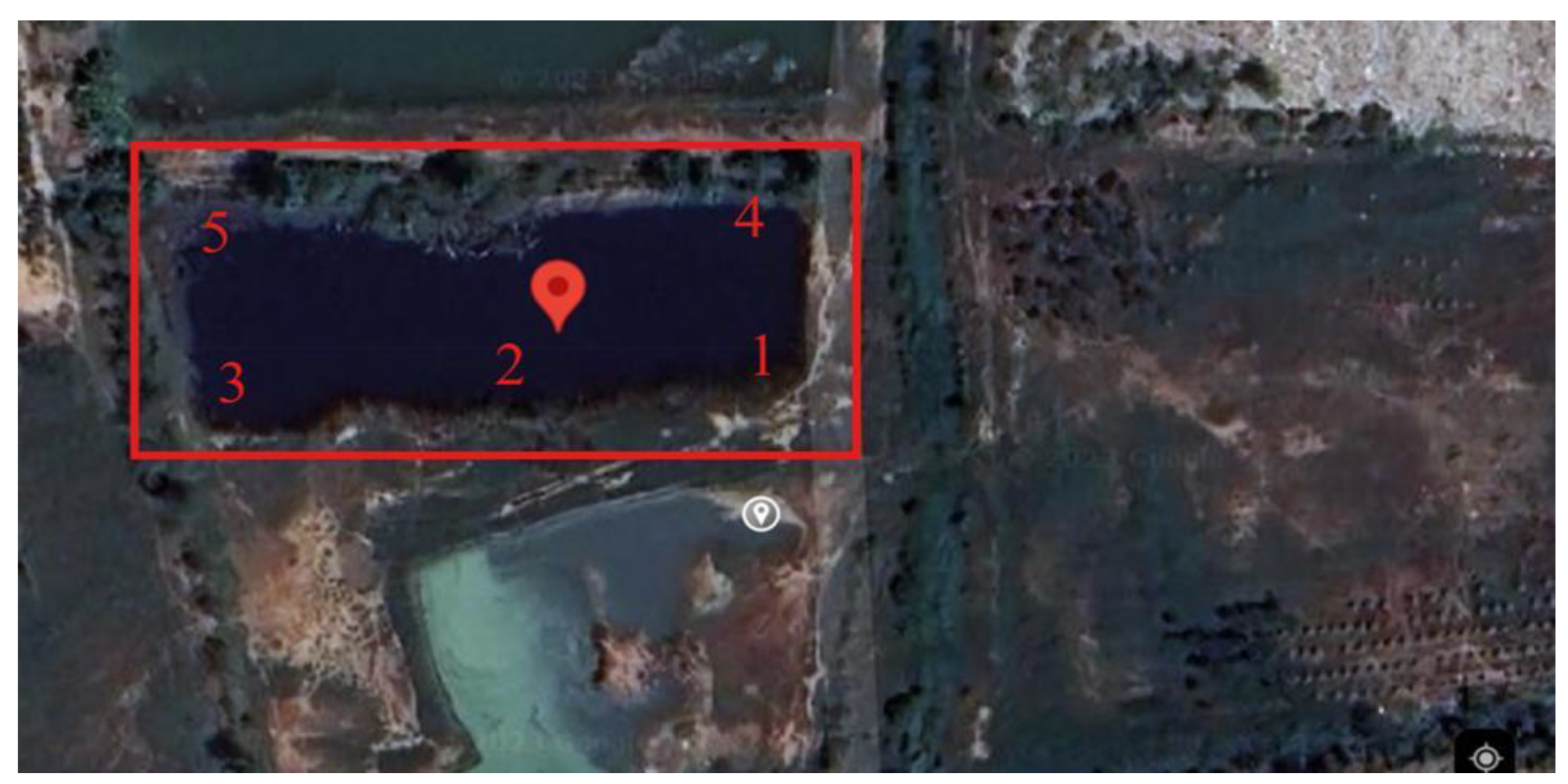
Figure 2.
The following examples of ISSR fingerprints are presented for the reference fish (1.1, 1.2, 1.3, 1.4, and 1.5) and the landfill fish (2.1, 2.2, 2.3, 2.4, and 2.5) from the specific primers CACACACACACAAG (a), AGAGAGAGAGAGAGAAA (b) and AGAGAGAGAGAGAGAAC (c) showing monomorphic bands.
Figure 2.
The following examples of ISSR fingerprints are presented for the reference fish (1.1, 1.2, 1.3, 1.4, and 1.5) and the landfill fish (2.1, 2.2, 2.3, 2.4, and 2.5) from the specific primers CACACACACACAAG (a), AGAGAGAGAGAGAGAAA (b) and AGAGAGAGAGAGAGAAC (c) showing monomorphic bands.
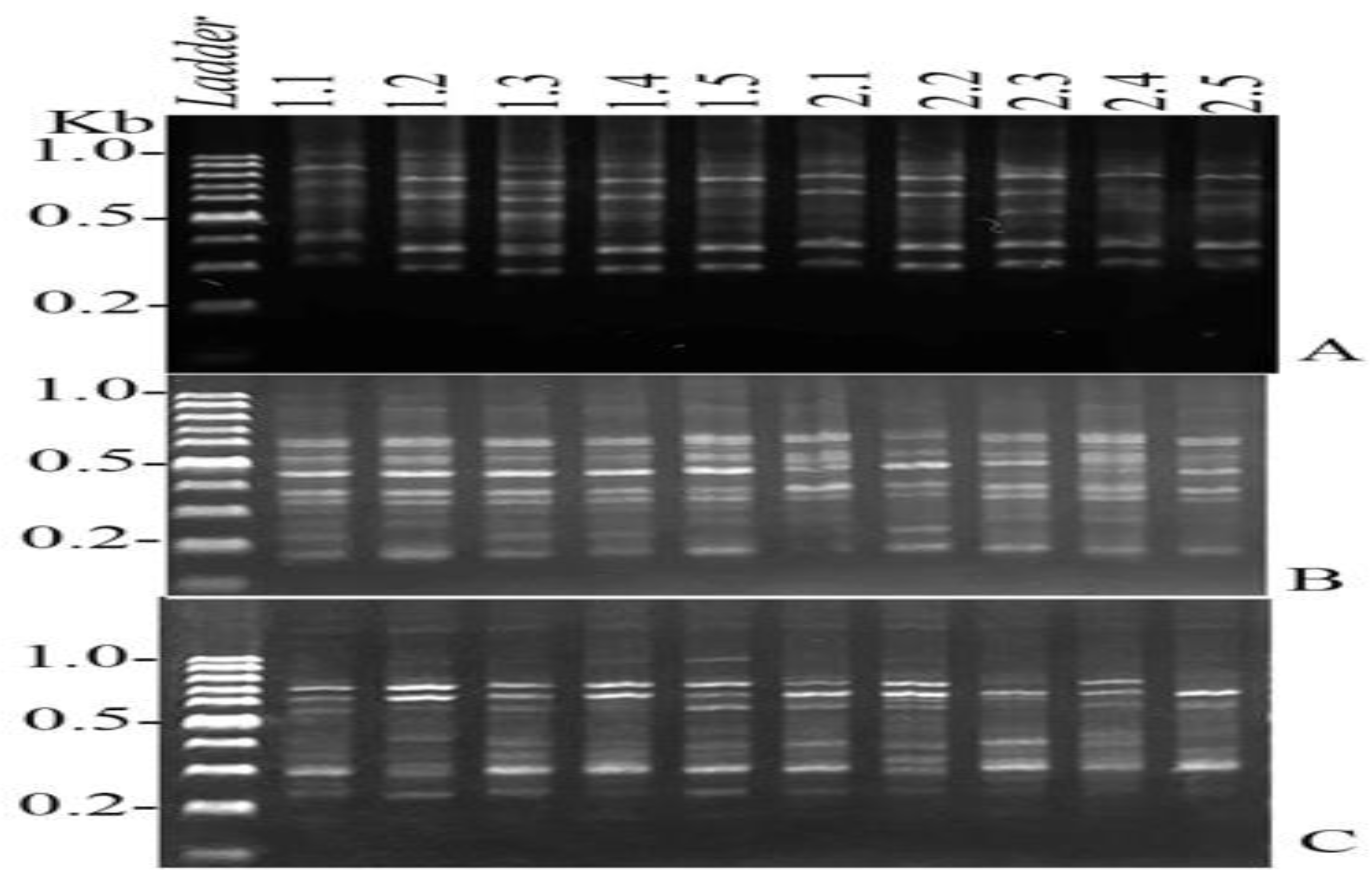
Figure 3.
A dendrogram was generated using 25 primers through the NTSYSpc2.10 software, illustrating the genetic relationships among the tilapia samples, which include both the reference tilapia (1.1-1.5) and the landfill tilapia (2.1-2.5).
Figure 3.
A dendrogram was generated using 25 primers through the NTSYSpc2.10 software, illustrating the genetic relationships among the tilapia samples, which include both the reference tilapia (1.1-1.5) and the landfill tilapia (2.1-2.5).
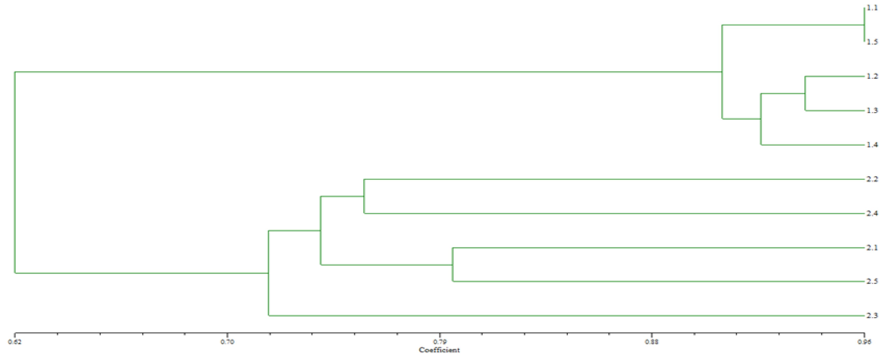
Table 2.
The 13 primer sequences demonstrated successful amplification of ISSRs through polymerase chain reaction (PCR) in this investigation.
Table 2.
The 13 primer sequences demonstrated successful amplification of ISSRs through polymerase chain reaction (PCR) in this investigation.
| No. | Nucleotide Sequences | Total bands | Monomorphic band | Polymorphic band |
|---|---|---|---|---|
| A4 | AGAGAGAGAGAGAGAA | 47 | 2 | 6 |
| A11 | AGAGAGAGAGAGAGAAA | 73 | 1 | 8 |
| A12 | AGAGAGAGAGAGAGAAC | 49 | 2 | 5 |
| A14 | AGAGAGAGAGAGAGAAT | 68 | 3 | 5 |
| P4 | CACACACACACAAC | 32 | 3 | 1 |
| P5 | CACACACACACAGT | 36 | 1 | 5 |
| P6 | CACACACACACAAG | 38 | 2 | 5 |
| P7 | CACACACACACAGG | 18 | 1 | 1 |
| P10 | GAGAGAGAGAGACC | 47 | 4 | 1 |
| P12 | CACCACCACGC | 52 | 2 | 4 |
| P13 | GAGGAGGAGGC | 43 | 1 | 5 |
| P14 | CTCCTCCTCGC | 50 | 2 | 4 |
| P15 | GTGGTGGTGGC | 68 | 3 | 6 |
| Total | 621 | 27 | 56 |
Table 3.
The water quality indicators of the reference and the landfill sites.
| Samples | Indicators | |||||
|---|---|---|---|---|---|---|
| Temperature (°C) | TDS (mg/L) | DO (mg/L) | EC (µs/cm) | pH | ||
| Reference | 31.36±0.72 | 0.47±0.03 | 6.57±0.24 | 317.62±6.08 | 7.65±0.46 | |
| Landfill | 29.06±0.65 | 0.51±0.06 | 4.51±0.38 | 456.72±4.88 | 7.16±0.61 | |
| Standard | N/A | N/A | ≥4.00 | N/A | 5-9 | |
Remarks: TDS = total dissolved solids; DO = dissolved oxygen; EC = electro-conductivity; N/A= not available; (numbers of measured individuals) = 5; Thailand’s wastewater quality standards (Thailand Pollution Control Department 2001) [29].
Table 4.
The concentrations of heavy metals in the water samples from the reference site and landfill site (mean ± standard deviation; n=5).
Table 4.
The concentrations of heavy metals in the water samples from the reference site and landfill site (mean ± standard deviation; n=5).
| Concentration (mg/L) | ||||
|---|---|---|---|---|
| Study Site | As | Cd | Cr | Pb |
| Reference | 0.0028±0.0008 | 0.03±0.006 | 0.015±0.011 | 0.031±0.007 |
| landfill | 0.0848±0.142 | 0.536±0.139 | 1.23±0.445 | 0.73±0.19 |
| P-value | 0.268 | 0.001* | 0.004* | 0.001* |
| Standard | 0.01 | 0.05 | 0.05 | 0.05 |
Remarks: The Ministry of Natural Resources and Environment of Thailand (PCD) established standards for water quality in surface sources in 1994 [30]. * Indicate a statistically significant difference (p<0.05) from the pooled standard error (p-value).
Table 5.
The concentrations of heavy metals in sediment samples from the reference site and the landfill site (mean ± standard deviation, n=5).
Table 5.
The concentrations of heavy metals in sediment samples from the reference site and the landfill site (mean ± standard deviation, n=5).
| Concentration (mg/kg) | ||||
|---|---|---|---|---|
| Study site | As | Cd | Cr | Pb |
| Reference | 0.556±0.236 | 0.0523±0.126 | 12.64±2.35 | 5.32±0.97 |
| landfill | 1.274±0.436 | 1.162±0.428 | 16.68±2.08 | 14.19±3.15 |
| p-value | 0.012* | 0.013* | 0.019* | 0.002* |
| Standard | 3.9 | 1 | 100 | 100 |
Remarks: The standard for soil quality, as established by the Pollution Control Department (PCD) of the Ministry of Natural Resources and Environment of Thailand (2004) [31]. * Indicate a statistically significant difference (p<0.05) from the pooled standard error (p-value).
Table 6.
The concentrations of heavy metals in fish sample from the reference site and the landfill site (mean ± standard deviation; n = 5).
Table 6.
The concentrations of heavy metals in fish sample from the reference site and the landfill site (mean ± standard deviation; n = 5).
| Concentration (mg/kg) | ||||
|---|---|---|---|---|
| Study Site | As | Cd | Cr | Pb |
| Reference | 0.0352±0.0069 | 0.0272±0.0024 | 1.17±0.36 | 0.094±0.08 |
| Landfill | 0.0758±0.0173 | 0.096±0.033 | 1.83±0.09 | 0.69±0.63 |
| p-value | 0.001* | 0.009* | 0.014* | 0.103* |
| Standard | 2 | 0.5 | 2.0 | 0.5 |
Remarks: The quality standards for food, as established by the Ministry of Public Health of Thailand [32]. *Indicate a statistically significant difference (p<0.05) from the pooled standard error (p-value).
Table 7.
HM BAFs in fish samples.
| Heavy metals | O. niloticus | |
|---|---|---|
| BAF values based on water | As | 0.282±1.83 |
| Cd | 0.179±0.112 | |
| Cr | 1.487±0.663 | |
| Pb | 0.945±1.557 |
Table 8.
Fish consumption's effects on heavy metal EDI, HRI, and CR.
| Heavy metals | EDI (μg/kg/day) | HRI | CR |
|---|---|---|---|
| O. niloticus | |||
| As | 0.0016297 | 0.00543 | 0.000001086 |
| Cd | 0.002064 | 0.00206 | - |
| Cr | 0.039345 | 0.013333 | - |
| Pb | 0.014835 | 0.0025 | 0.0011 |
| Unacceptable risk levels | - | >1 [20] | >1×10⁻⁴ [33] |
Table 9.
The genetic similarity indices for the tilapia collected from the reference (1.1-1.5) and the landfill (2.1-2.5) were analyzed using the NTSYSpc2.10 software.
Table 9.
The genetic similarity indices for the tilapia collected from the reference (1.1-1.5) and the landfill (2.1-2.5) were analyzed using the NTSYSpc2.10 software.
| Fish No. | 1.1 | 1.2 | 1.3 | 1.4 | 1.5 | 2.1 | 2.2 | 2.3 | 2.4 | 2.5 |
|---|---|---|---|---|---|---|---|---|---|---|
| 1.1 | 1.0000 | |||||||||
| 1.2 | 0.8765 | 1.0000 | ||||||||
| 1.3 | 0.9103 | 0.9383 | 1.0000 | |||||||
| 1.4 | 0.8734 | 0.9259 | 0.9125 | 1.0000 | ||||||
| 1.5 | 0.9600 | 0.9136 | 0.9487 | 0.8875 | 1.0000 | |||||
| 2.1 | 0.6184 | 0.6173 | 0.6000 | 0.6282 | 0.6364 | 1.0000 | ||||
| 2.2 | 0.4737 | 0.4634 | 0.4810 | 0.4684 | 0.4935 | 0.6250 | 1.0000 | |||
| 2.3 | 0.6316 | 0.6098 | 0.6125 | 0.6203 | 0.6076 | 0.6349 | 0.6140 | 1.0000 | ||
| 2.4 | 0.5946 | 0.5750 | 0.5769 | 0.5844 | 0.5921 | 0.6441 | 0.6226 | 0.6333 | 1.0000 | |
| 2.5 | 0.6757 | 0.6500 | 0.6538 | 0.6410 | 0.6933 | 0.7167 | 0.6140 | 0.6000 | 0.6333 | 1.0000 |
Table 10.
Oxidative stress biomarkers values of tilapia fish from the reference site and the landfill site (mean ± standard deviation, n = 5).
Table 10.
Oxidative stress biomarkers values of tilapia fish from the reference site and the landfill site (mean ± standard deviation, n = 5).
| Parameter | Reference | Landfill | p-Value |
|---|---|---|---|
| MDA (mmol/MDA/g liver) | 31.11±0.75 | 56.8±8.93 | 0.003* |
| H2O2 (mmol/g liver) | 13.54±0.906 | 20.65±3.12 | 0.001* |
| SOD (%) | 33.45±2.95 | 20.41±6.51 | 0.004* |
| CAT (Units/mg protein) | 23.51±2.96 | 14.85±3.48 | 0.003* |
Remark: * A statistically significant difference (p<0.05) is indicated by the pooled standard error (p-value).
Disclaimer/Publisher’s Note: The statements, opinions and data contained in all publications are solely those of the individual author(s) and contributor(s) and not of MDPI and/or the editor(s). MDPI and/or the editor(s) disclaim responsibility for any injury to people or property resulting from any ideas, methods, instructions or products referred to in the content. |
© 2025 by the authors. Licensee MDPI, Basel, Switzerland. This article is an open access article distributed under the terms and conditions of the Creative Commons Attribution (CC BY) license (http://creativecommons.org/licenses/by/4.0/).
Copyright: This open access article is published under a Creative Commons CC BY 4.0 license, which permit the free download, distribution, and reuse, provided that the author and preprint are cited in any reuse.


