
Submitted:
10 December 2025
Posted:
14 December 2025
You are already at the latest version
Abstract
Background: Frozen shoulder (FS) leads to pain, reduced shoulder function, and deficits in postural stability and sensorimotor control during upper-limb weight-bearing tasks. This study investigated how an eight-week Dynamic Neuromuscular Stabilization (DNS) program affected Center of Pressure (COP) control and pain in middle-aged women with FS. Methods: Twenty-two middle-aged women with FS were randomly assigned to a DNS group (DNSG, n = 11) or a control group (CG, n = 11). The DNSG performed DNS exercises twice weekly for eight weeks, while the CG performed dynamic stretching. COP variables (distance, velocity, and root mean square (RMS) in the anterior–posterior (AP) and medial–lateral (ML) directions) were measured using a force platform under affected-side single-hand support with visual input and bilateral hand support with and without visual input. Pain was assessed using the Visual Analog Scale (VAS). All variables were analyzed using a two-way mixed ANOVA. Results: Under the affected-side single-hand support condition, significant group-by-time interaction effects were found in AP distance, AP velocity, ML velocity, and ML RMS (p < 0.05). In the bilateral hand-support conditions with and without visual input, additional COP variables also showed significant interactions (p < 0.05), and VAS-measured pain decreased over time in both groups. Conclusions: The DNS intervention demonstrated positive changes in postural stability and pain during upper-limb weight-bearing tasks in middle-aged women with FS. These findings suggest that DNS has potential as an intervention strategy to support functional recovery and enhance sensorimotor control in individuals with FS.
Keywords:
dynamic neuromuscular stabilization
; frozen shoulder
; shoulder rehabilitation
; center of pressure
; postural stability
; middle-age
1. Introduction
FS is a disorder characterized by shoulder joint pain and limitation of range of motion [1], in which progressive restriction of movement occurs due to inflammation and fibrosis of the joint capsule [2,3]. This condition most commonly occurs after the age of 40, particularly between the ages of 50 and 60, and has been reported to show a higher prevalence in women [4]. As fibrosis and contracture of the shoulder capsule progress, pain is accompanied by limited active and passive movement of the shoulder, leading to increased stiffness [3,5]. Such painful stiffness of the shoulder causes discomfort in daily life and decreases quality of life [3,6], continuously emphasizing the need for evidence-based exercise interventions aimed at pain reduction and restoration of shoulder function and stability [7].
Particularly in middle-aged women, it is known that a decrease in estrogen after menopause leads to a reduction in muscle mass and deterioration in muscle fiber quality [8]. When lifestyle factors such as housework or repetitive use of the upper limbs are added, continuous mechanical loading is applied to the shoulder, exacerbating pain [9,10,11]. These mechanical factors reduce joint position sense, causing an imbalance in the normal muscle coordination that maintains joint stability [12], consequently leading to decreased postural stability of the shoulder and increased pain [13]. Therefore, middle-aged women can be considered a vulnerable group in which physiological and environmental factors act in combination in the development of frozen shoulder.
In general, exercise interventions such as stretching are utilized in the treatment of FS [6]. These treatments are effective for pain reduction and temporary improvement of range of motion; however, reports related to the recovery of proximal–distal coordination and enhancement of neuromuscular control ability remain limited [14]. That is, a purely passive approach alone is insufficient to fully restore the intermuscular coordination and postural control functions required to maintain shoulder stability. Therefore, to fundamentally improve shoulder stability and postural control ability, an active rehabilitation approach that strengthens neuromuscular control is necessary.
Pain is an unpleasant sensory and emotional experience associated with actual or potential tissue damage, and it acts as a major cause of musculoskeletal dysfunction [15,16]. Shoulder pain is known to increase during joint movement and tends to show heightened pain sensitivity at night [17,18]. Such pain is not merely a sensory stimulus but directly affects neuromuscular control [19]. The pain in FS occurs due to inflammation and fibrosis of the joint capsule [20], and when it becomes chronic, it has been reported to cause tension in the surrounding muscles and avoidance of movement [21,22]. As a result, joint stiffness develops, and imbalances in muscle activation and abnormal movement patterns are gradually reinforced [23]. Prolonged exposure to pain decreases the accuracy of shoulder proprioception, destabilizing intermuscular coordination and postural control, which in turn may lead to weakened dynamic stability and increased postural sway, ultimately reducing the efficiency of shoulder function [13,24]. Recently, DNS training has gained attention as an exercise method to restore impaired neuromuscular control [25,26].
DNS is an active rehabilitation approach that reorganizes intermuscular coordination and reactivates the movement control ability of the central nervous system through the integrated regulation of breathing, posture, and movement [27,28,29]. This neurological mechanism enhances the coordination of muscle activation between the deep stabilizing muscles and the upper-limb muscles, helping to maintain dynamic stability of the shoulder joint during upper-limb movements [28,30]. Furthermore, DNS training has been reported to be effective in improving mobility, reducing pain, and re-educating functional movement patterns in patients with joint pain [26,29]. Ultimately, by strengthening the central nervous system–based motor control ability, DNS can restore proximal–distal coordination and enable stable postural control during movement [31,32]. These characteristics complement the limitations of conventional passive therapeutic approaches and provide evidence-based intervention benefits for active neuromuscular recovery and improvement of shoulder stability.
Postural stability is considered to result from neuromuscular control and sensory integration [33,34]. COP analysis serves as an indicator that quantitatively evaluates this postural control ability [35]. Variables such as COP velocity, distance, and RMS are known to reflect subtle body sway and stabilization ability [35,36]. Edouard et al. (2012) proposed a procedure for measuring COP in an upper-limb support position as a method to assess shoulder postural control and sensorimotor control, and verified its reliability in healthy adults [37]. In particular, they reported high reproducibility of COP distance and velocity variables when the lower limbs were supported up to the anterior superior iliac spine (ASIS). This suggests that these parameters can reflect changes in shoulder stability and neuromuscular control ability. Although that study demonstrated the validity and reliability of the measurement method, it has not been applied in pathological shoulder conditions or clinical contexts such as rehabilitation interventions. The present study aimed to address these limitations by applying the measurement procedure proposed by Edouard et al. to patients with frozen shoulder and analyzing COP changes before and after DNS intervention, thereby expanding the clinical applicability of upper-limb support–based COP assessment. This approach holds academic significance in that it deepens the clinical application scope of COP as an objective indicator for quantitatively evaluating sensorimotor control of the shoulder.
In addition, previous studies have reported substantial evidence that DNS is effective in improving trunk stability [27,29]. However, no study has quantitatively verified the effects of DNS intervention on neuromuscular control and postural stability in middle-aged women with FS through COP analysis in a shoulder-supported posture. Therefore, the purpose of this study was to investigate the effects of an 8-week DNS training program on postural stability (COP) and pain (VAS) in middle-aged women with FS, and to provide clinical evidence for shoulder function recovery in this population.
2. Materials and Methods
2.1. Participants
Participants were recruited from a community welfare center and Busan University of Foreign Studies located in B metropolitan city. Before enrollment, all individuals were provided with a detailed explanation of the study objectives and procedures, and written informed consent was obtained from those who agreed to participate voluntarily. The study protocol was reviewed and approved by the Institutional Review Board (IRB approval number: P01-202509-01-052), and the trial was registered in the Clinical Research Information Service (CRIS) on November 20, 2025 (registration number: KCT0011186).
The target sample size was determined using the G*Power 3.1 program (Kiel University, Germany), referencing prior research with a comparable design. Parameters were set at an effect size of f = 0.25, an alpha level of 0.05, and a statistical power of 0.60 [38]. The calculation indicated that at least 20 participants were required, and to account for an anticipated dropout rate of approximately 20%, 24 participants were recruited.
Group allocation was performed using a random draw method, in which participants selected numbered balls from sealed containers. According to the assigned numbers, participants were placed into either the DNS group or the dynamic stretching group. During the intervention period, two participants discontinued participation due to health-related reasons, resulting in a final sample of 22 participants (11 per group) included in the analysis. All assessments were carried out by the same trained investigator following standardized measurement protocols, and all testing conditions were maintained consistently throughout the study to minimize procedural bias. Data analysis was conducted only after the intervention and post-assessments had been completed. To reduce the risk of bias, participants were blinded to their group allocation and were not informed about the detailed study hypotheses.
Inclusion Criteria
Participants were included in the study if they met all of the following criteria:
(1) Women aged between 40 and 64 years.
(2) Diagnosed with FS by an orthopedic or rehabilitation medicine specialist.
(3) In the chronic phase, with symptoms persisting for more than three months.
(4) Able to understand the study procedures and provided written informed consent.
(5) Able to participate in the entire intervention program consisting of two sessions per week for eight weeks (a total of 16 sessions), as well as all pre-, mid-, and post- intervention assessments.
Exclusion Criteria
Participants were excluded from the study if any of the following conditions applied:
(1) History of surgery involving the upper extremity or trunk within the past year.
(2) Presence of acute trauma, inflammation, or other medical conditions affecting the shoulder or upper limb.
(3) Inability to perform exercise due to severe pain or acute inflammatory symptoms.
In addition, to verify the baseline homogeneity between the experimental and control groups, independent t-tests were conducted for age, height, weight and Body Mass Index (BMI). The results showed no statistically significant differences between the groups in age (t = −3.712), height (t = 2.523), weight (t = 0.325), BMI (t = −1.141). The general characteristics of the participants are summarized in Table 1, and the recruitment and group allocation procedures are illustrated in Figure 1 as a flowchart.
2.2. Assessment of COP and Data Acquisition
In this study, the primary and secondary outcomes were evaluated separately. The primary outcome was the COP variable measured during eyes-open affected-side single-hand support. The secondary outcomes included COP variables measured under eyes-open bilateral support and eyes-closed double-hand support conditions, as well as the VAS score for pain intensity.
In this study, the COP was analyzed to quantitatively evaluate changes in postural stability following the DNS intervention. COP measurements were obtained using a force plate (AMTI OR6, Watertown, MA, USA) with a sampling frequency of 100 Hz. The assessment was conducted in the prone position, and to ensure consistent trunk alignment, both ASIS were positioned at the anterior edge of the treatment table. The upper limbs were extended forward to support body weight, and both palms were placed securely on the force plate.
The measurement procedure was adapted from the protocol of Edouard et al. [37], which evaluated COP changes during upper-limb weight-bearing tasks. The postural setup followed the criteria below: (1) the alignment from the shoulder joint to the wrist was adjusted to approximately 90°, (2) the wrists were maintained at approximately 15° of flexion, (3) the distance between both wrists was standardized to 4 cm, and (4) the examiner continuously monitored the participant’s posture throughout each trial to prevent compensatory trunk or scapular movements Figure 2.
The assessment was performed under three conditions: (1) eyes open with unilateral (affected-side) hand support. (2) eyes open with bilateral hand support and (3) eyes closed with bilateral hand support. Each condition was maintained for 30 seconds, with 30 seconds of rest between conditions and a 1-minute rest between sets.
The raw COP signals were extracted using AMTI NetForce software and smoothed using a fourth-order Butterworth low-pass filter with a cutoff frequency of 6 Hz. The displacement, velocity and RMS values were calculated for both the AP and ML directions. Each condition was measured five times, and the mean value of the middle three trials was used for analysis. All evaluations and data processing were performed by the same investigator to ensure measurement reliability.
2.3. Evaluation of Pain Intensity (VAS)
The VAS consisted of a 10-cm horizontal line, with the left end (0 point) indicating “no pain” and the right end (10 point) indicating “extreme pain.” The assessment was conducted immediately before the COP measurement at each time point (0, 4, and 8 weeks). Participants were instructed to mark the point on the line that best represented the intensity of pain they were experiencing at that moment. All evaluations were performed by the same examiner under identical environmental conditions and consistent procedures to minimize external factors and ensure the reliability and objectivity of the measurements.
2.4. Exercise Intervention
The experimental group participated in an exercise program based on the developmental kinesiological principles of DNS. The participants performed the program twice per week for eight weeks, completing a total of 16 sessions. Each session lasted approximately 50 minutes and consisted of a 10-minute warm-up, a 35-minute main exercise phase, and a 5-minute cool-down. Exercise intensity was progressively adjusted based on the number of repetitions, with each movement performed for 6 repetitions during Weeks 0–4 and 8 repetitions during Weeks 5–8. The mean adherence rate was 96%, corresponding to an average attendance of 15.5 out of 16 sessions.
Each session consisted of three phases: warm-up, main exercise, and cool-down. In the warm-up phase, participants were re-educated to form intra-abdominal pressure (IAP) and to reestablish basic trunk stabilization strategies based on the breathing–posture synergy. In the main exercise phase, closed-chain neuromuscular control training was performed using developmental positions such as supine, side-lying, quadruped, and modified prone. These exercises were designed to promote posterior–inferior stabilization of the scapula, regulate trunk stiffness, and enhance integrated strength and coordination during upper-limb support tasks [39]. The cool-down phase involved low-intensity postural readjustment and breathing recovery exercises aimed at stabilizing overall body tension.
The control group performed a dynamic stretching program with the same duration and frequency as the experimental group. Dynamic stretching is an active stretching technique that elongates the target muscle by voluntarily contracting its antagonist and repeatedly moving the associated joints [40]. This program focused on improving the mobility of the soft tissues surrounding the shoulder joint and restoring functional range of motion. Sequential stretching patterns incorporating active movements were applied to provide a control condition that ensured general joint mobility without specific neuromuscular stabilization training. The structure and detailed components of the training program are presented in Table 2 and Figure 3.
2.5. Statistical Analysis
All data collected in this study were analyzed using IBM SPSS Statistics for Windows, version 25.0 (IBM Corp., Armonk, NY, USA). Each variable was presented as mean ± standard deviation (SD). To examine baseline demographic characteristics between the two groups before the intervention, an independent t-test was conducted, and data normality was verified using the Shapiro–Wilk test.
A mixed-design two-way repeated-measures analysis of variance (ANOVA) was performed to evaluate the effects of the intervention. The group (experimental, control) was set as the between-subject factor, and time (pre-, mid-, and post-intervention) was set as the within-subject factor. When significant group × time interactions or main effects were observed, post-hoc analyses with Bonferroni correction were applied to identify differences between time points.
The effect size was calculated using partial eta squared (ηp²), interpreted according to Cohen’s criteria: ηp² = 0.01 (small), 0.06 (medium), and 0.14 (large). The level of statistical significance was set at p < 0.05 for all analyses.
3. Results
The analysis results of the COP variables and pain scores measured under each condition are presented in Table 3, Table 4, Table 5 and Table 6.
3.1. Changes in COP Under the Affected-Side Single-Hnad Support Condition
For the AP-direction COP variables, the assumption of sphericity was violated for all variables; therefore, the Greenhouse–Geisser correction was applied. The analysis revealed significant group × time interaction effects for AP-distance (p = 0.048, ηp2 = 0.160) and AP-velocity (p = 0.041, ηp2 = 0.168), whereas AP-RMS (p = 0.233, ηp2 = 0.071) was not significant. The main effect of time was significant for all variables [AP-distance (p < 0.001, ηp2 = 0.799), AP-velocity (p < 0.001, ηp2 = 0.770), and AP-RMS (p < 0.001, ηp2 = 0.742)]. For the AP-direction COP variables, both interaction effects showed large effect sizes and sufficient statistical power. AP-distance (ηp2 = 0.160, f = 0.44) and AP-velocity (ηp2 = 0.180, f = 0.47) demonstrated post hoc powers (1–β) of 0.97 and 0.98, respectively, exceeding the predetermined power of 0.60.
For the ML-direction COP variables, the assumption of sphericity was met; thus, the original degrees of freedom were used for the analysis. Significant group × time interaction effects were observed for ML-velocity (p = 0.030, ηp2 = 0.161) and ML-RMS (p = 0.009, ηp2 = 0.208), whereas ML-distance (p = 0.238, ηp2 = 0.069) was not significant. The main effect of time was significant for all variables [ML-distance (p < 0.001, ηp2 = 0.688), ML-velocity (p < 0.001, ηp2 = 0.706), and ML-RMS (p < 0.001, ηp2 = 0.840)]. For the ML-direction COP variables, both interaction effects showed large effect sizes and sufficient statistical power. ML-velocity (ηp2 = 0.161, f = 0.44) and ML-RMS (ηp2 = 0.208, f = 0.51) demonstrated post hoc powers (1–β) of 0.99 and 1.00, respectively, exceeding the predetermined power of 0.60.
Post-hoc analysis revealed that in the DNS group, all AP-direction COP variables (AP-distance, AP-velocity, and AP-RMS) demonstrated significant differences across time points (p < 0.05). Furthermore, all ML-related variables (ML-distance, ML-velocity, and ML-RMS) also showed significant time-dependent differences (p < 0.05).
In contrast, in the control group, AP-distance, AP-velocity, and AP-RMS showed significant changes only between 0 and 4 weeks (p < 0.05), with no additional changes observed between 4 and 8 weeks. Similarly, ML-distance, ML-velocity, and ML-RMS showed significant differences between 0 and 4 weeks but no significant changes from 4 to 8 weeks.
3.2. Changes in COP Under the Eyes-Open Bilateral Hand Support Condition
For the AP-direction COP variables, the assumption of sphericity was violated for AP-distance and AP-velocity; therefore, the Greenhouse–Geisser correction was applied, whereas AP-RMS met the assumption and was analyzed using the original degrees of freedom. The Time × Group interaction effects were observed for AP-distance (ηp2 = 0.016), AP-velocity (ηp2 = 0.189), and AP-RMS (ηp2 = 0.026). The main effect of Time showed effect sizes of AP-distance (ηp2 = 0.688), AP-velocity (ηp2 = 0.601), and AP-RMS (ηp2 = 0.697).
For the ML-direction COP variables, the assumption of sphericity was violated for ML-distance; therefore, the Greenhouse–Geisser correction was applied, whereas ML-velocity and ML-RMS met the assumption and were analyzed using the original degrees of freedom. The Time × Group interaction effects were found for ML-distance (ηp2 = 0.010), ML-velocity (ηp2 = 0.029), and ML-RMS (ηp2 = 0.326). The main effect of Time showed effect sizes of ML-distance (ηp2 = 0.782), ML-velocity (ηp2 = 0.719), and ML-RMS (ηp2 = 0.737).
3.3. Changes in COP Under the Eyes-Closed Bilateral Hand Support,Condition
For the AP-direction COP variables (AP-distance, AP-velocity, AP-RMS), the assumption of sphericity was met for AP-distance and AP-RMS, whereas AP-velocity violated the assumption; thus, the Greenhouse–Geisser correction was applied. The Time × Group interaction effects were observed for AP-distance (ηp2 = 0.028), AP-velocity (ηp2 = 0.166), and AP-RMS (ηp2 = 0.047). The main effect of Time showed effect sizes of AP-distance (ηp2 = 0.636), AP-velocity (ηp2 = 0.736), and AP-RMS (ηp2 = 0.688).
For the ML-direction COP variables (ML-distance, ML-velocity, ML-RMS) the assumption of sphericity was met for ML-distance and ML-RMS, whereas ML-velocity violated the assumption; thus, the Greenhouse–Geisser correction was applied. The Time × Group interaction effects were found for ML-distance (ηp2 = 0.007), ML-velocity (ηp2 = 0.164), and ML-RMS (ηp2 = 0.273). The main effect of Time showed effect sizes of ML-distance (ηp2 = 0.832), ML-velocity (ηp2 = 0.829), and ML-RMS (ηp2 = 0.584).
3.4. Changes in Pain Indicator (VAS)
The VAS scores satisfied the assumption of sphericity and were analyzed using the original degrees of freedom.
The Time × Group interaction showed an effect size of ηp2 = 0.081, while the main effect of Time demonstrated an effect size of ηp² = 0.756.
4. Discussion
This study aimed to analyze changes in the COP during upper-limb weight-bearing postures in middle-aged women with frozen shoulder and to determine the effects of a DNS intervention on shoulder stability and pain (VAS). The primary outcome of this study was the COP measured during the affected-side single-hand weight-bearing posture under eyes-open conditions, and the secondary outcomes were the COP measured during bilateral hand support under eyes-open and eyes-closed conditions, as well as pain (VAS).
The analysis revealed significant improvements in COP during the affected-side single-hand support posture, which was the primary outcome. These changes can be interpreted as indicating that the DNS intervention enhanced the load-control capacity and postural control efficiency of the shoulder complex, thereby enabling more stable maintenance and regulation of the body’s center during weight-bearing tasks. In other words, DNS suggests a positive effect on restoring shoulder stability and improving functional supportability in middle-aged women with frozen shoulder.
The COP results during the affected-side single-hand support posture in this study showed significant differences between groups. DNS is known to optimize the alignment of the scapula–upper limb–trunk in a closed-chain environment and to enhance proximal stability, thereby reducing unnecessary fluctuations of the body’s center during load transfer. These mechanisms are reflected in the significant reductions in COP variables observed in the affected-side single-hand support posture following the DNS intervention in this study. In a closed-chain condition, where the wrist and elbow are fixed, the coordination between the glenohumeral and scapulothoracic joints plays a key role in postural control [37]. DNS is interpreted to act by reorganizing such joint coordination and increasing coactivation of the muscles surrounding the shoulder, thereby enhancing the stability of the shoulder complex during weight-bearing tasks. In addition, the weight-bearing load generated during upper-limb support is transmitted through an integrated kinetic chain that extends to the trunk and lower limbs, and DNS activates this sequential movement, allowing the load to be redistributed throughout the body rather than concentrated on specific joints [41,42]. During this process, sensory feedback generated from each segment facilitates sensory–motor integration and helps the body reposition the COP within a more stable and efficient range in response to changes in ground reaction forces (GRF) [43,44,45].
Previous studies have reported that closed-chain exercises enhance dynamic stabilization by maintaining high joint compressive forces and joint congruency while strongly stimulating proprioceptors [46]. The upper-limb support environment provided by the DNS intervention in this study can likewise be interpreted as reinforcing such proprioceptive stimulation, thereby creating a sensory feedback–based environment in which the body must finely regulate load direction and balance. In this self–body-weight loading environment, muscles do not merely generate force but exhibit neuromuscular adaptations that actively control the direction of loading through the postural control system and demonstrate consistent regulatory patterns [47]. This process directly contributes to the reduction of small-amplitude COP fluctuations, and the decreased COP observed during affected-side support after the DNS intervention can be understood not as a simple change in local muscle strength but as the result of a more efficient reorganization of the mechanical interaction among GRF, center of mass (CoM), and COP in a self–body-weight loading environment. Therefore, the reduced variability of COP following the DNS intervention reflects the reorganization of the neuromuscular control system responsible for regulating load direction and the improvement of postural stability under weight-bearing conditions. This can be interpreted as a key mechanism by which DNS contributes to the restoration of dynamic stability of the shoulder complex in middle-aged women with frozen shoulder.
In the secondary outcome of this study, COP during eyes-open bilateral hand support showed between-group differences only in AP-velocity and ML-RMS, which demonstrates a somewhat different pattern from the changes observed under affected-side support or eyes-closed bilateral support conditions. The affected-side support or eyes-closed conditions involve either a narrower base of support or limited visual input, resulting in relatively higher postural control demands on the shoulder–scapula–trunk complex [30,48,49], and thus the effects of the DNS intervention tend to appear more clearly. In contrast, the eyes-open bilateral support task provides a wider base of support and visual feedback, making it a comparatively stable task [50], and therefore subtle neuromuscular control changes induced by DNS may be less likely to immediately manifest in COP variables. Particularly, individuals with shoulder pain tend to utilize automated compensatory strategies—such as contralateral weight shifting in stable conditions, increased visual dependence, and fixation strategies through bilateral support—which may act to reduce sway. As a result, these strategies may have limited the extent to which the effects of the DNS intervention were fully reflected in the COP outcomes.
In the secondary outcome of this study, COP during eyes-closed bilateral hand support showed changes that suggested between-group differences in AP velocity, ML velocity, and RMS. When visual cues are removed, the available information for detecting postural sway becomes limited, leading the body to naturally increase its reliance on proprioceptive input from muscles and joints to maintain balance [29]. Because DNS intervention is characterized by reorganizing coordination and synchronization of muscle activation [29], it is plausible that proprioception-based postural control strategies required for upper-limb support operated more efficiently during this sensory shift. In upper-limb support tasks, even slight changes in the base of support can easily compromise the stability of the upper-limb joints, and it is known that proprioceptive dependence becomes further reinforced to compensate for this instability [51]. Proprioceptive input assists in detecting subtle shifts in the body’s center and in modulating muscle activation accordingly [52], and this process may have contributed to regulating COP displacement within a certain range. As proprioception functions more effectively, the demand for corrective movements decreases, leading to a tendency for COP velocity to become more attenuated. Therefore, the COP changes observed in this study can be interpreted as partially reflecting the improved efficiency of sensory-based postural control following the DNS intervention.
The secondary outcome of pain (VAS) in this study did not show differences between groups, and both groups exhibited a decreasing trend. This indicates that changes in pain may have been influenced not by the specific characteristics of the intervention but by nonspecific factors such as the passage of time or increased general movement, which cannot be ruled out. Considering these pathological characteristics, it is possible that both DNS and stretching contributed to pain reduction through shared mechanisms, including the restoration of tissue flexibility, enhanced periarticular circulation, and reduced movement avoidance. DNS may help reduce unnecessary muscle tension and compressive loading during support tasks by reorganizing the coordination of breathing, posture, and muscle activation [29,32], whereas stretching has been reported to decrease pain sensitivity by increasing joint range of motion and elongating soft tissues [53,54]. Therefore, the reduction in pain observed in this study may have been influenced not by specific effects unique to each intervention method, but rather by the shared movement stimuli and mechanical or physiological changes in the tissues provided by both interventions.
The limitations of this study are as follows:
1. The intervention period was limited to eight weeks, which may not have been sufficient to evaluate the long-term neuromuscular adaptations or sustained pain-reduction effects that may result from DNS training.
2. This study was conducted with a relatively small sample size, which may have limited the statistical power for certain outcomes; therefore, caution is needed when generalizing the findings.
3. Pain assessment relied solely on the subjective VAS scale, and the evaluation of neuromuscular function was focused on COP-based analysis. The absence of objective physiological indicators such as EMG or muscle fatigue measures presents a limitation in fully elucidating the mechanisms underlying DNS.
4. The study population was restricted to middle-aged women, and given potential physiological differences by sex, the applicability of the findings to men may be limited.
The reduction in COP variability under the affected-side single-hand support condition, the primary outcome of this study, demonstrates that the DNS intervention distinctly improved neuromuscular control and dynamic stability of the shoulder complex during weight-bearing tasks. Partial changes associated with the DNS intervention were also observed in the bilateral hand support tasks included as secondary outcomes. In particular, under the eyes-closed bilateral support condition—where reliance on proprioception increases due to the absence of visual cues—it is likely that the DNS-induced changes in muscle activation coordination were partially reflected in this condition. Meanwhile, the reduction in pain may be interpreted as a general physiological and mechanical effect resulting from increased movement in both the DNS and stretching groups. Future studies should analyze EMG activity of the shoulder and trunk muscles during upper-limb support tasks in conjunction with COP variables to more clearly elucidate how the DNS intervention contributes to the regulation of dynamic stability of the shoulder complex.
5. Conclusions
This study confirmed that an eight-week DNS intervention may produce certain positive effects on postural stability and pain changes during upper-limb support postures in middle-aged women with frozen shoulder. In particular, reductions in several COP variables were observed under the affected-side single-hand support condition, suggesting that DNS may have improved neuromuscular control among the trunk, scapula, and upper limb, thereby contributing to load transmission and postural regulation. In contrast, changes in the control group were relatively limited, indicating that the DNS intervention may have acted more effectively in specific tasks. These findings support the potential applicability of DNS as a useful intervention strategy for assisting pain management and functional recovery of the shoulder in patients with frozen shoulder.
Author Contributions
Conceptualization, C.K.L., I.B.P., and H.Ji.K; Methodology, I.B.P., and H.Ji.K; Software, H.Ji.K; Validation, C.K.L., I.B.P., H.Ju.K, and H.Ji.K; Formal analysis, H.Ji.K; Investigation, H.Ji.K; Data curation, H.Ji.K; Writing—original draft preparation, H.Ji.K; Writing—review and editing, C.K.L., I.B.P., H.Ju.K, and H.Ji.K; Visualization, H.Ji.K; Supervision, C.K.L. and I.B.P. All authors have read and agreed to the published version of the manuscript. .
Funding
This work was supported by the research grant of Busan University of Foreign Studies in 2025.
Institutional Review Board Statement
The study was conducted in accordance with the Declaration of Helsinki, and approved by the Institutional Review Board of the Public Institutional Review Board (IRB approval number: P01-202509-01-052, approval date: 16 September 2025).
Informed Consent Statement
Informed consent was obtained from all subjects involved in the study.
Data Availability Statement
The data used in this study are available upon reasonable request and will be deposited in a public repository upon publication.
Acknowledgments
This study was conducted under the supervision of Chae-Kwan Lee during the master’s program at Busan University of Foreign Studies. I extend my sincere gratitude to Lee for his continuous professional guidance and encouragement throughout all stages of the research, from planning to completion. I would also like to thank Hyunju Kim for her assistance with data collection and experimental equipment. In addition, I express my deep appreciation to Il-Bong Park for sharing his academic insight and expertise during the research process. Finally, I am grateful to Busan University of Foreign Studies for providing administrative support throughout the study.
Conflicts of Interest
The authors declare no conflicts of interest.
References
- Date, A.; Rahman, L. Frozen shoulder: Overview of clinical presentation and review of the current evidence base for management strategies. Future Sci. OA 2020, 6, FSO647. [Google Scholar] [CrossRef]
- Bunker, T.D. Frozen shoulder: Unravelling the enigma. Ann. R. Coll. Surg. Engl. 1997, 79, 210–213. [Google Scholar]
- Le, H.V.; Lee, S.J.; Nazarian, A.; Rodriguez, E.K. Adhesive capsulitis of the shoulder: Review of pathophysiology and current clinical treatments. Shoulder & Elbow 2017, 9, 75–84. [Google Scholar] [CrossRef]
- DePalma, A.F. Loss of scapulohumeral motion (frozen shoulder). Clin. Orthop. Relat. Res. 2008, 466, 552–560. [Google Scholar] [CrossRef] [PubMed]
- Neviaser, A.S.; Hannafin, J.A. Adhesive capsulitis: A review of current treatment. Am. J. Sports Med. 2010, 38, 2346–2356. [Google Scholar] [CrossRef]
- Choi, J.-M.; Cho, E.-Y.; Lee, B.-H. Effects of dynamic stretching combined with manual therapy on pain, ROM, function, and quality of life of adhesive capsulitis. Healthcare 2024, 12, 45. [Google Scholar] [CrossRef] [PubMed]
- Gulwani, A.H. A Study to Find Out the Effect of Scapular Stabilization Exercises on Shoulder ROM and Functional Outcome in Diabetic Patients with Stage 2 Adhesive Capsulitis of the Shoulder Joint—An Interventional Study. Int. J. Sci. Healthc. Res. 2020, 5, 320–333. [Google Scholar]
- Collins, B.C.; Laakkonen, E.K.; Lowe, D.A. Aging of the musculoskeletal system: How the loss of estrogen impacts muscle strength. Bone 2019, 123, 137–144. [Google Scholar] [CrossRef]
- Lu, J.; Chen, Y.; Lv, Y. The effect of housework, psychosocial stress and residential environment on musculoskeletal disorders for Chinese women. SSM Popul. Health 2023, 23, 101545. [Google Scholar] [CrossRef] [PubMed]
- Leclerc, A.; Chastang, J.-F.; Niedhammer, I.; Landre, M.-F.; Roquelaure, Y. Incidence of shoulder pain in repetitive work. Occup. Environ. Med. 2004, 61, 39–44. [Google Scholar]
- Pehkonen, I.; Miranda, H.; Haukka, E.; Luukkonen, R.; Takala, E.-P.; Ketola, R.; Leino-Arjas, P.; Riihimäki, H.; Viikari-Juntura, E. Prospective study on shoulder symptoms among kitchen workers in relation to self-perceived and observed work load. Occup. Environ. Med. 2009, 66, 416–423. [Google Scholar] [CrossRef] [PubMed]
- Carpenter, J.E.; Blasier, R.B.; Pellizzon, G.G. The effects of muscle fatigue on shoulder joint position sense. Am. J. Sports Med. 1998, 26, 262–265. [Google Scholar] [CrossRef] [PubMed]
- Alfaya, F.F.; Reddy, R.S.; Alkhamis, B.A.; Kandakurti, P.K.; Mukherjee, D. Shoulder proprioception and its correlation with pain intensity and functional disability in individuals with subacromial impingement syndrome—A cross-sectional study. Diagnostics 2023, 13, 2099. [Google Scholar] [CrossRef] [PubMed]
- Balci, N.C.; Yuruk, Z.O.; Zeybek, A.; Gulsen, M.; Tekindal, M.A. Acute effect of scapular proprioceptive neuromuscular facilitation (PNF) techniques and classic exercises in adhesive capsulitis: A randomized controlled trial. J. Phys. Ther. Sci. 2016, 28, 1219–1227. [Google Scholar] [CrossRef]
- International Association for the Study of Pain (IASP). IASP revises its definition of pain for the first time since 1979. Available online: https://www.iasp-pain.org/wp-content/uploads/2022/04/revised-definition-flysheet_R2-1-1-1.pdf (accessed on 16 November 2025).
- Mallick-Searle, T.; Sharma, K.; Toal, P.; Gutman, A. Pain and function in chronic musculoskeletal pain—Treating the whole person. J. Multidiscip. Healthc. 2021, 14, 335–347. [Google Scholar] [CrossRef]
- Longo, U.G.; Facchinetti, G.; Marchetti, A.; Candela, V.; Risi Ambrogioni, L.; Faldetta, A.; De Marinis, M.G.; Denaro, V. Sleep Disturbance and Rotator Cuff Tears: A Systematic Review. Medicina 2019, 55, 453. [Google Scholar] [CrossRef]
- Dupuis, F.; Sole, G.; Wassinger, C.A.; Osborne, H.; Beilmann, M.; Mercier, C.; Campeau-Lecours, A.; Bouyer, L.J.; Roy, J.-S. The Impact of Experimental Pain on Shoulder Movement during an Arm Elevated Reaching Task in a Virtual Reality Environment. Physiol. Rep. 2021, 9, e15025. [Google Scholar] [CrossRef]
- Sterling, M.; Jull, G.; Wright, A. The effect of musculoskeletal pain on motor activity and control. The Journal of Pain 2001, 2(3), 135–145. [Google Scholar] [CrossRef]
- Tamai, K.; Hamada, J.; Nagase, Y.; Morishige, M.; Naito, M.; Asai, H.; Tanaka, S. Frozen shoulder: An overview of pathology and biology with hopes to novel drug therapies. Mod. Rheumatol. 2024, 34, 439–443. [Google Scholar] [CrossRef]
- Vlaeyen, J.W.S. Learning to predict and control harmful events: Chronic pain and conditioning. Pain 2015, 156, S86–S93. [Google Scholar] [CrossRef]
- Falla, D.; Arendt-Nielsen, L.; Farina, D. The pain-induced change in relative activation of upper trapezius muscle regions is independent of the site of noxious stimulation. Clin. Neurophysiol. 2008, 119, 2433–2439. [Google Scholar] [CrossRef]
- McGinnis, K.; Snyder-Mackler, L.; Flowers, P.; Zeni, J. Dynamic joint stiffness and co-contraction in subjects after total knee arthroplasty. Clin. Biomech. 2013, 28, 205–210. [Google Scholar] [CrossRef]
- Ager, A.L.; Borms, D.; Deschepper, L.; Dhooghe, R.; Dijkhuis, J.; Roy, J.-S.; Cools, A. Proprioception: How is it affected by shoulder pain? A systematic review. J. Hand Ther. 2019, xxx, 1–9. [Google Scholar] [CrossRef] [PubMed]
- Hei, P.; Zhang, Z.; Wei, J.; Lan, C.; Wang, X.; Jing, X.; Chen, X.; Wu, Z. The effect of dynamic neuromuscular stabilization technique combined with Kinesio taping on neuromuscular function and pain self-efficacy in individuals with chronic nonspecific low back pain: A randomized trial. Medicine 2025, 104, e41265. [Google Scholar] [CrossRef]
- Sakinepoor, A.; Mazidi, M. Neck stabilization exercise and dynamic neuromuscular stabilization reduce pain intensity, forward head angle and muscle activity of employees with chronic nonspecific neck pain: A retrospective study. J. Exp. Orthop. 2025, 12, e70188. [Google Scholar] [CrossRef]
- Frank, C.; Kobesova, A.; Kolar, P. Dynamic neuromuscular stabilization & sports rehabilitation. Int. J. Sports Phys. Ther. 2013, 8, 62–73. [Google Scholar] [PubMed]
- Kobesova, A.; Kolar, P. Developmental kinesiology: Three levels of motor control in the assessment and treatment of the motor system. J. Bodyw. Mov. Ther. 2014, 18, 496–505. [Google Scholar] [CrossRef] [PubMed]
- Rabieezadeh, A.; Mahdavinejad, R.; Sedehi, M.; Adimi, M. The effects of an 8-week dynamic neuromuscular stabilization exercise on pain, functional disability, and quality of life in individuals with non-specific chronic low back pain: A randomized clinical trial with a two-month follow-up study. BMC Sports Sci. Med. Rehabil. 2024, 16, 161. [Google Scholar] [CrossRef]
- Huang, H.; Xie, H.; Zhang, G.; Xiao, W.; Ge, L.; Chen, S.; Zeng, Y.; Wang, C.; Li, H. Effects of dynamic neuromuscular stabilization training on the core muscle contractility and standing postural control in patients with chronic low back pain: A randomized controlled trial. BMC Musculoskelet. Disord. 2025, 26, 213. [Google Scholar] [CrossRef]
- Karartı, C.; Özsoy, İ.; Özyurt, F.; Basat, H.Ç.; Özsoy, G.; Özüdoğru, A. The effects of dynamic neuromuscular stabilization approach on clinical outcomes in older patients with chronic nonspecific low back pain: A randomized, controlled clinical trial. Somatosens. Mot. Res. 2023. [Google Scholar] [CrossRef]
- Kobesova, A.; Nørgaard, I.; Kolar, P. Dynamic Neuromuscular Stabilization – DNS Neurorehab Chapter. Unpublished educational document, 2014. Available online: https://rehabps.com/DATA/DNS_Neurorehab_Chapter.pdf (accessed on 16 November 2025).
- Teng, Y.-L.; Chen, C.-L.; Lou, S.-Z.; Wang, W.-T.; Wu, J.-Y.; Ma, H.-I.; Chen, V.C.-H. Postural stability of patients with schizophrenia during challenging sensory conditions: Implication of sensory integration for postural control. PLoS ONE 2016, 11, e0158219. [Google Scholar] [CrossRef]
- Munoz-Martel, V.; Santuz, A.; Ekizos, A.; Arampatzis, A. Neuromuscular organisation and robustness of postural control in the presence of perturbations. Sci. Rep. 2019, 9, 10591. [Google Scholar] [CrossRef] [PubMed]
- Quijoux, F.; Nicolaï, A.; Chairi, I.; Bargiotas, I.; Ricard, D.; Yelnik, A.; Oudre, L.; Bertin-Hugault, F.; Vidal, P.-P.; Vayatis, N.; Buffat, S.; Audiffren, J. A review of center of pressure (COP) variables to quantify standing balance in elderly people: Algorithms and open-access code. Physiol. Rep. 2021, 9, e15067. [Google Scholar] [CrossRef]
- Lin, D.; Seol, H.; Nussbaum, M.A.; Madigan, M.L. Reliability of COP-based postural sway measures and age-related differences. Gait Posture 2008, 28, 337–342. [Google Scholar] [CrossRef]
- Edouard, P.; Gasq, D.; Calmels, P.; Ducrot, S.; Degache, F. Shoulder Sensorimotor Control Assessment by Force Platform: Feasibility and Reliability. Clin. Physiol. Funct. Imaging 2012, 32, 409–413. [Google Scholar] [CrossRef]
- Kang, S.; Park, I.; Ha, M.-S. Effect of dynamic neuromuscular stabilization training using the inertial load of water on functional movement and postural sway in middle-aged women: A randomized controlled trial. BMC Womens Health 2024, 24, 2972. [Google Scholar] [CrossRef] [PubMed]
- Sharma, K.; Yadav, A. Dynamic Neuromuscular Stabilization: A Narrative Review. Int. J. Health Sci. Res. 2020, 10(9), 221–231. [Google Scholar]
- Yamaguchi, T.; Ishii, K. Effects of Static Stretching for 30 Seconds and Dynamic Stretching on Leg Extension Power. J. Strength Cond. Res. 2005, 19(3), 677–683. [Google Scholar] [PubMed]
- Kerwin, D.G.; Trewartha, G. Strategies for Maintaining a Handstand in the Anterior–Posterior Direction. Med. Sci. Sports Exerc. 2001, 33, 1182–1188. [Google Scholar] [CrossRef]
- Richardson, E.; Lewis, J.S.; Halaki, M.; Ginn, K.; Yeowell, G.; Gibson, J.; Morgan, C. Role of the Kinetic Chain in Shoulder Rehabilitation: Does Incorporating the Trunk and Lower Limb into Shoulder Exercise Regimes Influence Shoulder Muscle Recruitment Patterns? A Systematic Review of Electromyography Studies. BMJ Open Sport Exerc. Med. 2020, 6, e000683. [Google Scholar] [CrossRef]
- Sozzi, S.; Ghai, S.; Schieppati, M. The ‘Postural Rhythm’ of the Ground Reaction Force during Upright Stance and Its Conversion to Body Sway—The Effect of Vision, Support Surface and Adaptation to Repeated Trials. Brain Sci. 2023, 13, 978. [Google Scholar] [CrossRef]
- Lin, P.E.; Sigward, S.M. Subtle Alterations in Whole Body Mechanics during Gait Following Anterior Cruciate Ligament Reconstruction. Gait Posture 2019, 68, 494–499. [Google Scholar] [CrossRef]
- Winter, D.A. Human balance and posture control during standing and walking. Gait Posture 1995, 3, 193–214. [Google Scholar] [CrossRef]
- Lephart, S. M.; Henry, T. J. The physiological basis for open and closed kinetic chain rehabilitation for the upper extremity. Journal of Sport Rehabilitation 1996, 5(1), 71–87. [Google Scholar] [CrossRef]
- Pasluosta, C.F.; Steib, S.; Klamroth, S.; Gaßner, H.; Goßler, J.; Hannink, J.; von Tscharner, V.; Pfeifer, K.; Winkler, J.; Klucken, J.; Eskofier, B.M. Acute neuromuscular adaptations in the postural control of patients with Parkinson’s disease after perturbed walking. Front. Aging Neurosci. 2017, 9, 316. [Google Scholar] [CrossRef]
- Oleksy, Ł.; Mika, A.; Kuchciak, M.; Bril, G.; Sopa, M.; Adamska, O.; Kielnar, R.; Zyznawska, J.; Dzi˛ecioł-Anikiej, Z.; Stolarczyk, A.; Witkowski, J.; Deszczy´nski, J.M. Reliability study of weight-bearing upper extremity sway test performed on a force plate in the one-handed plank position. Appl. Sci. 2024, 14, 11945. [Google Scholar] [CrossRef]
- Schmidt, D.; Carpes, F.P.; Milani, T.L.; Germano, A.M.C. Different visual manipulations have similar effects on quasi-static and dynamic balance responses of young and older people. PeerJ 2021, 9, e11221. [Google Scholar] [CrossRef]
- Albertsen, I.M.; Ghédira, M.; Gracies, J.-M.; Hutin, É. Postural stability in young healthy subjects: Impact of reduced base of support, visual deprivation, dual tasking. J. Electromyogr. Kinesiol. 2017, 33, 27–33. [Google Scholar] [CrossRef]
- Pontillo, M.; Orishimo, K.F.; Kremenic, I.J.; McHugh, M.P.; Mullaney, M.J.; Tyler, T. Shoulder musculature activity and stabilization during upper extremity weight-bearing activities. N. Am. J. Sports Phys. Ther. 2007, 2(2), 90–96. [Google Scholar] [PubMed]
- Craig, C.E.; Goble, D.J.; Doumas, M. Proprioceptive acuity predicts muscle co-contraction of the tibialis anterior and gastrocnemius medialis in older adults’ dynamic postural control. Neuroscience 2016, 322, 251–261. [Google Scholar] [CrossRef] [PubMed]
- Konrad, A.; Nakamura, M.; Sardroodian, M.; Aboozari, N.; Anvar, S.H.; Behm, D.G. The effects of chronic stretch training on musculoskeletal pain. Eur. J. Appl. Physiol. 2025, 125, 2037–2048. [Google Scholar] [CrossRef] [PubMed]
- Støve, M.P.; Thomsen, J.L.; Magnusson, S.P.; Riis, A. The effect of six-week regular stretching exercises on regional and distant pain sensitivity: An experimental longitudinal study on healthy adults. BMC Sports Sci. Med. Rehabil. 2024, 16, 202. [Google Scholar] [CrossRef] [PubMed]
Figure 1.
Flow diagram of participant recruitment and allocation.
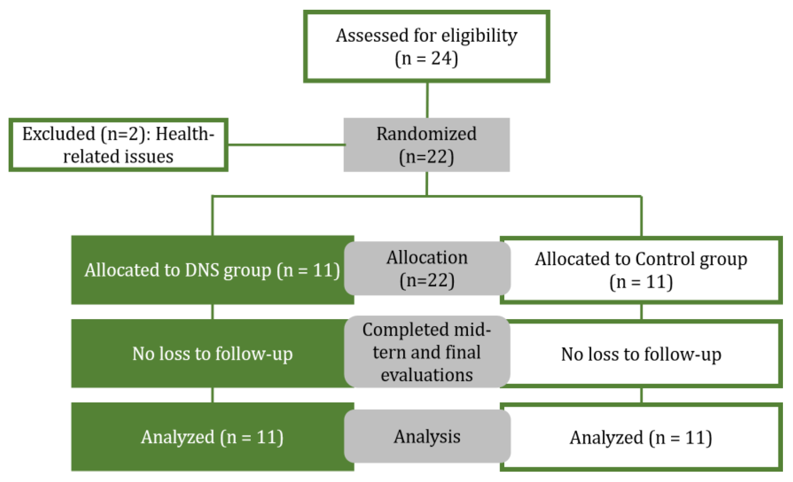
Figure 2.
(a) Affected-side single-hand support position; (b) Hand placement on the force plate during single-hand support; (c) Bilateral support position; (d) Hand placement on the force plate during bilateral support.
Figure 2.
(a) Affected-side single-hand support position; (b) Hand placement on the force plate during single-hand support; (c) Bilateral support position; (d) Hand placement on the force plate during bilateral support.
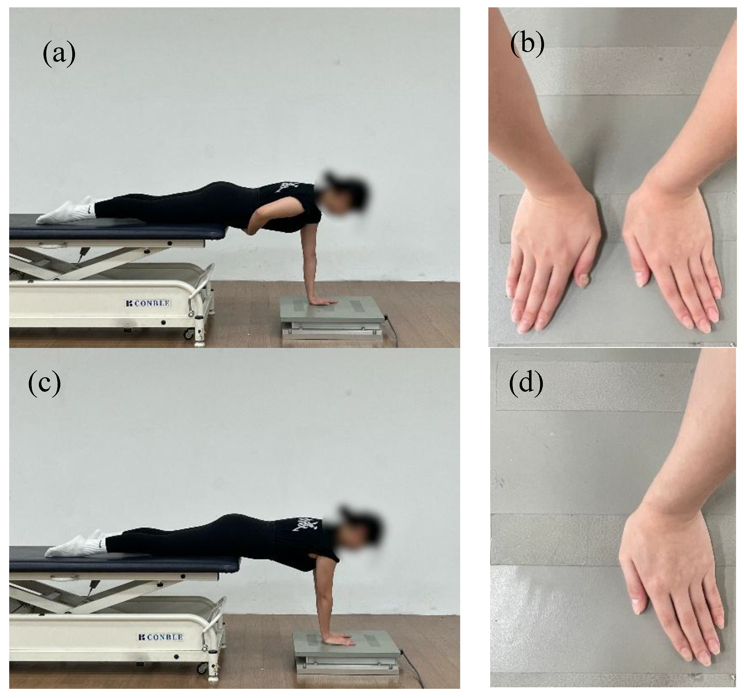
Figure 3.
DNS exercise : (a) Prone and quadruped rocking; (b) Bear position; (c) Contralateral hip extension in quadruped; (d) Prone position with head and chest elevation; (e) Oblique sitting with trunk rotation; (f) Three-point transition to tall kneeling with contralateral reach.
Figure 3.
DNS exercise : (a) Prone and quadruped rocking; (b) Bear position; (c) Contralateral hip extension in quadruped; (d) Prone position with head and chest elevation; (e) Oblique sitting with trunk rotation; (f) Three-point transition to tall kneeling with contralateral reach.
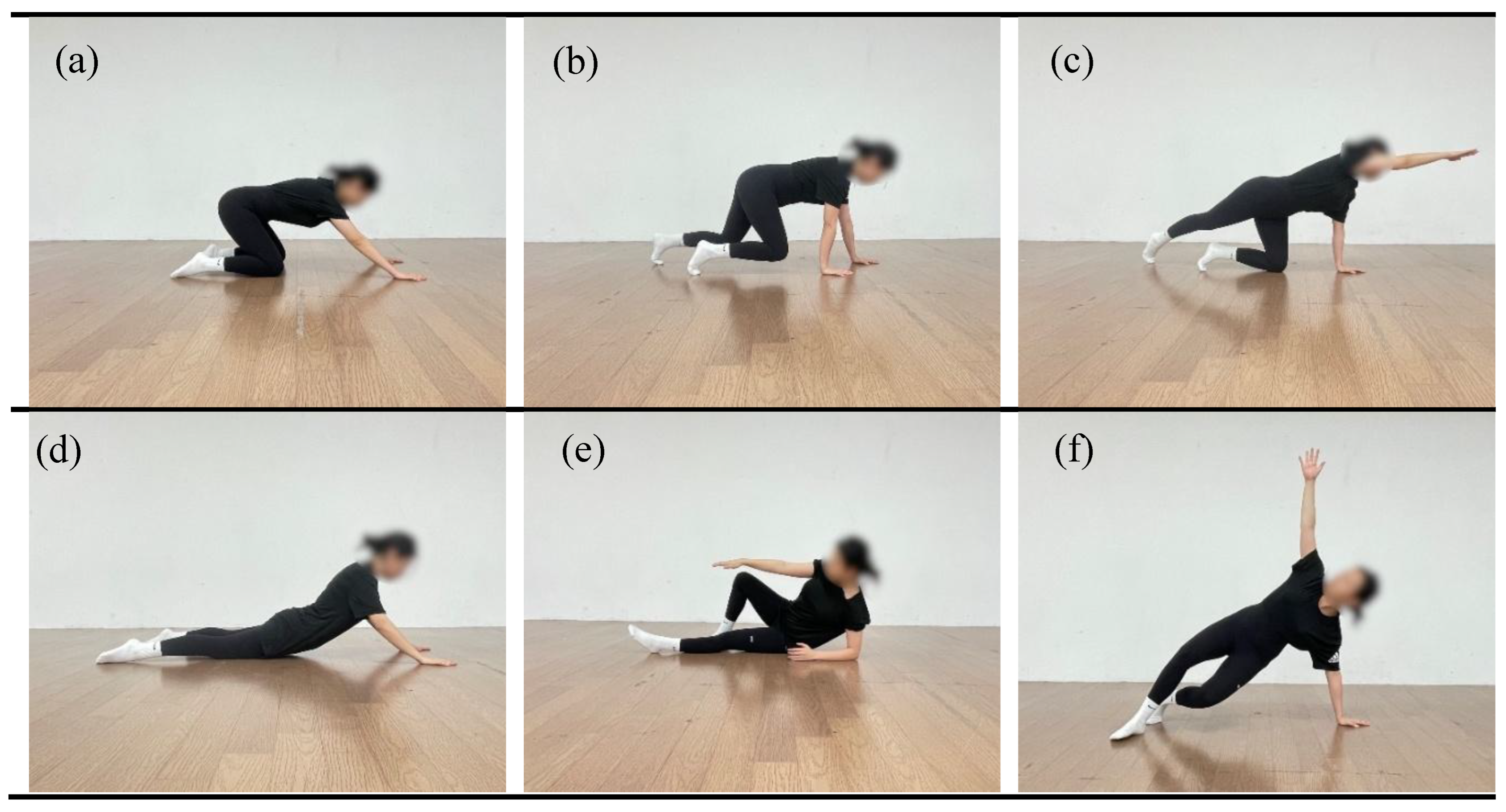

Table 1.
Participant characteristics summary (n=22).
| DNSG (n = 11) | CG (n = 11) | |
|---|---|---|
| Age (years) | 54.63 ± 4.27 | 60.09 ± 2.34 |
| Height (cm) | 161.42 ± 6.08 | 156.20 ± 3.19 |
| Weight (kg) | 56.66 ± 3.87 | 57.99 ± 12.97 |
| BMI (kg/m²) | 21.82 ± 2.28 | 23.73 ± 5.03 |
Note. Values are presented as mean ± standard deviation. DNSG, DNS group; CG: Control group.
Table 2.
Dynamic neuromuscular stabilization training program.
| Training | DNSG | CG | Time/Reps |
|---|---|---|---|
| Warm-up | 1. Supine breathing 2. Supine cross-pattern activation |
1. Standing shoulder circles 2. Arm swing (forward/backward & lateral) 3. Upper thoracic extension stretch (wall-supported) 4. Shoulder internal rotation stretch 5. Cross-body posterior capsule stretch |
10min /8reps |
| Exercise | 1. Side-lying rolling 2. Prone & quadruped rocking 3. Prone position with head and chest elevation 4. Contralateral hip extension in quadruped 5. Transition from quadruped to oblique sitting via lower extremity loading 6. 3-Point transition to tall kneeling with contralateral reach 7. Quadruped Locomotion Pattern 8. Transition from Quadruped to High Side Support 9. Bear 10. Bear with alternating upper limb unloading |
1. Shoulder external & internal rotation (standing / supine) 2. Scapular retraction exercise 3. Scapular protraction/elevation control (“Y” & “T”) 4. Quadruped push-up plus ("Camel") 5. Lawn mower pull (bodyweight version) 6. Doorway pectoral stretch 7. Supine cane flexion (without equipment) 8. Cross-body shoulder stretch |
35min /6reps -8 reps |
| Cool-down | 1. Resting position for deep stabilization 2. Prone resting with diaphragmatic control |
1. Repeat upper thoracic stretch 2. Arm and shoulder swing |
5min /8reps |
Note. DNSG, DNS group; CG: Control group.
Table 3.
Changes in COP parameters under eyes-open affected-side single-hand support condition.
| Task | Variables | Group | 0 Weeks | 4 Weeks | 8 Weeks | Source | p | ηp2 | Post Hoc |
|---|---|---|---|---|---|---|---|---|---|
| EO-AH | AP-Distance (mm) | DNSG | 86.62±14.08 | 66.59±11.36 | 49.44±8.96 | Group × Time | 0.048 | 0.160 | 0–4weeks: 0.013 |
| 4–8weeks: 0.000 | |||||||||
| 0–8weeks: 0.000 | |||||||||
| CG | 86.84±10.16 | 61.39±5.45 | 58.75±7.79 | Time | 0.000 | 0.799 | 0–4weeks: 0.000 |
||
| 4–8weeks: 0.555 | |||||||||
| 0–8weeks: 0.000 | |||||||||
| AP-velocity (mm/s) | DNSG | 2.88±0.46 | 2.21±0.37 | 1.71±0.28 | Group × Time | 0.041 | 0.168 | 0–4weeks: 0.013 |
|
| 4–8weeks: 0.001 | |||||||||
| 0–8weeks: 0.000 | |||||||||
| CG | 2.83±0.39 | 1.96±0.37 | 1.97±0.32 | Time | 0.000 | 0.770 | 0–4weeks: 0.001 |
||
| 4–8weeks: 1.000 | |||||||||
| 0–8weeks: 0.001 | |||||||||
| AP-RMS (mm) | DNSG | 0.21±0.02 | 0.15±0.01 | 0.12±0.01 | Group × Time | 0.233 | 0.071 | 0–4weeks: 0.001 |
|
| 4–8weeks: 0.005 | |||||||||
| 0–8weeks: 0.000 | |||||||||
| CG | 0.21±0.05 | 0.14±0.02 | 0.14±0.04 | Time | 0.000 | 0.742 | 0–4weeks: 0.001 |
||
| 4–8weeks: 1.000 | |||||||||
| 0–8weeks: 0.003 | |||||||||
| ML-Distance (mm) | DNSG | 87.24±13.57 | 69.55±10.65 | 57.81±13.82 | Group × Time | 0.238 | 0.069 | 0–4weeks: 0.016 |
|
| 4–8weeks: 0.002 | |||||||||
| 0–8weeks: 0.002 | |||||||||
| CG | 84.75±20.41 | 58.96±11.58 | 57.59±12.38 | Time | 0.000 | 0.688 | 0–4weeks: 0.001 |
||
| 4–8weeks: 1.000 | |||||||||
| 0–8weeks: 0.000 | |||||||||
| ML-velocity (mm/s) | DNSG | 2.90±0.45 | 2.31±0.35 | 1.91±0.30 | Group × Time | 0.030 | 0.161 | 0–4weeks: 0.016 |
|
| 4–8weeks: 0.020 | |||||||||
| 0–8weeks: 0.000 | |||||||||
| CG | 2.82±0.31 | 2.02±0.33 | 2.12±0.15 | Time | 0.000 | 0.706 | 0–4weeks: 0.000 |
||
| 4–8weeks: 0.947 | |||||||||
| 0–8weeks: 0.000 | |||||||||
| ML-RMS (mm) | DNSG | 0.20±0.03 | 0.17±0.03 | 0.13±0.02 | Group × Time | 0.009 | 0.208 | 0–4weeks: 0.000 |
|
| 4–8weeks: 0.002 | |||||||||
| 0–8weeks: 0.000 | |||||||||
| CG | 0.20±0.02 | 0.16±0.01 | 0.15±0.02 | Time | 0.000 | 0.840 | 0–4weeks: 0.000 |
||
| 4–8weeks: 0.081 | |||||||||
| 0–8weeks: 0.000 |
Note: Data are presented as mean ± standard deviation.DNSG, DNS group; CG: Control group.p-values are based on mixed-design two-way ANOVA.Post hoc comparisons were adjusted using the Bonferroni correction. ηp² = Partial eta squared (effect size): small = 0.01, medium = 0.06, large = 0.14.AP = anterior–posterior; ML = medial–lateral.EO: eyes open; AH: affected hand.
Table 4.
Changes in COP parameters under eyes-open bilateral support condition.
| Task | Variables | Group | 0 Weeks | 4 Weeks | 8 Weeks | Source | p | ηp2 | Post Hoc |
| EO-BH | AP-Distance (mm) | DNSG | 58.32±9.14 | 49.65±8.24 | 40.35±7.04 | Group × Time | 0.730 | 0.016 | 0–4weeks: 0.103 |
| 4–8weeks: 0.000 | |||||||||
| 0–8weeks: 0.001 | |||||||||
| CG | 57.87±13.51 | 46.13±11.86 | 38.95±5.88 | Time | 0.000 | 0.688 | 0–4weeks: 0.001 |
||
| 4–8weeks: 0.116 | |||||||||
| 0–8weeks: 0.000 | |||||||||
| AP-velocity (mm/s) | DNSG | 1.94±0.30 | 1.58±0.21 | 1.23±0.18 | Group × Time | 0.029 | 0.189 | 0–4weeks: 0.010 |
|
| 4–8weeks: 0.000 | |||||||||
| 0–8weeks: 0.000 | |||||||||
| CG | 1.89±0.52 | 1.55±0.36 | 1.56±0.26 | Time | 0.000 | 0.601 | 0–4weeks: 0.001 |
||
| 4–8weeks: 0.942 | |||||||||
| 0–8weeks: 0.001 | |||||||||
| AP-RMS (mm) | DNSG | 0.17±0.03 | 0.12±0.02 | 0.09±0.01 | Group × Time | 0.590 | 0.026 | 0–4weeks: 0.009 |
|
| 4–8weeks: 0.000 | |||||||||
| 0–8weeks: 0.000 | |||||||||
| CG | 0.20±0.04 | 0.14±0.04 | 0.13±0.02 | Time | 0.000 | 0.697 | 0–4weeks: 0.001 |
||
| 4–8weeks: 0.936 | |||||||||
| 0–8weeks: 0.001 | |||||||||
| ML-Distance (mm) | DNSG | 65.19±13.62 | 45.25±6.79 | 37.86±4.30 | Group × Time | 0.754 | 0.010 | 0–4weeks: 0.003 |
|
| 4–8weeks: 0.001 | |||||||||
| 0–8weeks: 0.000 | |||||||||
| CG | 67.20±11.37 | 45.31±8.75 | 40.83±3.60 | Time | 0.000 | 0.782 | 0–4weeks: 0.000 |
||
| 4–8weeks: 0.371 | |||||||||
| 0–8weeks: 0.000 | |||||||||
| ML-velocity (mm/s) | DNSG | 2.17±0.45 | 1.50±0.22 | 1.33±0.22 | Group × Time | 0.557 | 0.029 | 0–4weeks: 0.003 |
|
| 4–8weeks: 0.189 | |||||||||
| 0–8weeks: 0.000 | |||||||||
| CG | 2.22±0.74 | 1.35±0.34 | 1.23±0.24 | Time | 0.000 | 0.719 | 0–4weeks: 0.001 |
||
| 4–8weeks: 0.889 | |||||||||
| 0–8weeks: 0.001 | |||||||||
| ML-RMS (mm) | DNSG | 0.18±0.03 | 0.15±0.04 | 0.09±0.00 | Group × Time | 0.000 | 0.326 | 0–4weeks: 0.127 |
|
| 4–8weeks: 0.001 | |||||||||
| 0–8weeks: 0.000 | |||||||||
| CG | 0.17±0.03 | 0.12±0.02 | 0.12±0.02 | Time | 0.000 | 0.737 | 0–4weeks: 0.000 |
||
| 4–8weeks: 1.000 | |||||||||
| 0–8weeks: 0.000 |
Note: Data are presented as mean ± standard deviation.DNSG, DNS group; CG: Control group.p-values are based on mixed-design two-way ANOVA.Post hoc comparisons were adjusted using the Bonferroni correction. ηp² = Partial eta squared (effect size): small = 0.01, medium = 0.06, large = 0.14.AP = anterior–posterior; ML = medial–lateral.EO: eyes open; BH: bilateral hand support.
Table 5.
Changes in COP parameters under eyes-closed bilateral support condition.
| Task | Variables | Group | 0 Weeks | 4 Weeks | 8 Weeks | Source | p | ηp2 | Post Hoc |
|---|---|---|---|---|---|---|---|---|---|
| EC-BH | AP-Distance (mm) | DNSG | 59.25±9.00 | 50.88±8.38 | 42.04±6.98 | Group × Time | 0.562 | 0.028 | 0–4weeks: 0.157 |
| 4–8weeks: 0.008 | |||||||||
| 0–8weeks: 0.000 | |||||||||
| CG | 57.10±6.05 | 46.25±8.41 | 41.64±6.44 | Time | 0.000 | 0.636 | 0–4weeks: 0.002 |
||
| 4–8weeks: 0.453 | |||||||||
| 0–8weeks: 0.000 | |||||||||
| AP-velocity (mm/s) | DNSG | 1.97±0.30 | 1.62±0.31 | 1.33±0.21 | Group × Time | 0.040 | 0.166 | 0–4weeks: 0.024 |
|
| 4–8weeks: 0.002 | |||||||||
| 0–8weeks: 0.000 | |||||||||
| CG | 1.96±0.30 | 1.54±0.36 | 1.53±0.26 | Time | 0.000 | 0.736 | 0–4weeks: 0.001 |
||
| 4–8weeks: 1.000 | |||||||||
| 0–8weeks: 0.000 | |||||||||
| AP-RMS (mm) | DNSG | 0.15±0.02 | 0.12±0.01 | 0.10±0.02 | Group × Time | 0.382 | 0.047 | 0–4weeks: 0.007 |
|
| 4–8weeks: 0.012 | |||||||||
| 0–8weeks: 0.000 | |||||||||
| CG | 0.18±0.03 | 0.14±0.01 | 0.12±0.01 | Time | 0.000 | 0.688 | 0–4weeks: 0.003 |
||
| 4–8weeks: 0.090 | |||||||||
| 0–8weeks: 0.002 | |||||||||
| ML-Distance (mm) | DNSG | 65.97±9.61 | 45.27±6.48 | 39.53±6.35 | Group × Time | 0.877 | 0.007 | 0–4weeks: 0.000 |
|
| 4–8weeks: 0.015 | |||||||||
| 0–8weeks: 0.000 | |||||||||
| CG | 67.70±8.67 | 45.07±9.68 | 40.94±8.07 | Time | 0.000 | 0.832 | 0–4weeks: 0.000 |
||
| 4–8weeks: 0.588 | |||||||||
| 0–8weeks: 0.000 | |||||||||
| ML-velocity (mm/s) | DNSG | 2.25±0.20 | 1.51±0.20 | 1.12±0.15 | Group × Time | 0.045 | 0.164 | 0–4weeks: 0.000 |
|
| 4–8weeks: 0.000 | |||||||||
| 0–8weeks: 0.000 | |||||||||
| CG | 2.24±0.51 | 1.53±0.41 | 1.55±0.34 | Time | 0.000 | 0.829 | 0–4weeks: 0.001 |
||
| 4–8weeks: 1.000 | |||||||||
| 0–8weeks: 0.001 | |||||||||
| ML-RMS (mm) | DNSG | 0.16±0.02 | 0.13±0.03 | 0.09±0.01 | Group × Time | 0.002 | 0.273 | 0–4weeks: 0.001 |
|
| 4–8weeks: 0.001 | |||||||||
| 0–8weeks: 0.000 | |||||||||
| CG | 0.15±0.02 | 0.13±0.03 | 0.13±0.04 | Time | 0.000 | 0.584 | 0–4weeks: 0.317 |
||
| 4–8weeks: 1.000 | |||||||||
| 0–8weeks: 0.214 |
Note: Data are presented as mean ± standard deviation.DNSG, DNS group; CG: Control group.p-values are based on mixed-design two-way ANOVA.Post hoc comparisons were adjusted using the Bonferroni correction. ηp² = Partial eta squared (effect size): small = 0.01, medium = 0.06, large = 0.14.AP = anterior–posterior; ML = medial–lateral.EC: eyes closed; BH: bilateral hand support.
Table 6.
Changes in VAS according to time and side.
| Task | Group | 0 Weeks | 4 Weeks | 8 Weeks | Source | p | ηp2 |
|---|---|---|---|---|---|---|---|
| AH | DNSG | 6.27±1.42 | 3.63±1.62 | 1.90±1.37 | Group × Time | 0.186 | 0.081 |
| CG | 5.36±0.80 | 3.72±1.00 | 2.18±1.60 | Time | 0.000 | 0.756 |
Note: Data are presented as mean ± standard deviation.DNSG, DNS group; CG: Control group.p-values are based on mixed-design two-way ANOVA.Post hoc comparisons were adjusted using the Bonferroni correction. ηp2: Partial eta squared (small=0.01, medium=0.06, large=0.14).AP = anterior–posterior; ML = medial–lateral.AH: affected hand.
Disclaimer/Publisher’s Note: The statements, opinions and data contained in all publications are solely those of the individual author(s) and contributor(s) and not of MDPI and/or the editor(s). MDPI and/or the editor(s) disclaim responsibility for any injury to people or property resulting from any ideas, methods, instructions or products referred to in the content. |
© 2025 by the authors. Licensee MDPI, Basel, Switzerland. This article is an open access article distributed under the terms and conditions of the Creative Commons Attribution (CC BY) license (http://creativecommons.org/licenses/by/4.0/).
Copyright: This open access article is published under a Creative Commons CC BY 4.0 license, which permit the free download, distribution, and reuse, provided that the author and preprint are cited in any reuse.


